Tissue engineering composite stent for osteochondral repair and preparation method thereof
A tissue engineering and composite scaffold technology, applied in tissue regeneration, medical science, prosthesis, etc., can solve the problems of slow integration of the two interfaces, repair failure, instability, etc., and achieve beneficial technical effects, wide application prospects, good biological degradability effect
- Summary
- Abstract
- Description
- Claims
- Application Information
AI Technical Summary
Problems solved by technology
Method used
Image
Examples
preparation example Construction
[0025] Its concrete preparation method is:
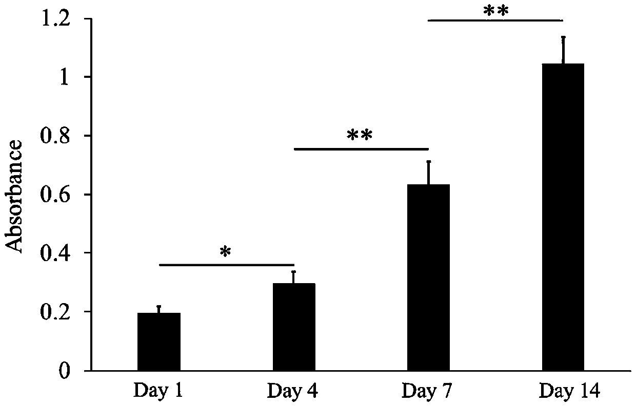
[0026] Step 1. Add the osteoblast suspension or stem cell suspension to the cell-adhesive porous scaffold material, incubate in a 37°C incubator for 0.5-2h, add the corresponding cell culture medium for 14-28 days, and replace it every other day A culture medium to obtain a cell-porous material complex;
[0027] The cell-adhesive porous scaffold material is a porous scaffold containing collagen, gelatin, fibronectin or cell-adhesive polypeptide; for example, gelatin sponge, hyaluronic acid-gelatin composite porous scaffold, fibronectin-based porous scaffold, and the like.
[0028] Osteoblasts are MC3T3-E1, hFOB1.19, etc.; stem cells are mesenchymal stem cells (MSC), embryonic stem cells (ESC) or induced pluripotent stem cells (iPSC), etc.;
[0029] Step 2, resuspending chondrocytes in a hydrogel macromer solution to obtain a cell suspension; wherein the hydrogel macromer is methacrylated hyaluronic acid, acrylated hyaluronic acid, ...
Embodiment 1
[0035] Step 1: Cut the gelatin porous material (gelatin sponge) into small pieces of 0.5cmx0.5cmx0.5cm, place them in a sterile petri dish, and irradiate with ultraviolet light overnight. MC3T3-E1 cells were trypsinized and resuspended in proliferation medium (DMEM plus 10% serum) at a cell density of 10 7 pieces / ml. Add the cell suspension evenly to the gelatin porous material that has been irradiated with UV rays before, squeeze the sponge with a pipette tip to make the gelatin porous material fully absorb the cell suspension, and put the gelatin porous material containing the cell suspension in a 37°C incubator for 2 hours , and then transferred to a 24-well plate; add osteoinductive medium (DMEM medium plus 10% serum, 100nM dexamethasone, 50ug / mL vitamin C, 10mM β-sodium glycerophosphate) for a total of 14 days, and change it every other day Culture solution to obtain MC3T3-E1-gelatin porous material complex;
[0036] Step 2, resuspending the P1 generation chondrocytes i...
Embodiment 2
[0041] Step 1: Cut the gelatin porous material (gelatin sponge) into small pieces of 0.5cmx0.5cmx0.5cm, place them in a sterile petri dish, and irradiate with ultraviolet light overnight. MC3T3-E1 cells were trypsinized and resuspended in proliferation medium (DMEM plus 10% serum) at a cell density of 10 7pieces / ml. The cell suspension was evenly added to the gelatin porous material that had been irradiated by UV light before, and the gelatin porous material containing the cell suspension was placed in a 37°C incubator for 0.5h, and then transferred to a 24-well plate; osteoinductive medium (DMEM 10% serum, 100nM dexamethasone, 50ug / mL vitamin C, 10mM β-sodium glycerol phosphate) were added to the culture medium for a total of 18 days, and the culture medium was changed every other day to obtain the MC3T3-E1-gelatin porous material complex;
[0042] Step 2, resuspending the P1 generation chondrocytes in a 2% (w / v) methacrylated hyaluronic acid solution containing a photoiniti...
PUM
 Abstract
Description
Claims
Application Information
 - R&D
- Intellectual Property
- Life Sciences
- Materials
- Tech Scout
- Unparalleled Data Quality
- Higher Quality Content
- 60% Fewer Hallucinations
Browse by: Latest US Patents, China's latest patents, Technical Efficacy Thesaurus, Application Domain, Technology Topic, Popular Technical Reports.
© 2025 PatSnap. All rights reserved.Legal|Privacy policy|Modern Slavery Act Transparency Statement|Sitemap|About US| Contact US: help@patsnap.com


