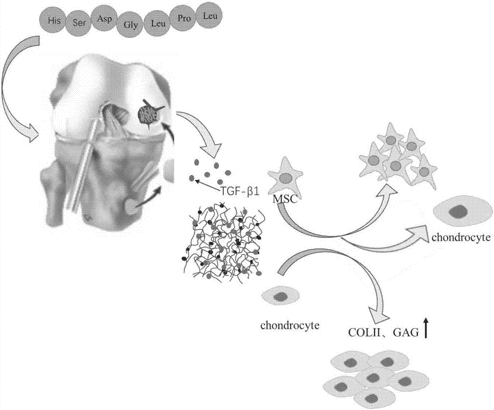
Preparation method of co-crosslinked double-network hydrogel support for promoting cartilage injury repair
A cartilage injury and co-crosslinking technology, used in medical science, prosthesis, tissue regeneration, etc., can solve the problems of decreased hydrogel strength and fast drug release, and can promote adhesion, promote cartilage defect healing, and enhance adsorption and aggregation. effect of function
- Summary
- Abstract
- Description
- Claims
- Application Information
AI Technical Summary
Problems solved by technology
Method used
Image
Examples
Embodiment 1
[0049] Example 1 Preparation of Co-crosslinked Double Network Hydrogel Scaffold for Cartilage Damage Repair
[0050] The specific steps are:
[0051] 1) Preparation of methacrylate-modified gelatin
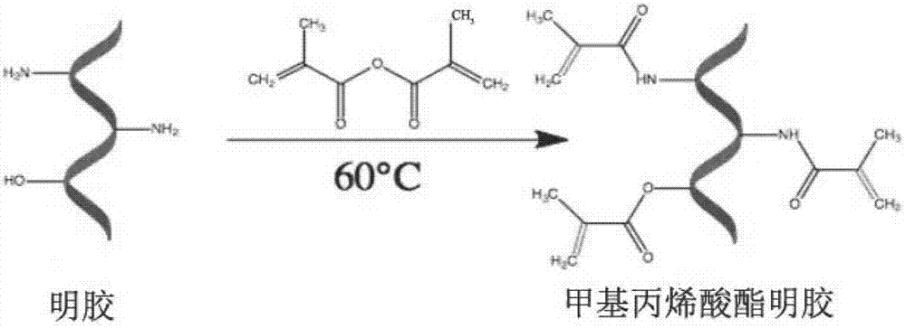
[0052] figure 2 This is the flow chart for the preparation of methacrylate-modified gelatin. Add 200 mL of phosphate buffer to 20 g of gelatin, and keep stirring at 60°C for 2 hours; Add 1mL methacrylic anhydride to the gelatin mixture every 4 minutes while stirring, add 16 times in total, and keep stirring for 2 hours to form methacrylate-modified gelatin; the prepared methacrylate-modified Place the gelatin solution in 800mL preheated phosphate buffer solution for dilution, and continue to stir slowly for 15 minutes, place the diluted solution in a dialysis bag, the molecular weight cut-off of the dialysis bag is 10KD, soak in pure aqueous solution, Change the dialysate twice a day to remove unreacted methacrylic anhydride, and continue dialysis for one week; after dialysis,...
Embodiment 2
[0059] Example 2 GelMA, GelMA-HSNGLPL biocompatibility detection and observation
[0060] Experimental grouping: GelMA scaffold group, GelMA-HSNGLPL scaffold group.
[0061] Put the GelMA and GelMA-HSNGLPL scaffolds into 96-well plates first, then resuspend hMSCs, count them, and plant them on the scaffolds in two sets of 96-well plates (2×10 per well 3 Cells), five replicate wells in each group, add the prepared DMEM-L medium, at 37°C, 5% CO 2 They were cultured in an incubator, and the DMEM-L medium was replaced every two days, and the cells were cultured for 5 days to stain live and dead cells and observe under a fluorescent microscope.
[0062] The staining steps for living and dead cells are: suck out the culture medium → wash twice with PBS → add 50 ul of the mixture to each well (1μL A + 1μL B + 2mL PBS to form the mixture) → place in the incubator for 30 minutes → under the fluorescence microscope Observed.
[0063] After the cells were stained in the dark for 30 m...
Embodiment 3
[0064] Example 3 Detection of hMSCs proliferation activity
[0065] Experimental groups: control group, GelMA stent group, GelMA-HSNGLPL stent group.
[0066] Resuspend hMSCs and seed into 96-well plates (3 plates, 2×10 per well 3 Cells), add DMEM-L medium or GelMA group or GelMA-HSNGLPL group leaching solution, and culture in an incubator until the cells fully adhere to the wall and grow. The 3 plates were tested for CCK-8 on the 1st, 3rd, 5th, and 7th day after cell attachment.
[0067] CCK-8 detection steps: Add 10ul CCK-8 reaction solution to the wells of the 96-well plate (3 groups, 3 replicate wells in each group) (avoid air bubbles in the wells) → incubate in an incubator for 2 hours → use a microplate reader Measure the absorbance (450nm).
[0068] CCK-8 detected the effect of GelMA and GelMA-HSNGLPL materials on the proliferation of hMSCs. The results show( Figure 8 ), GelMA, GelMA-HSNGLPL material leachate did not affect the growth of cells, and the material h...
PUM
 Abstract
Description
Claims
Application Information
 - R&D
- Intellectual Property
- Life Sciences
- Materials
- Tech Scout
- Unparalleled Data Quality
- Higher Quality Content
- 60% Fewer Hallucinations
Browse by: Latest US Patents, China's latest patents, Technical Efficacy Thesaurus, Application Domain, Technology Topic, Popular Technical Reports.
© 2025 PatSnap. All rights reserved.Legal|Privacy policy|Modern Slavery Act Transparency Statement|Sitemap|About US| Contact US: help@patsnap.com


