
Preparation method and use of poly(lactic-co-glycolic acid) (PLGA) microspheres as nucleic acid vaccine vectors

A nucleic acid vaccine and microsphere technology, applied in the field of vaccine carrier system and preparation, can solve the problems of limited size of loaded foreign genes, lack of tissue specificity, low gene transfer efficiency, etc., and achieves convenient preparation, strong versatility, The effect of simple ingredients

Inactive Publication Date: 2012-06-06
JILIN UNIV
View PDF0 Cites 37 Cited by
  • Summary
  • Abstract
  • Description
  • Claims
  • Application Information

AI Technical Summary

Problems solved by technology

Viral vectors have many defects, such as immunogenicity and cytotoxicity, lack of tissue specificity, limitations on loading foreign gene size, potential tumorigenicity, and the ability to produce active virus particles during recombination.
Most of the traditional non-viral vectors are derivatives of polycationic polymers and liposomes, the biggest defect of which is the low efficiency of gene transfer, especially in vivo

Method used


Image

  • Preparation method and use of poly(lactic-co-glycolic acid) (PLGA) microspheres as nucleic acid vaccine vectors
  • Preparation method and use of poly(lactic-co-glycolic acid) (PLGA) microspheres as nucleic acid vaccine vectors
  • Preparation method and use of poly(lactic-co-glycolic acid) (PLGA) microspheres as nucleic acid vaccine vectors

Examples


Embodiment 1

[0041] Preparation of PLGA magnetic microspheres

[0042] FeCl 3 and FeCl 2 The solution is mixed according to the molar ratio of 1:2 (Fe 2+ Slight excess), under nitrogen atmosphere, after 30min in 60°C water bath, quickly add a certain amount of ammonia water to the mixture under vigorous stirring, and continue stirring for 30min under nitrogen atmosphere. Stir. After the reaction is completed, use a commercial permanent magnet to absorb the generated Fe 3 o 4 For black particles, pour off the supernatant and wash with high-purity water several times. Its reaction equation: Fe 2+ +2Fe 3+ +8OH - → Fe 3 o 4 +4H 2 o

[0043] Add an aqueous solution containing a polymer (PEI, PLGA, chitosan or dextran) to the obtained magnetic fluid precipitate, resuspend the magnetic fluid, place it for 30 minutes, and then magnetically separate. After repeating 3 times, dilute in PBS with pH 7.2 and set aside.

Embodiment 2

[0045] Characterization of PLGA magnetic microspheres

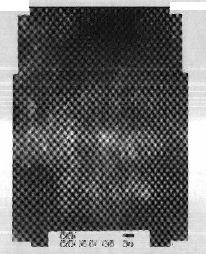
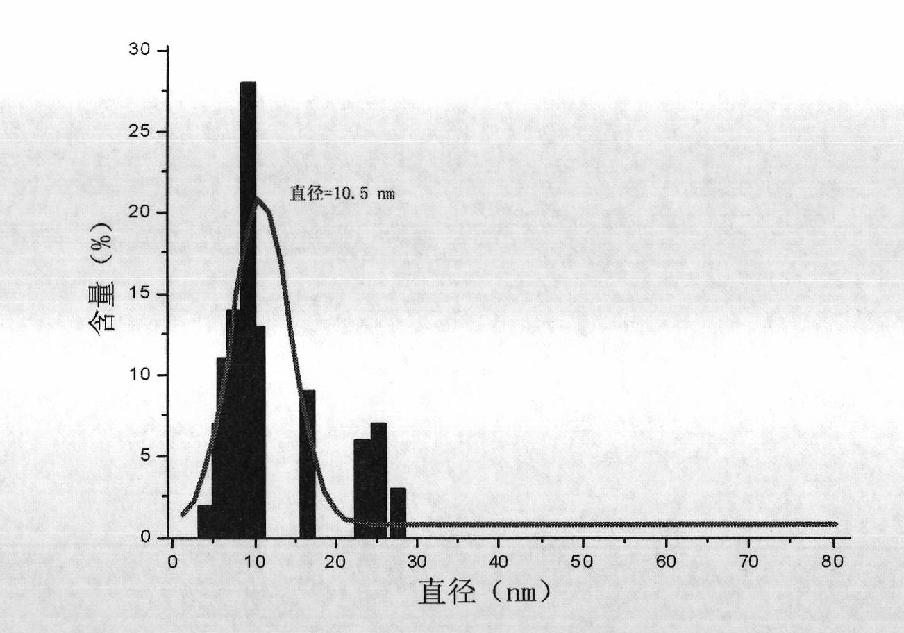
[0046] Fix the pH value and ionic strength of the system, and use the method of rapidly dropping ammonia water to ensure that Fe 3 o 4 The rapid nucleation and stable growth of nanoparticles are beneficial to reduce the size distribution of nanoparticles. The morphology of nanoparticles can be observed and their size can be determined by transmission electron microscopy (TEM).

[0047] like figure 2 As shown, Fe 3 o 4 Nanoparticles have good monodispersity, and the particle size distribution conforms to Gaussian distribution;

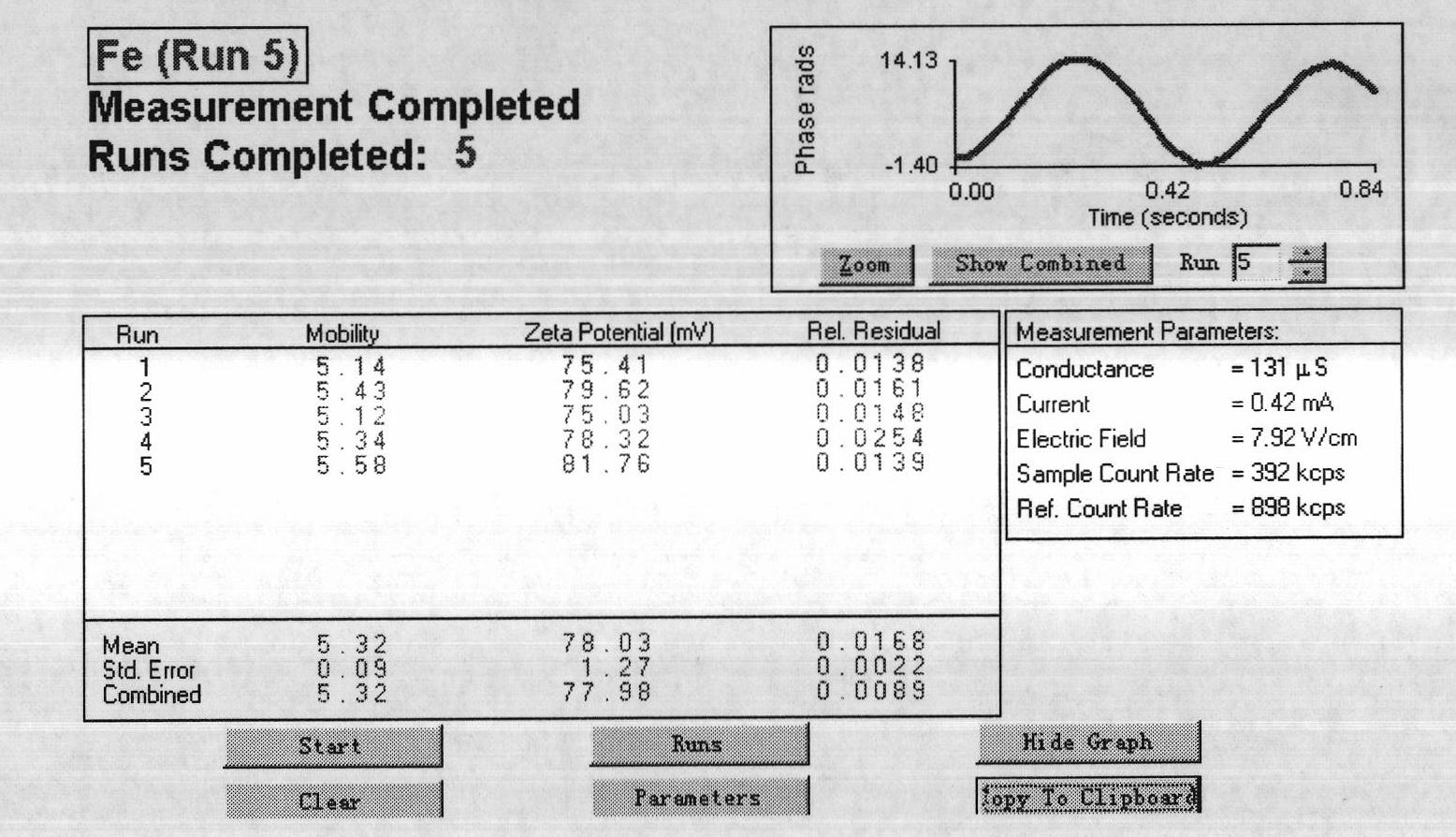
[0048] like image 3 As shown, Fe 3 o 4 The surface potential of nanoparticles has a high positive charge, which can bind and concentrate genetic vaccines.

Embodiment 3

[0050] Gene Vaccine Binding Experiment of PLGA Magnetic Microspheres

[0051] In a fixed buffer system (1×TAE), the quality of the fixed DNA vaccine is constant, and magnetic nanoparticles of different masses (w / w=0.25, 0.5~3.0, 3.5, 4.0) are added to it, and after standing at 4°C for 30 minutes, The results of agarose gel electrophoresis were used to screen for the best (magnetic particle / DNA) ratio. When the magnetic nanoparticles / DNA (w / w) is greater than 3:1, the DNA is completely blocked in the sample hole (such as Figure 4 ). By carrying out electrophoresis analysis to DNA and using computer simulation, the in vitro binding curve (such as Figure 5 ).


PUM

PropertyMeasurementUnit
particle sizeaaaaaaaaaa

Abstract

The invention discloses a preparation method and a use of poly(lactic-co-glycolic acid) (PLGA) microspheres as nucleic acid vaccine vectors. A result of an animal immunization experiment shows that the PLGA microspheres can be utilized as gene vaccine vectors. Principles of the PLGA microspheres comprise that 1, the PLGA microspheres have core-shell structures; surface polymers comprise polymine, PLGA, glucose, chitosan, polylysine, FeCl3 and FeCl2; and through static electricity, dewatering interaction and hydrogen bond-nucleic acid vaccine interaction, a nucleic acid vaccine is concentrated to form a compact nucleic acid vaccine so that nucleic acid vaccine degradation is reduced in vivo; 2, the PLGA microspheres have magnetism and thus after immunization injection, in a strong magnetic field, the distribution of the PLGA microspheres in muscular tissue is improved and the defect of limited contact between the PLGA microspheres and target cells is overcome; and 3, through long-term strong magnetic field induction, a magnetic PLGA microsphere / nucleic acid vaccine complex can enter into the skin; and because of rich antigen presenting cells in the skin, a strong and fast immune response can be induced.

Description

technical field [0001] The invention relates to the field of biotechnology immune preparations, to a vaccine carrier system, preparation method and application, in particular to using PLGA microspheres as gene vaccine carriers to induce strong humoral and cellular immune responses for the prevention and treatment of infectious diseases. Background technique [0002] Gene vaccines can induce both humoral immunity and cellular immunity, which is called the third revolution of vaccines. Genetic vaccines have been used to elicit protective antibodies and cell-mediated immune responses in preclinical animal models of viruses, bacteria, and parasites, and have been tested in the prevention and treatment of cancer, allergy, and autoimmune diseases. Research. However, naked DNA is unstable in and out of cells and in vivo, and is easily degraded by nucleases; its inherent negative charge limits contact with negatively charged cell membranes for internalization. As a result, the imm...

Claims


Application Information

Patent Timeline
Patent Type & Authority Applications(China)
IPC IPC(8): A61K47/34A61K47/36A61K47/04A61K48/00A61K39/00A61K39/002A61K39/02A61K39/12A61K9/10A61K9/16A61P31/04A61P31/12A61P33/02A61P35/00A61P31/18
CPCY02A50/30
Inventor 孔维吴永革陈妍周现峰
Owner JILIN UNIV
Learn More