Photochemical therapy to affect mechanical and/or chemical properties of body tissue
A tissue and compound technology, applied in the direction of drug devices, drug devices, other medical devices, etc.
- Summary
- Abstract
- Description
- Claims
- Application Information
AI Technical Summary
Problems solved by technology
Method used
Image
Examples
Embodiment 1
[0166] Following the teachings of US Published Application No. 20050271590, the present inventors used Eosin Y / TEOA as a selective photoinitiator for PEGDM crosslinking for reinforcement of scleral tissue. It was surprisingly found that Eosin Y / TEOA was comparable effective in enhancing the strength of scleral tissue in the absence of the target crosslinker (eg PEGDM). It is particularly unexpected that Type II photoinitiators are able to strengthen the sclera to a therapeutically useful extent with irradiation times of about 30 minutes or less. Without wishing to be bound by any theory, it is believed that Eosin Y may be sufficient to promote the direct cross-linking of collagen and other scleral components to directly increase the strength of scleral tissue. While this does not rule out the co-action of cross-linkers such as PEGDM, the results for Eosin Y suggest that simpler formulations are possible utilizing reagents already approved by the US Food and Drug Administration...
Embodiment 2
Dose response and mechanical properties of treated tissues
[0167] A number of different techniques have been used to initially assess scleral tissue enhancement, including tensile testing, oscillatory shear measurements, and eye dilation analysis discussed more fully below.
[0168] The ability of the initiators with and without crosslinking compounds to strengthen the sclera was initially assessed using rheological analysis of tissue sections. Scleral sections (8 mm diameter) were excised from porcine eyes and tested on a TA Instruments AR1000 rheometer (using a tethered parallel plate geometry) to quantify the change in modulus before and after treatment. Two example photoinitiators were chosen for initial evaluation. 2959 (2-Hydroxy-1-[4-(2-hydroxyethoxy)phenyl]-2-methyl-1-propanone and Eosin Y with triethanolamine (Eosin Y / TEOA). 2959 (12959) is a UV photoactivatable photoinitiator that has shown low toxicity in cell encapsulation studies. Eosin Y / TEOA was chosen a...
Embodiment 3
Eye shape in an inflated model of myopia and keratoconus
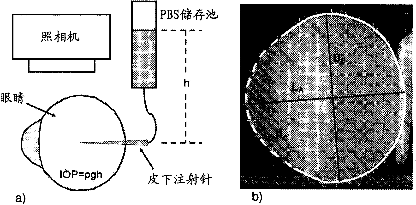
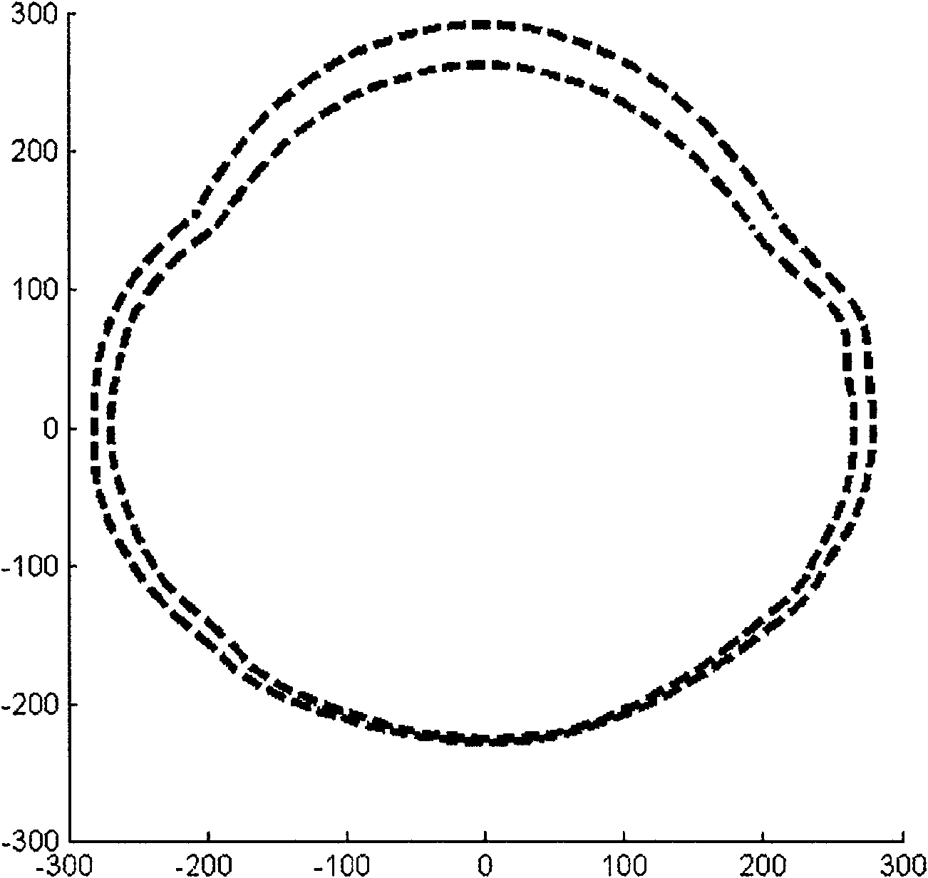
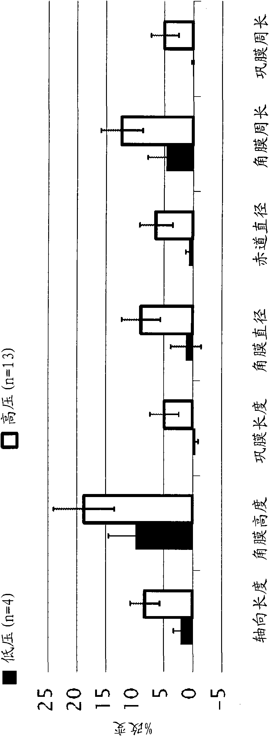
[0169] Stabilization of eye shape was assessed in vitro using pairs of eyes enucleated from young (1-2 weeks) New Zealand white rabbits, one eye being treated and the companion eye serving as a control. In order to simulate and accelerate the loading geometry that exists in vivo, intact eyeballs are mechanically tested by imposing elevated intraocular pressure: a syringe connects the intraocular fluid to the elevated reservoir of Dulbecco's (DPBS), while the exterior of the eye is immersed in the In DPBS ( figure 1 ). Eyes enucleated from young rabbits provide a model of expandable corneal and scleral tissue: when subjected to elevated intraocular pressure in vitro, the cornea progressively bulges outward and the scleral wall expands ( figure 2 and 3 ).
[0170] Eyeball size and shape were recorded over time from two perpendicular viewpoints. To prepare the eye for testing, the epithelium is removed from the co...
PUM
 Abstract
Description
Claims
Application Information
 - R&D
- Intellectual Property
- Life Sciences
- Materials
- Tech Scout
- Unparalleled Data Quality
- Higher Quality Content
- 60% Fewer Hallucinations
Browse by: Latest US Patents, China's latest patents, Technical Efficacy Thesaurus, Application Domain, Technology Topic, Popular Technical Reports.
© 2025 PatSnap. All rights reserved.Legal|Privacy policy|Modern Slavery Act Transparency Statement|Sitemap|About US| Contact US: help@patsnap.com


