
Agents for the molecular imaging of serine-protease in human pathologies

a technology of molecular imaging and serine proteases, which is applied in the direction of peptides, enzyme stabilisation, therapy, etc., can solve the problems of irreversible inhibitors not possessing the required parameters, and no investigation has been carried out to use irreversible ligands for the molecular imaging of serine proteases

Inactive Publication Date: 2016-03-01
PHARMALEADS INC +3
View PDF7 Cites 0 Cited by
  • Summary
  • Abstract
  • Description
  • Claims
  • Application Information

AI Technical Summary

Benefits of technology

[0011]The inventors have indeed demonstrated that peptide irreversible ligands of the selected serine proteases can be used for the molecular imaging of these serine proteases in physiological and physiopathological conditions by introducing at the N-terminal position of the irreversible cmk peptide ligand a detectable moiety (DOTA or DTPA) via a small spacer, in order to minimize a possible steric hindrance between these two moieties.
[0042]SPECT and PET acquire information on the concentration of radionuclides introduced into a subject's body. PET generates images by detecting pairs of gamma rays emitted indirectly by a positron-emitting radionuclide. A PET analysis results in a series of thin slice images of the body over the region of interest (e.g., brain, breast, liver). These thin slice images can be assembled into a three dimensional representation of the examined area. SPECT is similar to PET, but the radioactive substances used in SPECT have longer decay times than those used in PET and emit single instead of double gamma rays. Although SPECT images exhibit less sensitivity and are less detailed than PET images, the SPECT technique is much less expensive than PET and offers the advantage of not requiring the proximity of a particle accelerator. Planar scintigraphy (PS) is similar to SPECT in that it uses the same radionuclides. However, PS only generates 2D-information.
[0058]These methods can also be used to monitor the response of a patient to a treatment for a pathological condition associated with serine proteases selected from the group consisting of leukocyte elastase, thrombin, tissue plasminogen activator (t-PA) and plasmin. For example, an image of part of the patient's body that contains tissue “abnormal” serine proteases is generated before and after submitting the patient to a treatment. Comparison of the “before” and “after” images allows the response of the patient to that particular treatment to be monitored.

Problems solved by technology

Consequently, due to the restricted selectivity of the P1 residue, some of these irreversible inhibitors do not possess the required parameters of affinity and selectivity needed for a specific labelling of each protease.
Moreover, no investigations have been carried out to use these irreversible ligands for the molecular imaging of serine proteases.

Method used


Image

  • Agents for the molecular imaging of serine-protease in human pathologies
  • Agents for the molecular imaging of serine-protease in human pathologies
  • Agents for the molecular imaging of serine-protease in human pathologies

Examples


example 1

Design of a Peptide Ligand for Plasmin Molecular Imaging

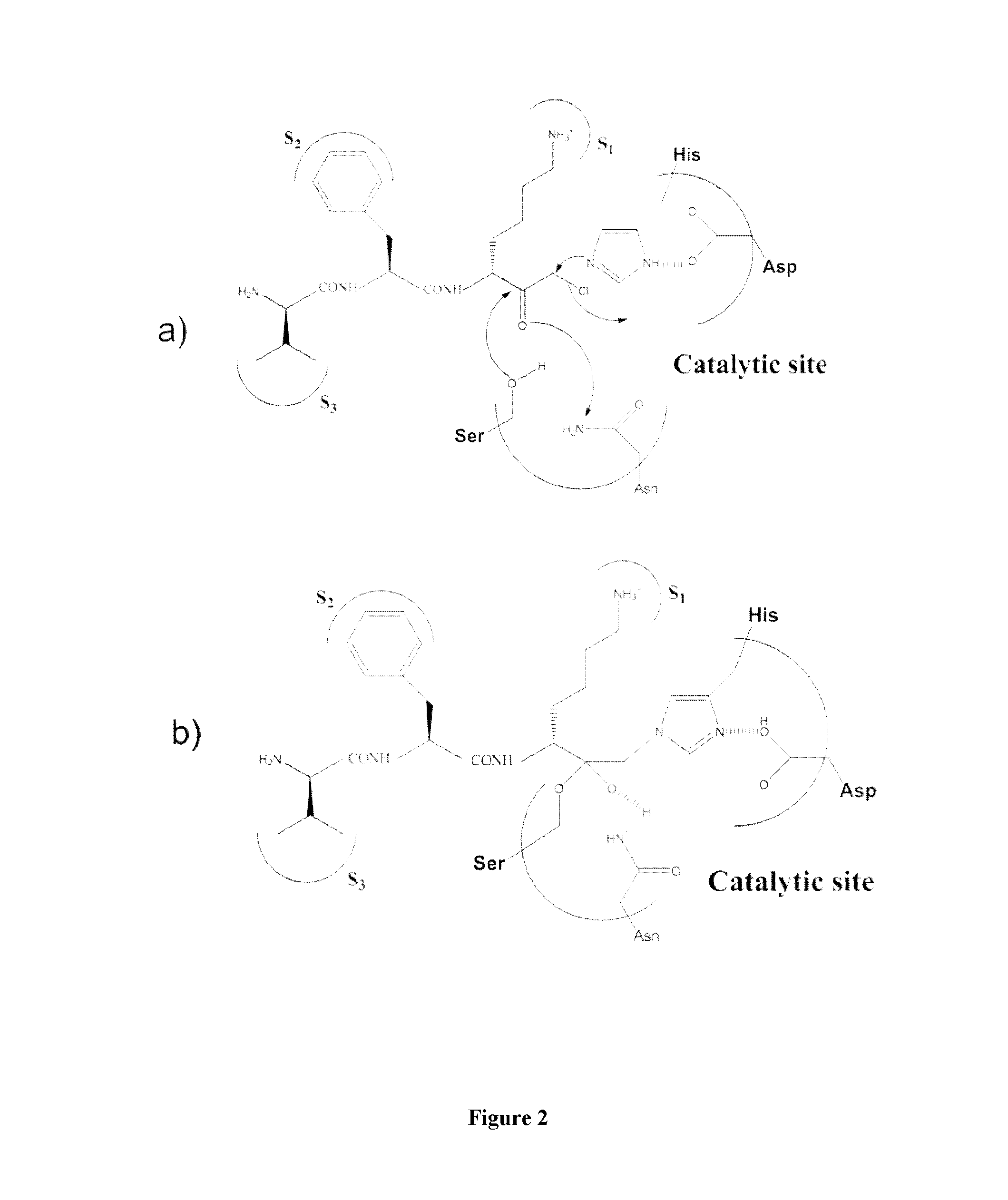
[0082]Plasmin catalyses the cleavage of Lys-X or Arg-X bonds with specificity similar to that of trypsin, but with a lesser efficiency for hydrolysis of these bonds in proteins. In this family of proteases the selectivity of the enzyme is essentially ensured by the amino acid interacting with the S1 subsite and, consequently plasmin is able to cleave esters or amides of Lys and Arg as well as small peptide substrate recognizing the S3 to S1 subsites of the enzyme and containing a C-terminal lysine or arginine {Lottenberg, 1981}. Likewise, small three amino acids peptide analogues, such as leupeptin (Ac-Leu-Leu-Arg-H), are efficient competitive inhibitors with Ki values in the 10−7 molar range {Chi, 1989}.

[0083]However, to obtain an efficient labeling of the peptidase for molecular imaging, an irreversible inhibitor has been chosen as starting material for the design of such a marker. Indeed, the replacement of the C-terminal ca...

example 2

In Vitro Ligation of the Plasmin Active Site by PL704

[0105]Material and Methods:

[0106]Determination of Plasmin Activity:

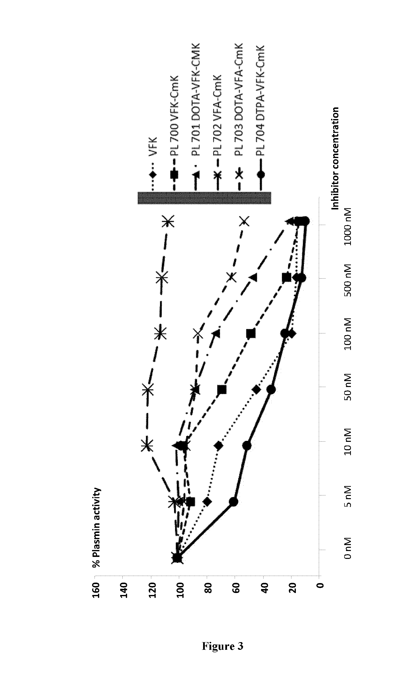
[0107]10 nM of plasmin from human plasma (calbiochem) was diluted in Tris-HCl buffer pH 7.4 50 mM, NaCl 100 mM and Tween-20 0.01%. The samples were then incubated or not with the different inhibitors (see below) for 15 minutes at room temperature. Then, the fluorescent substrate (Suc-Ala-Phe-Lys-AMC, 40 mM) was added just before reading (λ excitation 390 nm, λ emission 460 nm) on the Fluoroskan Ascent plate reader (Thermo Fisher). The fluorescence intensity was evaluated every 10 minutes for 2 hours at 37° C.—The integration time for each measure was 20 ms.

[0108]Each inhibitor was tested in a range of concentrations from 5 nM up to 1000 nM. All the inhibitors were tested in duplicates for each concentration and 4 independent experiments were performed.

[0109]Statistical Analysis:

[0110]Results are presented as % of active plasmin. The differences between the concentr...

example 3

Ex Vivo and In Vivo Scintigraphy

[0115]Material and Methods:

[0116]Experimental Models:

[0117]we have used several experimental models of endovascular thrombus formation in rats, which have been already developed for molecular molecular imaging of platelet activation and fibrin formation, including aneurysm of the abdominal aorta {Sarda-Mantel, 2006}, endocarditic vegetations {Rouzet, 2008}. Aneurysm of the aorta was induced by matrix decellularized xenograft in rats {Allaire, 1996}, and endocarditis by left or right ventricle catheterization followed by provoked bacteraemia {Rouzet, 2008} as previously described. These thrombus models were completed by a model of stroke, provoked by cerebral autologous thrombus emboli in rats (reference).

[0118]These experimental models were also compared to 99m Tc aprotinin signal ex vivo on human aneurymal thrombus.

[0119]Radiolabelling Procedures:

[0120]Aprotinin Labelling with 99mTechnetium:

[0121]Aprotinin labelling was performed according to a proce...


PUM

PropertyMeasurementUnit
thicknessaaaaaaaaaa
thicknessaaaaaaaaaa
temperatureaaaaaaaaaa

Abstract

The present invention is directed to the use of an irreversible ligand of a serine protease selected from the group consisting of leukocyte elastase, thrombin, tissue plasminogen activator (t-PA) and plasmin for the molecular imaging of said serine protease and the diagnosis of pathophysiological conditions associated with said serine protease activity.

Description

FIELD OF THE INVENTION[0001]The present invention is directed to the use of irreversible ligands of the serine proteases, leukocyte elastase, thrombin, tissue plasminogen activator (t-PA) and plasmin, for the molecular imaging of said serine proteases in pathophysiological conditions associated with said serine protease activities.BACKGROUND OF THE INVENTION[0002]Serine proteases have been shown to play a role in diverse physiological functions, many of which can play important roles in disease processes such as cardiovascular disease (Kohler, H. T., et al., (2000) N. Engl. J. Med. 342(24):1792-801; Hamsten, A., et al., (2000) Thromb. Haemost. 83(3):397403; Califf, R. M., et al., (2000) Circulation, 101(19):2231-8; Krendel, S., et al., (2000) Ann. Emerg. Med. 35(5):502-5), cancer (Schmidt, M., et al., (1999) Acta Otolaryngol. 119(8):949-53; Raigoso, P., et al., (2000) Int. J. Biol. Markers, 15(1):44-50; Haese, A., et al., (2000) J. Urol. 163(5):1491-7; Hoopera, J. D., et al., (2000)...

Claims


Application Information

Patent Timeline
Patent Type & Authority Patents(United States)
IPC IPC(8): A61K51/00A61K49/00A61M36/14A61K51/08A61K49/14
CPCA61K49/0004A61K49/14A61K51/08A61K51/088A61K51/048A61K51/0482C07K5/1024
Inventor MICHEL, JEAN-BAPTISTEROUZET, FRANCOISLEGULUDEC, DOMINIQUEPORAS, HERVEFOURNIE-ZALUSKI, MARIE-CLAUDEROGUES, BERNARD
Owner PHARMALEADS INC
Learn More