
Method for extracting quantitative information relating to an influence on a cellular response

a quantitative information and cellular response technology, applied in the field of methods and tools for extracting quantitative information relating to an influence, can solve the problems of cumbersome highly invasive techniques, difficult to obtain detailed knowledge on e.g. exact timing and spatial characteristics of signalling events, and inapplicability to large-scale screening of biologically active substances

Inactive Publication Date: 2011-11-15
FISHER BIOIMAGE
View PDF29 Cites 0 Cited by
  • Summary
  • Abstract
  • Description
  • Claims
  • Application Information

AI Technical Summary

Benefits of technology

[0014]The present invention provides an important new dimension in the investigation of cellular systems involving redistribution in that the invention provides quantification of the redistribution responses or events caused by an influence, typically contact with a chemical substance or mixture of chemical substances, but also changes in the physical environment. The quantification makes it possible to set up meaningful relationships, expressed numerically, or as curves or graphs, between the influences (or the degree of influences) on cellular systems and the redistribution response. This is highly advantageous because, as has been found, the quantification can be achieved in both a fast and reproducible manner, and—what is perhaps even more important—the systems which become quantifiable utilizing the method of the invention are systems from which enormous amounts of new information and insight can be derived.
[0015]The present screening assays have the distinct advantage over other screening assays, e.g., receptor binding assays, enzymatic assays, and reporter gene assays, in providing a system in which biologically active substances with completely novel modes of action, e.g. inhibition or promotion of redistribution / translocation of a biologically active polypeptide as a way of regulating its action rather than inhibition / activation of enzymatic activity, can be identified in a way that insures very high selectivity to the particular isoform of the biologically active polypeptide and further development of compound selectivity versus other isoforms of the same biologically active polypeptide or other components of the same signalling pathway.
[0061]In addition to gene-specific sequences, the primers contain at least one recognition sequence for a restriction enzyme, to allow subsequent cloning of the PCR product. The sites are chosen so that they are unique in the PCR product and compatible with sites in the cloning vector. Furthermore, it may be necessary to include an exact number of nucleotides between the restriction enzyme site and the gene-specific sequence in order to establish the correct reading frame of the fusion gene and / or a translation initiation consensus sequence. Lastly, the primers always contain a few nucleotides in front of the restriction enzyme site to allow efficient digestion with the enzyme.

Problems solved by technology

Although the involvement of protein kinases in cellular signalling and regulation have been subjected to extensive studies, detailed knowledge on e.g. the exact timing and spatial characteristics of signalling events is often difficult to obtain due to lack of a convenient technology.
A disadvantage of this technology is that the labelled protein kinase A has to be microinjected into the cells of interest.
This highly invasive technique is cumbersome and not applicable to large scale screening of biologically active substances.
A further disadvantage of this technique, as compared to the presented invention, is that the labelled protein kinase A cannot be inserted into organisms / animals as a transgene.
However, examples of such fused proteins are not provided, and the use of fusion proteins with GFP for detection or quantitation of translocation or redistribution of biologically active polypeptides affecting intracellular processes upon activation, such as proteins involved in signalling pathways, e.g. protein kinases or phosphatases, has not been suggested.

Method used


Image

  • Method for extracting quantitative information relating to an influence on a cellular response
  • Method for extracting quantitative information relating to an influence on a cellular response
  • Method for extracting quantitative information relating to an influence on a cellular response

Examples


example 2

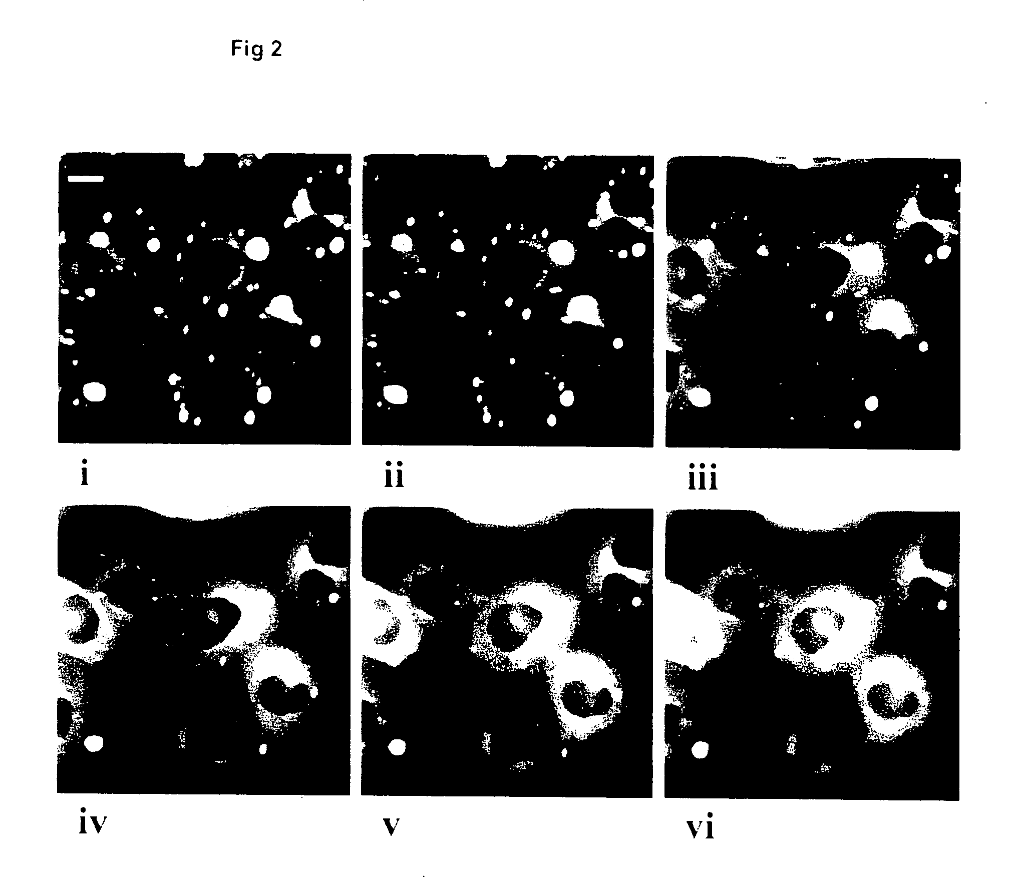
Quantitation of Redistribution in Real-Time within Living Cells

Probe for Detection of PKC Activity in Real Time within Living Cells:

Construction of PKC-GFP Fusion:

[0153]The probe was constructed by ligating two restriction enzyme treated polymerase chain reaction (PCR) amplification products of the cDNA for murine PKCα (GenBank Accession number: M25811) and F64L-S6ST-GFP (sequence disclosed in WO 97 / 11094) respectively. Taq® polymerase and the following oligonucleotide primers were used for PCR;

[0154]

5′mPKCa:                             (SEQ ID NO: 5)TTggACACAAgCTTTggACACCCTCAggATATggCTgACgTTTACCCggC                                            CAACg,3′mPKCa:                             (SEQ ID NO: 6)gTCATCTTCTCgAgTCTTTCAggCgCgCCCTACTgCACTTTgCAAgATTg                                            ggTgC,5′F64L-S65T-GFP:                     (SEQ ID NO: 1)TTggACACAAgCTTTggACACggCgCgCCATgAgTAAAggAgAAgAACTT                                              TTC,3′F64L-S65T-GFP:                     (...

example 3

Probes for Detection of Mitogen Activated Protein Kinase Erk1 Redistribution

[0161]Useful for monitoring signalling pathways involving MAPK, e.g. to identify compounds which modulate the activity of the pathway in living cells.

[0162]Erk1, a serine / threonine protein kinase, is a component of a signalling pathway which is activated by e.g. many growth factors.

Probes for Detection of ERK-1 Activity in Real Time within Living Cells:

[0163]The extracellular signal regulated kinase (ERK-1, a mitogen activated protein kinase, MAPK) is fused N- or C-terminally to a derivative of GFP. The resulting fusions expressed in different mammalian cells are used for monitoring in vivo the nuclear translocation, and thereby the activation, of ERK1 in response to stimuli that activate the MAPK pathway.

a) Construction of Murine ERK1-F64L-S65T-GFP Fusion:

[0164]Convenient restriction endonuclease sites are introduced into the cDNAs encoding murine ERK1 (GenBank Accession number: Z14249) and F64L-S65T-GFP (s...

example 4

Probes for Detection of Erk2 Redistribution

[0173]Useful for monitoring signalling pathways involving MAPK, e.g. to identify compounds which modulate the activity of the pathway in living cells.

[0174]Erk2, a serine / threonine protein kinase, is closely related to Erk1 but not identical; it is a component of a signalling pathway which is activated by e.g. many growth factors.

[0175]a) The rat Erk2 gene (GenBank Accession number: M64300) was amplified using PCR according to standard protocols with primers Erk2-top (SEQ ID NO:11) and Erk2-bottom / +stop (SEQ ID NO:13) The PCR product was digested with restriction enzymes Xho1 and BamH1, and ligated into pEGFP-C1 (Clontech, Palo Alto; GenBank Accession number U55763) digested with Xho1 and BamH1. This produces an EGFP-Erk2 fusion (SEQ ID NO:40 &41) under the control of a CMV promoter.

[0176]b) The rat Erk2 gene (GenBank Accession number: M64300) was amplified using PCR according to standard protocols with primers (SEQ ID NO:11) Erk2-top and E...


PUM

PropertyMeasurementUnit
Lifetime stabilityaaaaaaaaaa
Resonance energyaaaaaaaaaa
Biological propertiesaaaaaaaaaa

Abstract

Cells are genetically modified to express a luminophore, e.g., a modified (F64L, S65T, Y66H) Green Fluorescent Protein (GFP, EGFP) coupled to a component of an intracellular signalling pathway such as a transcription factor, a cGMP- or cAMP-dependent protein kinase, a cyclin-, calmodulin- or phospholipid-dependent or mitogen-activated serine / threonin protein kinase, a tyrosine protein kinase, or a protein phosphatase (e.g. PKA, PKC, Erk, Smad, VASP, actin, p38, Jnk1, PKG, IkappaB, CDK2, Grk5, Zap70, p85, protein-tyrosine phosphatase 1C, Stat5, NFAT, NFkappaB, RhoA, PKB). An influence modulates the intracellular signalling pathway in such a way that the luminophore is being redistributed or translocated with the component in living cells in a manner experimentally determined to be correlated to the degree of the influence. Measurement of redistribution is performed by recording of light intensity, fluorescence lifetime, polarization, wavelength shift, resonance energy transfer, or other properties by an apparatus consisting of e.g. a fluorescence microscope and a CCD camera. Data stored as digital images are processed to numbers representing the degree of redistribution. The method can be used as a screening program for identifying a compound that modulates a component and is capable of treating a disease related to the function of the component.

Description

[0001]This application is a divisional of application Ser. No. 09 / 417,197, now U.S. Pat. No. 6,518,021, filed on Oct. 7, 1999 and for which priority is claimed under 35 U.S.C. §120. Application Ser. No. 09 / 417,197 is the continuation of PCT International Application No. PCT / DK98 / 00145 filed on Apr. 7, 1998. The entire contents of each of the above-identified applications are hereby incorporated by reference. This application also claims priority of Application No. PA 00392 filed in Denmark on Apr. 7, 1997 under 35 U.S.C. §119.FIELD OF INVENTION[0002]The present invention relates to a method and tools for extracting quantitative information relating to an influence, on a cellular response, in particular an influence caused by contacting or incubating the cell with a substance influencing a cellular response, where the cellular response is manifested in redistribution of at least one component in the cell. In particular, the invention relates to a method for extracting quantitative in...

Claims


Application Information

Patent Timeline
IPC IPC(8): G01N33/53G01N21/76C12P21/02C12Q1/25C12Q1/48G01N33/50G01N33/533G01N33/58
CPCG01N33/5005G01N33/5008G01N33/5014G01N33/5035G01N33/5041G01N33/533G01N33/582G01N2333/43595G01N2333/9121
Inventor THASTRUP, OLEBJORN, SARA PETERSENTULLIN, SORENALMHOLT, KASPERSCUDDER, KURT
Owner FISHER BIOIMAGE
Learn More