
Hepatitis virus core proteins as vaccine platforms and methods of use thereof

a technology of core proteins and vaccines, applied in the field of hepatitis virus core proteins and nucleic acids, can solve the problems of unfulfilled promise of hapten-like technology, unable inability to meet the requirements of clinical trials,

Inactive Publication Date: 2011-02-08
VLP BIOTECH
View PDF34 Cites 10 Cited by
  • Summary
  • Abstract
  • Description
  • Claims
  • Application Information

AI Technical Summary

Benefits of technology

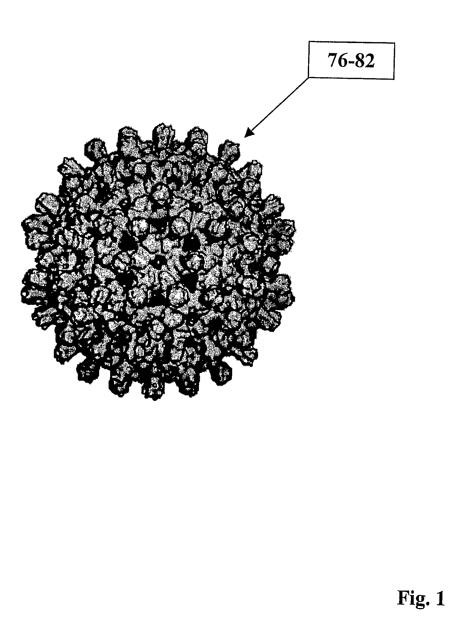
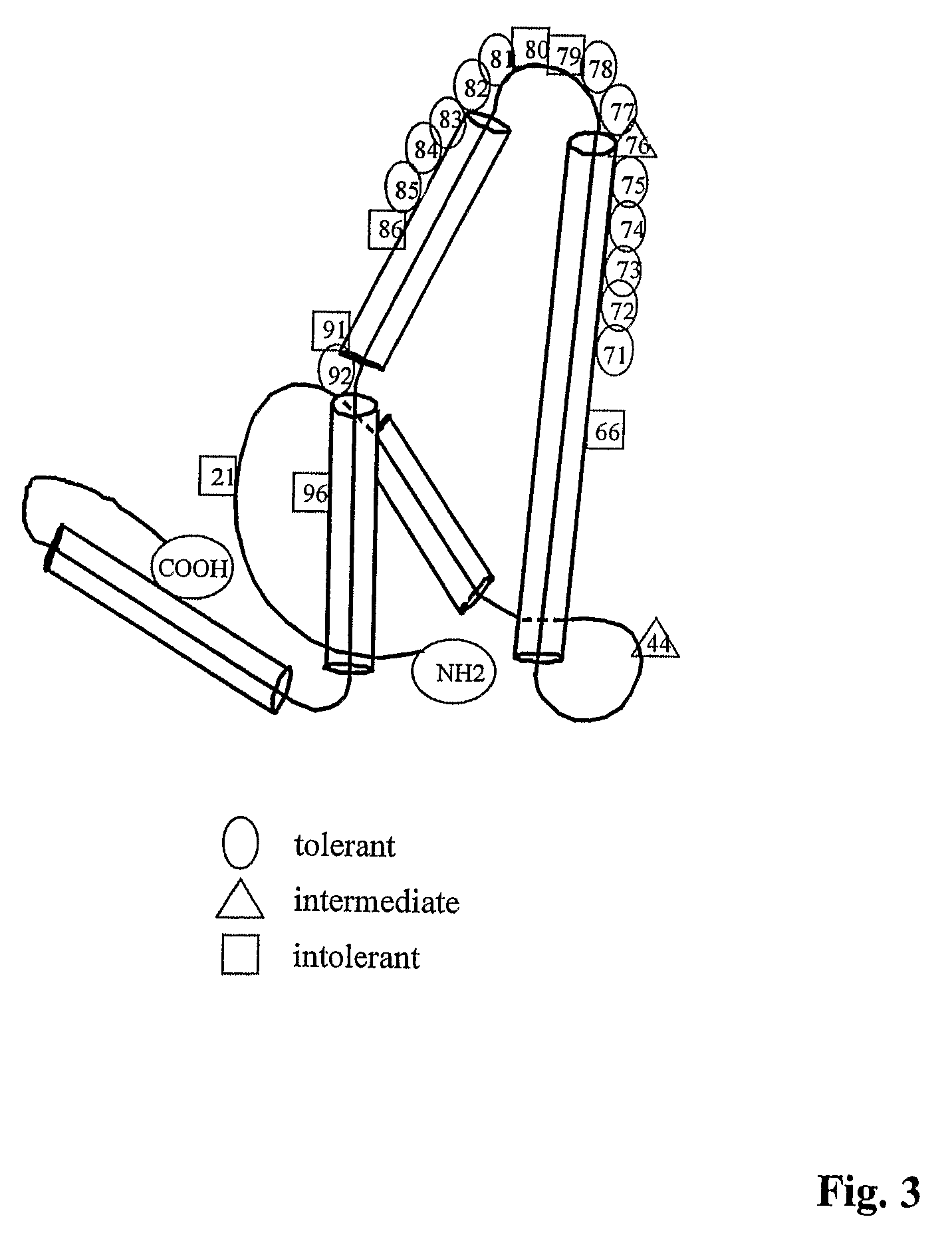
[0091]As disclosed herein, one advantage of the invention's combinatorial modification that includes epitope insertion at preferred locations in a hepadnavirus core antigen as well as preferred C-terminal modifications of the core antigen is that inclusion of the C-terminal modification rescues assembly of the resulting virus particles that contain the inserted epitope (e.g., Tables 12-14). Conversely, non-permissive C-terminal modifications to hepadnavirus core antigens may be rescued by altering the insertion position of the epitope into the hepadnavirus core antigen (e.g., Tables 13, 14). Moreover, substitution of amino acids with acidic amino acids and / or the use of acidic amino acid linkers in the hepadnavirus core antigen rescue assembly of the inserted epitope (e.g., Table 18).
[0092]The term “gene” refers to a nucleic acid (e.g., DNA) sequence that comprises coding sequences necessary for the production of a polypeptide or precursor or RNA (e.g., tRNA, siRNA, rRNA, etc.). The polypeptide can be encoded by a full length coding sequence or by any portion of the coding sequence so long as the desired activity or functional properties (e.g., enzymatic activity, ligand binding, signal transduction, etc.) of the full-length or fragment are retained. The term also encompasses the coding region of a structural gene and the sequences located adjacent to the coding region on both the 5′ and 3′ ends, such that the gene corresponds to the length of the full-length mRNA. The sequences that are located 5′ of the coding region and which are present on the mRNA are referred to as 5′ untranslated sequences. The sequences that are located 3′ or downstream of the coding region and that are present on the mRNA are referred to as 3′ untranslated sequences. The term “gene” encompasses both cDNA and genomic forms of a gene. A genomic form or clone of a gene contains the coding region, which may be interrupted with non-coding sequences termed “introns” or “intervening regions” or “intervening sequences.” Introns are removed or “spliced out” from the nuclear or primary transcript, and are therefore absent in the messenger RNA (mRNA) transcript. The mRNA functions during translation to specify the sequence or order of amino acids in a nascent polypeptide.
[0093]In particular, the term “WHcAg gene” refers to the full-length WHcAg nucleotide sequence (e.g., contained in SEQ ID NO:37). However, it is also intended that the term encompass fragments of the WHcAg sequence, and / or other domains within the full-length WHcAg nucleotide sequence. Furthermore, the terms “WHcAg nucleotide sequence” or “WHcAg polynucleotide sequence” encompasses DNA, cDNA, and RNA (e.g., mRNA) sequences.
[0094]The term “plasmid” as used herein, refers to a small, independently replicating, piece of DNA. Similarly, the term “naked plasmid” refers to plasmid DNA devoid of extraneous material typically used to affect transfection. As used herein, a “naked plasmid” refers to a plasmid substantially free of calcium-phosphate, DEAE-dextran, liposomes, and / or polyamines. As used herein, the term “purified” refers to molecules (polynucleotides or polypeptides) that are removed from their natural environment, isolated or separated. “Purified” molecules are at least 50% free, preferably at least 75% free, and more preferably at least 90% free from other components with which they are naturally associated.
[0095]The term “recombinant DNA” refers to a DNA molecule that is comprised of segments of DNA joined together by means of molecular biology techniques. Similarly, the term “recombinant protein” refers to a protein molecule that is expressed from recombinant DNA. The term “fusion protein” as used herein refers to a protein formed by expression of a hybrid gene made by combining two gene sequences. Typically this is accomplished by cloning a cDNA into an expression vector in frame with an existing gene. The fusion partner may act as a reporter (e.g., βgal) or may provide a tool for isolation purposes (e.g., GST).
[0096]Suitable systems for production of recombinant proteins include but are not limited to prokaryotic (e.g., Escherichia coli), yeast (e.g., Saccaromyces cerevisiae), insect (e.g., baculovirus), mammalian (e.g., Chinese hamster ovary), plant (e.g., safflower), and cell-free systems (e.g., rabbit reticulocyte).

Problems solved by technology

The promise of the hapten-like technology hasn't been fully realized because efficient and reproducible methods for the delivery of these small epitopes to the immune system are lacking.
Although the HBcAg is highly immunogenic, the existing HBcAg-based platform technology has a number of serious theoretical and practical limitations.
For example, less than 50% of foreign epitopes can be accommodated by the HBcAg platform because of adverse effects on particle assembly (Jegerlehner et al., Vaccine, 20:3104, 2002 and PCT / US01 / 25625); use of the HBcAg compromises the use of the anti-HBc diagnostic assay; pre-existing anti-HBc antibody is present in all HBV chronically infected patients and in most previously infected and recovered patients, which may limit the effectiveness of the HBcAg as a vaccine carrier; and immune tolerance to HBcAg in individuals chronically infected with HBV (300-400 million worldwide) limits immunogenicity in this population.

Method used


Image

  • Hepatitis virus core proteins as vaccine platforms and methods of use thereof
  • Hepatitis virus core proteins as vaccine platforms and methods of use thereof
  • Hepatitis virus core proteins as vaccine platforms and methods of use thereof

Examples


example 1

Immunization of Mice

[0329]Groups of 3-5 female mice, approximately 6-8 weeks old of various strains (either bred at the Vaccine Research Institute of San Diego, San Diego, Calif. or obtained from Jackson Laboratories, Bar Harbor, Me.) were immunized intraperitoneally for antibody assays and subcutaneously for T cell assays. Antigens were injected in saline, or absorbed in 0.1% (w / v) AlPO4 suspension, or emulsified in IFA or the squalene water-in-oil adjuvant Montanide ISA 720 (Seppic, Paris) depending on the experiment. Mice were bled pre-immunization and at various times after primary and booster immunizations for anti-insert / PS and anti-WHcAg antibody determinations. A larger number of mice / group (at least 10) were used to perform mouse potency (dose) studies because at limiting antigen doses, less than 100% of mice produce antibody, and the limiting dose was typically defined as the dose at which 50% of the mice produce antibody.

example 2

Antibody Assays

[0330]Anti-WHcAg or peptide antibodies were measured in pooled or individual, murine sera by indirect solid phase ELISA using solid phase WT WHcAg (50 ng / well) or insert peptide (0.5 μg / well) and goat anti-mouse Ig (or IgG isotype-specific) antibodies were used as the secondary antibody. The ELISA was developed with a peroxidase-labelled, affinity-purified swine anti-goat Ig. The data were expressed as antibody titer representing the highest dilution yielding three times (3×) the optical density of the pre-immunization sera. Anti-PS antibodies were measured in an identical manner on solid phase purified PSs (10 μg / ml), except that PolySorp plates (Nunc, Rosklide, Denmark) were used to coat PS antigens. Fifty micrograms of pneumococcal cell wall polysaccharide (C—PS) per ml of sera were added to absorb any anti-C—PS antibodies.

example 3

T Cell Assays

[0331]To measure T cell proliferation, groups of 3-5 mice were primed with either 10 μg of WT core, hybrid core or PS-core conjugates by hind footpad injection. Approximately, 7-10 days after immunization, draining popliteal lymph node (LN) cells were harvested, and 5×10⁵ cells in 0.1 ml of Click's medium were cultured with 0.1 ml of medium containing WT core, hybrid core or PS-core conjugates, various synthetic peptides, or medium alone. Cells were cultured for 96 hr at 37° C. in a humidified 5% CO2 atmosphere, and during the final 16 hr, 1 μCi of 3H-thymidine (3H-TDR; at 6.7 Ci / mmol, New England Nuclear, Boston, Mass.) was added to each well. The cells were then harvested onto filter strips for determination of 3H-TdR incorporation. The data were expressed as counts per minute corrected for background proliferation in the absence of antigen (Acpm). The T cell nature of the proliferation was confirmed by analyzing nylon-wool column-enriched T cells in selected experime...


PUM


Abstract

The present invention relates to hepatitis virus core proteins and nucleic acids. In particular, the present invention provides compositions and methods comprising recombinant hepatitis virus core proteins or nucleic acids for use in vaccine formulations.

Description

[0001]This application is a U.S. national entry of International Application No. PCT / US2004 / 023391, filed on Jul. 19, 2004, which is a continuation-in-part of, and claims priority to, application Ser. No. 10 / 630,070, filed Jul. 30, 2003, now U.S. Pat. No. 7,320,795, and application Ser. No. 10 / 630,074, filed Jul. 30, 2003, now U.S. Pat. No. 7,144,712.[0002]The invention was made in part with Government support by the National Institutes of Health, Grants RO1 AI020720 and RO1 AI049730. As such, the Government has certain rights in the invention.FIELD OF THE INVENTION[0003]The present invention relates to hepatitis virus core proteins and nucleic acids. In particular, the present invention provides compositions and methods comprising recombinant hepatitis virus core proteins or nucleic acids for use in vaccine formulations.BACKGROUND OF THE INVENTION[0004]The ability to map neutralizing B cell epitopes on protein and carbohydrate antigens has created much interest in the potential use...

Claims


Application Information

Patent Timeline
Patent Type & Authority Patents(United States)
IPC IPC(8): C12Q1/70A61K39/29A61K39/12A61KA61K39/385C07K14/02
CPCA61K39/385C07K14/005A61K2039/6075C12N2730/10022C12N2730/10122A61K39/00C12N2730/10023C12N2730/10123A61P37/00A61P43/00Y02A50/30
Inventor MILICH, DAVID R.BILLAUD, JEAN-NOEL
Owner VLP BIOTECH
Learn More