Rapid immunochromatographic lateral flow assay for early zika disease detection
a lateral flow assay and immunochromatographic technology, applied in the field of rapid immunochromatographic lateral flow assay for early zika disease detection, can solve the problems of inability to accurately and quickly determine the early, intermediate, or late zika virus infection status of the patient, and no vaccines or drugs are available to prevent or treat infection, etc., to achieve rapid detection and rapid detection. , the effect of rapid detection
- Summary
- Abstract
- Description
- Claims
- Application Information
AI Technical Summary
Benefits of technology
Problems solved by technology
Method used
Image
Examples
experiment i and ii
Devices
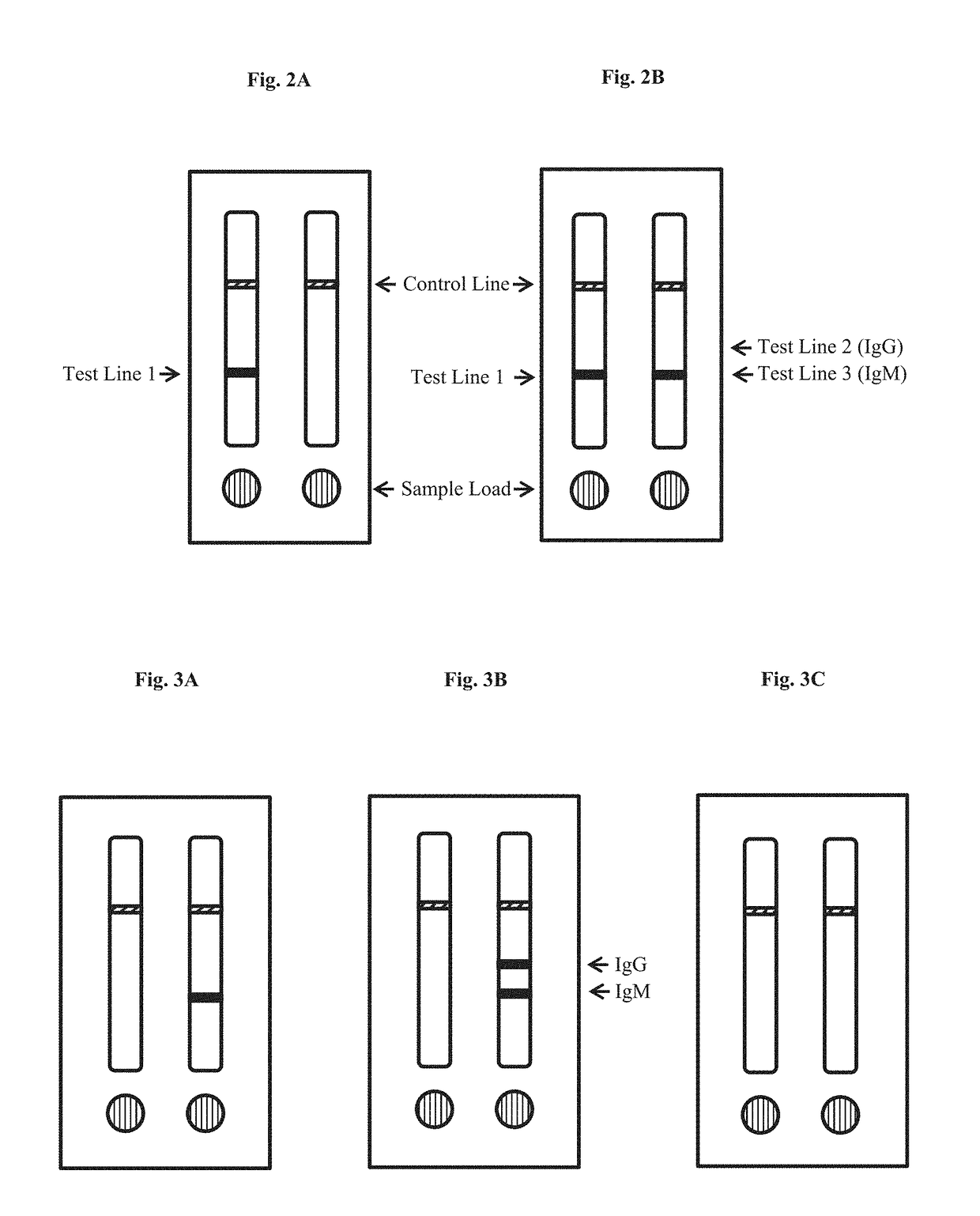
[0138]For each of Experiments I and II, described in Example 2, two-strip immunochromatographic lateral flow anti-Zika IgG assay device was constructed.
First Test Strip—Antibody Sandwich Assay
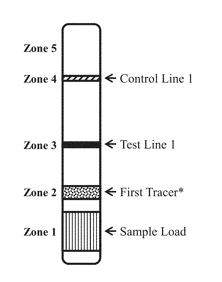
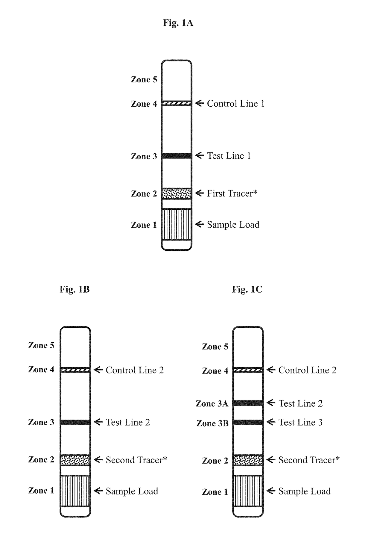
[0139]The first test strip, formatted as generally set forth in FIG. 1A, was made for detecting anti-Zika antigen in a test sample.
[0140]Conjugate Pad (Zone 2): Gold-labeled Zika NS1 protein was sprayed and dried onto a glass fiber conjugate pad of the first test strip for use as the tracer reagent. The Zika NS1 protein (Ross Southern Laboratories, Utah) was labeled with 60 nm gold particles prepared by adding 50 ng particles in 10 ml pH 7.4 phosphate buffer, mixing well, followed by centrifugation at 12,000 rpm. The resulting pellet was dissolved in phosphate buffer pH 7.2 containing 1% BSA, 1% Milk, and 0.05% Tween 20. This solution was sprayed onto the conjugate pad and vacuum dried.
[0141]Test Line 1 (Zone 3): Zika NS1 protein (from Ross Southern Laboratories, Utah) was immobilized on...
example 2
IgG Sandwich Assay Serum Sample Analysis—Preliminary Testing
[0147]In two separate experiments (Experiments I and II), described below, test strips constructed as described in Example 1 were used to test serum samples determined to be anti-Zika IgM positive or anti-Zika IgM negative by an ELISA (Euroimmun US, New Jersey). The presence of IgG was not tested by the ELISA. In each experiments, 20 uL of serum was loaded into sample well (sample pad, Zone 1) of the first test strip in the device, followed by 40 uL of chase buffer (PBS). 5 uL serum was loaded into the sample well of the second test strip in the device, followed by 60 uL of chase buffer (PBS). Test results for both strips were read at 20 minutes.
PUM
 Abstract
Description
Claims
Application Information
 - R&D
- Intellectual Property
- Life Sciences
- Materials
- Tech Scout
- Unparalleled Data Quality
- Higher Quality Content
- 60% Fewer Hallucinations
Browse by: Latest US Patents, China's latest patents, Technical Efficacy Thesaurus, Application Domain, Technology Topic, Popular Technical Reports.
© 2025 PatSnap. All rights reserved.Legal|Privacy policy|Modern Slavery Act Transparency Statement|Sitemap|About US| Contact US: help@patsnap.com


