
MRI contrast agents for cell labeling

a contrast agent and cell technology, applied in the field of porphyrin compounds, can solve the problems of limited time window, low sensitivity, limited in terms of lack of anatomical information, etc., and achieve the effect of less prone to movement and less able to move across the cell membran

Inactive Publication Date: 2016-08-18
THE GOVERNINIG COUNCIL OF THE UNIV OF TORANTO +1
View PDF0 Cites 0 Cited by
  • Summary
  • Abstract
  • Description
  • Claims
  • Application Information

AI Technical Summary

Benefits of technology

The patent describes a new type of magnetic resonance imaging (MRI) contrast agent called paramagnetic porphyrins, which can be used to label and track cells in the body. These porphyrins are free of toxic heavy metals and have high relaxivity (signal-to-noise ratio) at high magnetic fields. They can be easily taken up by cells and retained inside them for long periods of time, making them ideal for in vivo applications. The porphyrins can be modified to have high T1 relaxivity and high biocompatibility for in vivo use. The patent also describes a mechanism for the porphyrins to enter cells and release their polar, cell-permeable form. The contrast agent can be in monomeric, dimeric, oligomeric, or polymeric forms, with the latter two having increased sensitivity for MRI cell imaging. The contrast agent can also includes a hydrophilic group that increases its sensitivity when cleaved by an intracellular enzyme.

Problems solved by technology

Among these widely used techniques, CT has relatively low resolution and low sensitivity.
PET and SPECT provide excellent sensitivity and information about cell function but are limited in terms of lack of anatomical information, restricted time window due to decay of radioactive nuclides, and exposure to ionizing radiation.
For the differentiation of normal and diseased tissues however it is often necessary to use a contrast agent (CA) because the difference in native relaxation times are too small to detect.
There are however, several major limitations associated with T2-based CAs and IONs in particular.
The need for transfection agents (TAs) complicates the labeling procedure because each TA will require FDA approval.
There are also limitations for determination of the cell volume and quantitative analysis of the signal because the large magnetic susceptibility of the particles induces strong image artifacts that affect an area that extends far beyond the volume of the labeled cells.
This blooming affect is also detrimental for visualization of the fine delineation of the labeled cells from the surrounding tissue and leads to loss of anatomical information in the vicinity of the labeled cells.
T2 agents also suffer in terms of specificity since they generate signal voids on the image which are ambiguous because they can also be generated from blood vessels, air spaces, hemorrhages, tissue interfaces or other imaging artifacts associated with magnetic susceptibility.10 One method to overcome these limitations has been the development of pulse sequences that generate hyperintense contrast from SPIOs such as GRASP.32 However, these pulse sequences require specialized hardware and there are safety concerns with the use of multiple high-energy pulses.
Furthermore, some of the normal anatomical background of the image may be lost.
Cells must therefore internalize significant numbers of PFC's and only a large number of cells in close proximity can be visualized (>200 cells / voxel).10 This is due to a lower signal to noise ratio (SNR) and also results in substantially longer image acquisition times. Heteronuclear MRI also requires extra hardware limiting the widespread use.
A limitation shared by all of these contrast agents is the previously mentioned lack of cell permeability.
Despite the progress made towards synthesis of cell permeable and trappable Gd-based CAs for cell labeling, these CAs have not found widespread use for a number of reasons.
Low relaxivity of Gd-based CAs at fields above 1.0 T is an issue.
Furthermore, intracellular biocompatibility has been raised as a concern for long term cell tracking experiments since these complexes are less stable at lower pH such as in lysosomes and free Gd(III) is a toxic heavy metal.
Taken together, these drawbacks have limited the scope of MnCl2 for cell labeling and long-term tracking in vivo.

Method used


Image

  • MRI contrast agents for cell labeling
  • MRI contrast agents for cell labeling
  • MRI contrast agents for cell labeling

Examples


examples of experimental procedures

[0113]All reagents and solvents were of commercial reagent grade and were used without further purification except where noted. All reactions were carried out with oven dried glassware, anhydrous solvents and under argon atmosphere unless stated otherwise. Column Chromatography was carried out using Caledon Silica Gel 60; 50-200 microns 70-300 mesh, or using Sephadex™ LH-20 with dry bead size of 18-111 □m from GE Health Care. Dialysis was performed with spectrumlabs Float-A-Lyzer™ G2 500 MWCO. Cation ion exchange was performed using an Amberlite® IR120, H resin. Phosphate Buffer Saline was purchased from Sigma® Life Science, sterile filtered and endotoxin tested. The cell line was obtained from ATCC (American Tissue Culture Collection Manassas, Va., USA). 1640-RPMI medium was purchased from Sigma-Aldrich, trypsin EDTA was purchased from Gibco (Carlsbad, Calif., USA). All the spectroscopy data for structural characterizations were obtained using the research facilities at University ...


PUM


Abstract

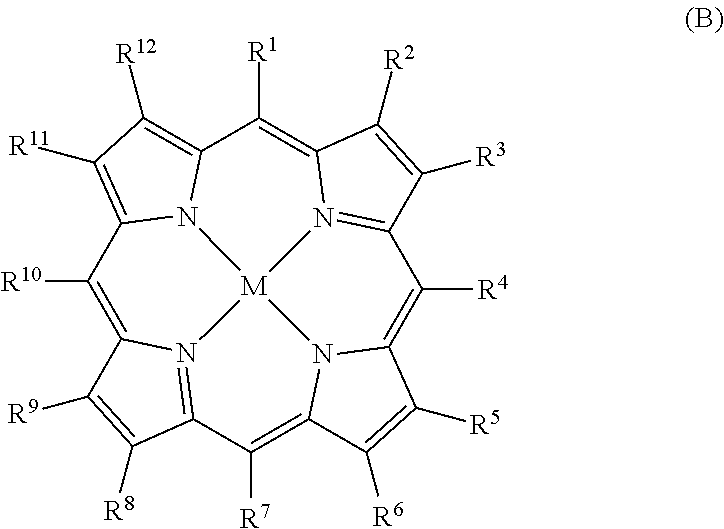
Porphyrin compounds useful in the field of magnetic resonance imaging (MRI) as contrast agents. The compounds are relatively lipophilic porphyrins, include one or more enzyme-reactive functional groups, and are cell membrane permeable. Relatively lipophilic group(s) can be enzymatically released within a cell to produce a relatively hydrophilic porphyrin compound.

Description

FIELD OF THE INVENTION[0001]The present invention relates to porphyrin compounds useful in the field of magnetic resonance imaging (MRI) as contrast agents, particularly as cellular contrast agents.BACKGROUND OF THE INVENTION[0002]Cellular imaging has been critical in monitoring the function of endogenous or implanted cells under both physiological and pathological conditions. In recent years, there are increasing demands for new techniques to noninvasively monitor and track cells in vivo, as the field of cell transplantation, especially immune-cell and stem-cell therapy, is very rapidly expanding. In vivo cellular imaging technique is unique in its ability to visualize biological processes in real time at the cellular level and in an intact living subject.1 Optical imaging (OI) has been at the forefront of the research field and is well-established with a large variety of fluorescent probes2-6 and fluorescent proteins7 available for labeling of specific cell structures, as well as ...

Claims


Application Information

Patent Timeline
IPC IPC(8): A61K49/10C07F13/00G01N33/58A61K49/08
CPCC07D519/00C07D487/22A61K49/106G01N33/581A61K49/128A61K49/085C07F13/005A61K49/124
Inventor ZHANG, XIAO-ANHAEDICKE, INGA E.CHENG, HAI-LING MARGARET
Owner THE GOVERNINIG COUNCIL OF THE UNIV OF TORANTO
Learn More