
Chitosan biomimetic scaffolds and methods for preparing the same

a biomimetic and scaffold technology, applied in the field of biopolymer development, can solve the problems of inability to adsorb large quantities of exudate from wounds, difficult electropinning of chitosan, and inability to permit three-dimensional growth of granulation tissu

Inactive Publication Date: 2014-02-13
UNIV LIEGE
View PDF1 Cites 36 Cited by
  • Summary
  • Abstract
  • Description
  • Claims
  • Application Information

AI Technical Summary

Benefits of technology

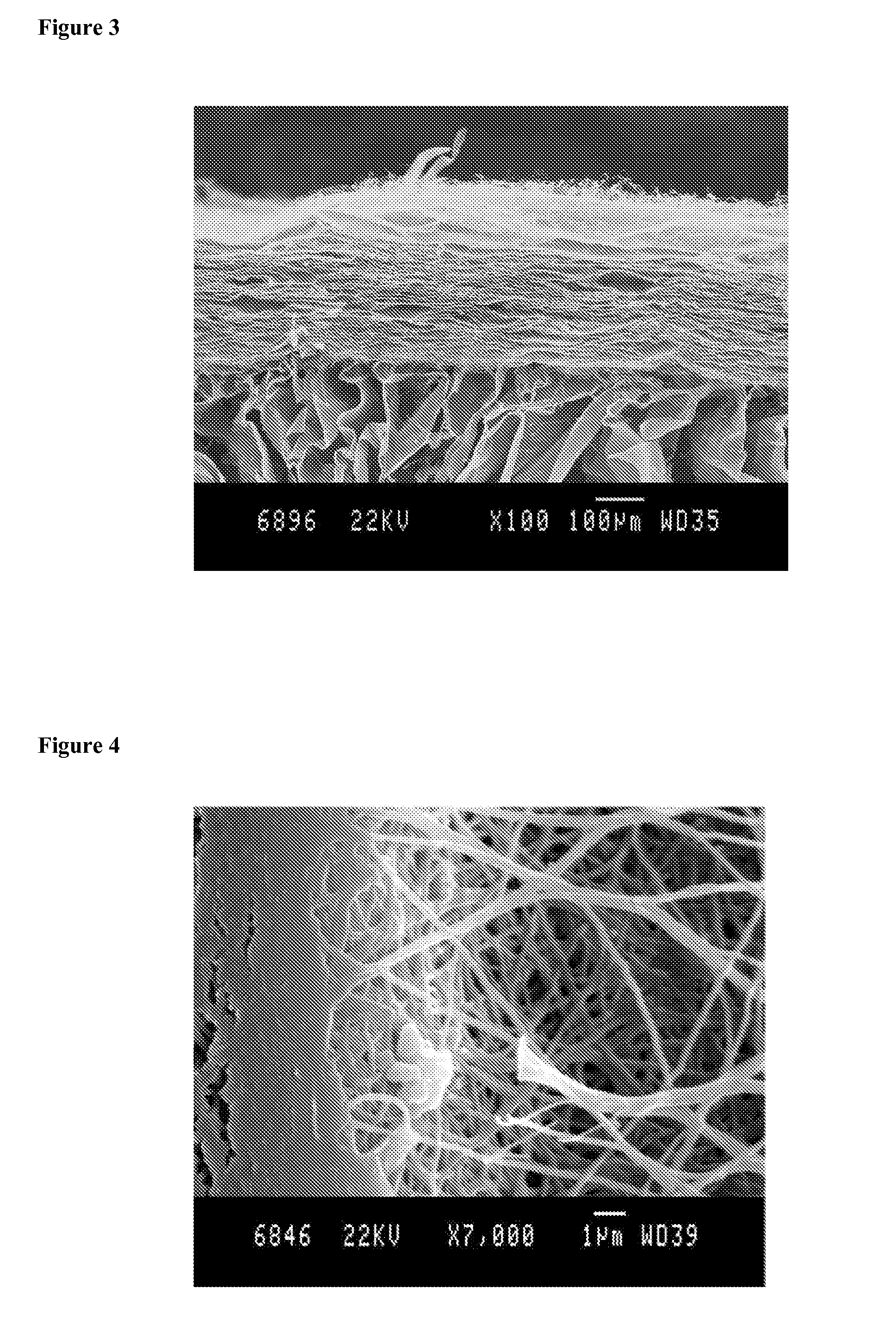
The present invention solves the technical problem of providing a layered chitosan scaffold that has good adhesion between the porous and nanofiber layers, a tuneable porosity of the nanofiber layer, and a stable nanofibers and porous morphology even when immersed in water or other solvents. The process for the preparation of the layered chitosan scaffold includes the steps of preparing a solution of chitosan, feeding the solution in a syringe, covering a collector by a porous chitosan scaffold support layer, applying an electrospinning voltage, and collecting the layered chitosan scaffold. The present invention provides the use of the layered electrospun chitosan scaffold as a wound dressing, in tissue engineering, or for biomedical applications.

Problems solved by technology

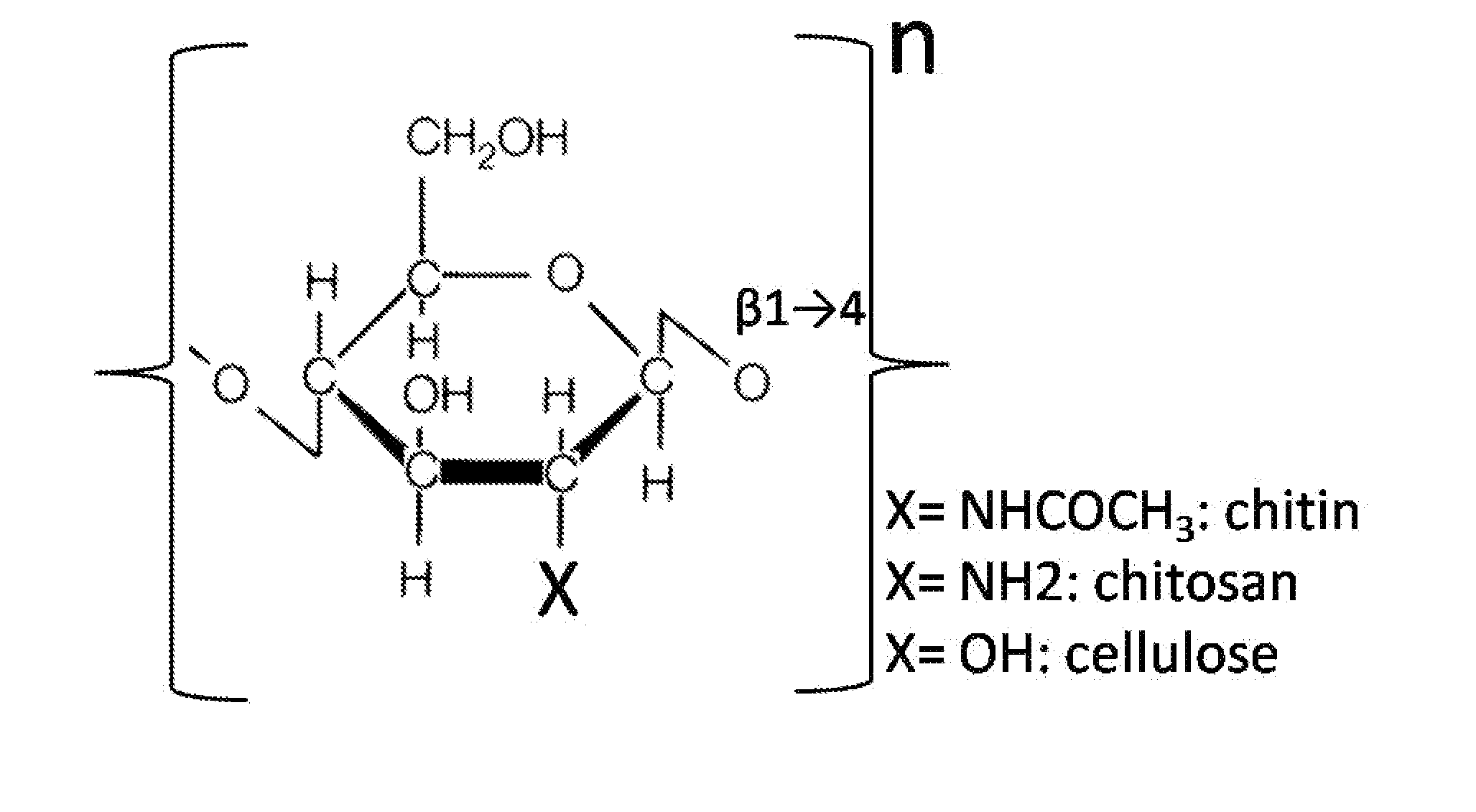
Electrospinning of chitosan has proven to be difficult, pure chitosan fibres being obtained only from specific chitosan solutions such as in acetic acid or in trifluoroacetic acid (TFA) [4] for example.
The problem of membranes and films is that they only have pores that are too small for the migration of cells (fibroblasts, keratinocytes, etc.) and do not permit three-dimensional growth of granulation tissue.
In addition, owing to its low porosity and reduced volume, this type of material is not able to adsorb large quantities of exudates from the wound.
Porous sponges have better absorption properties (U.S. Pat. No. 5,116,824,1992; US 2002161440, 2002; DE 101 17234 A1, 2002), but are usually not an appropriate substrate for cell growth.
Although this improves the biological properties of the dressings in vitro and in preclinical studies in animal, it makes them less attractive for large scale clinical applications in patients because of potential problems linked to the availability, potential presence of pathogens (such as, among others, virus, BSE), immunogenicity and cost of these extracted biologicals.
Long term shelve storage could also be problematic in many cases.
However, major limitations for its use as a wound dressing accelerating the healing of deep ulcer and potentially the tri-dimensional repair of connective tissues, are the facts that a chitosan film does not provide a tri-dimensional scaffold temporally replacing the lost tissue and that a chitosan sponge is a poor substrate for many cell types including keratinocytes, fibroblasts and endothelial cells.
This explains why newly developed chitosan dressings contain “additives” such as collagen or gelatin from animal origin (for example ChitoSkin from Sangui BioTech), again facing problems linked to the potential presence of pathogens and immunogenicity of these extracted biologicals.
Moreover, due to its crustacean origin, anaphylactic reactions for some patients can not be totally excluded.
Another limitation for commercialization of currently developed chitosan dressing results from their activation and / or stabilization through the use of potentially harmful chemicals such as cross-linkers for example.
For instance, pure chitosan produced as a film is not considered to be an appropriate wound dressing because of its poor tensile strength and elasticity.
Chitosan can also be protected from degradation by glutaraldehyde or formaldehyde cross-linking, although this reduces its mucoadhesive properties and is expected to modify other biophysical and biological properties.
The two major problems are directly related to the electrospinning of proteins: denaturation or aggregation of proteins during the electrospinning process and maintainance of the biological properties of the proteins in the electrospun product.
Classical dressings (such as gauze for example) are not expensive but do not favour the healing of ulcers.
New “bio-dressings”, containing proteins of animal or human origin and / or living cells, are potentially more efficient but are expensive and face other problems such as innocuousness of their components and short shelve-life.
However, these dressings are produced by a “freeze-dry” procedure and do not contain chitosan nanofibers.
However, it is now recognized that chitosan, under the form of gel, granules, film and sponge, can be only considered as non toxic but poor substrate for cell growth.

Method used


Image

  • Chitosan biomimetic scaffolds and methods for preparing the same
  • Chitosan biomimetic scaffolds and methods for preparing the same
  • Chitosan biomimetic scaffolds and methods for preparing the same

Examples


example 1

Electrospinning of Chitosan Solution

1.1 Material:

[0154]Medical grade chitosan with acetylation degree 19.6%, viscosity (1% solution in 1% acetic acid) 21 mPa·s, Mw 77 kDa (L08049), from Kitozyme, Belgium and PEO high molecular weight (HMW, 900000 g / mol) purchased from Aldrich were used as received. Acetic acid, ethanol and sodium hydroxide (Sigma) were analytical grade and used as received.

1.2 Method:

1.2.1 Electrospinning of Chitosan Membranes

[0155]Solutions of 10.5% chitosan, 4% HMW PEO were prepared by dissolving the appropriate amount of chitosan (in 6.5% acetic acid solution) and PEO (in distilled water), by stirring overnight. The next day, the chitosan and PEO solutions were then mixed to obtain mixtures with weight ratios of chitosan:PEO of 90:10. Two ml of the well homogenized resulting mixture were fed into 5 ml plastic syringes fitted with blunt tipped stainless steel needles (gauge 18 and 21). The solution feed was driven using a syringe pump (Razel Scientific Instruments...

example 2

Engineering of the Dressing

[0159]As reported above, bandages and dressings for treating wounds have to satisfy various requirements. This explains why the conventional fibrous matrix scaffold obtained so far by electrospinning of chitosan is not used today. Some of these disadvantages are that:[0160]a) a fibrous scaffold composed of chitosan nano fibers has poor mechanical properties that prevent easy handling and cause rapid destruction in situ when placed on wounds.[0161]b) nano fibrous scaffolds having only 2-dimensional structure are limited in applications since they do not provide protection against outside mechanical influences.[0162]c) thin nanofibrous scaffolds are not appropriate to maintain the moisture equilibrium promoting the wound healing process.[0163]d) The pores in scaffold made of cross-linked nanofibers are too small to permit cell invasion, which, in certain circumstances, is a disadvantage.

[0164]The present invention overcomes all of these problems by providing...

example 3

Co-Electrospinning of Chitosan with Proteins and Chemical Molecules Having the Ability to Maintain and to Preserve the Biological Activity of Proteins

3.1. Materials:

[0170]The β-lactamase BlaP of Bacillus licheniformis were used as protein models in co-electrospinning experiments. This protein corresponds to a bacterial enzyme with a globular 3D-structure. The recombinant protein was overproduced overproduced in E. coli and purified to homogeneity by using affinity chromatography methods.

3. 2. Methods:

3.2.1 Co-Electrospun Chitosan-Proteins Based Membranes:

[0171]For co-electrospinning experiments, the BlaP protein was added in the chitosan / PEO mixture (see Example 1) at a final concentration of 3 mg / ml and electrospun using the same procedure described in Example 1.2.1. In some cases, the mixture was also supplemented with a derivative of β-cyclodextrin.

3.2.2 Detection of Proteins in Co-Electrospun Chitosan Membranes

[0172]For the β-lactamase assays performed with BlaP, the co-electros...


PUM

PropertyMeasurementUnit
Fractionaaaaaaaaaa
Fractionaaaaaaaaaa
Sizeaaaaaaaaaa

Abstract

The present invention relates to a layered chitosan scaffold wherein said layered scaffold comprises at least two fused layers, wherein at least one of the fused layers comprises a chitosan nanofiber membrane and the other fused layer comprises a porous chitosan support layer. Moreover, the present invention provides a layered chitosan scaffold characterized by (i) a good adhesion between the porous and nanofiber layers, (ii) a tuneable porosity of the nanofiber layer by tuning the distance between the nanofibers, (iii) a stable nanofibers and porous morphology even when immersed in water or other solvents and a process for the preparation of such layered chitosan scaffold. The present invention also provides a process for the preparation of the layered chitosan scaffold.

Description

[0001]The invention concerns chitosan biomimetic scaffolds and methods for modulating their intrinsic properties such as rigidity, elasticity, resistance to mechanical stress, porosity, biodegradation and absorbance of exudates. Therefore, the present invention relates to a layered chitosan scaffold comprising at least two fused layers, wherein at least one of the fused layer comprises a chitosan nano fiber membrane and the other fused layer comprises a porous chitosan support layer. Moreover, the present invention provides a layered chitosan scaffold characterized by (i) a good adhesion between the porous and nano fiber layers, (ii) a tuneable porosity of the nano fiber layer by tuning the distance between the nanofibers, (iii) a stable nanofibers and porous morphology even when immersed in water or other solvents and a process for the preparation of such layered chitosan scaffold. Finally, the present invention provides the use of the layered chitosan scaffold of the invention or ...

Claims


Application Information

Patent Timeline
IPC IPC(8): A61F13/00
CPCA61F13/00012A61F13/00008A61F13/00063A61L15/28A61L15/425A61L15/64A61L27/20A61L27/48A61L27/56A61L27/58A61L27/60A61L2400/12C08L5/08A61F13/01008A61F13/01012
Inventor FILEE, PATRICEFREICHELS, ASTRIDJEROME, CHRISTINEAQUIL, ABDELHAFIDCOLIGE, ALAINTCHEMTCHOUA TATEU, VICTOR
Owner UNIV LIEGE
Learn More