In vivo flow cytometry based on cellular autofluorescence

a flow cytometry and autofluorescence technology, applied in the field of flow cytometry based on cellular autofluorescence, can solve the problems of invasive blood withdrawal, inability to monitor the circulation in real time, and difficulty in blood withdrawal in certain patient populations

Inactive Publication Date: 2011-02-24
THE GENERAL HOSPITAL CORP
View PDF32 Cites 33 Cited by
  • Summary
  • Abstract
  • Description
  • Claims
  • Application Information

AI Technical Summary

Benefits of technology

In some embodiments, the exogenous fluorophore is tetracycline, an FDA approved antibiotic that can be orally administered to a patient. Within the blood, tetracycline molecules can selectively label leukocytes via accumulation in their mitochondria. Tetracycline is a fluorescent molecule that can absorb radiation in the ultraviolet (UV) region at about 376 nm and can emit fluorescence radiation at a wavelength of about 516 nm. Rather than using UV radiation to excite tetracycline in the circulating leukocytes, in many embodiments, multi-photon excitation (e.g., two-photon excitation) of tetracycline, for example, two-photon excitation at about 752 nm, can be employed to avoid exposing the subject to damaging UV radiation.

Problems solved by technology

These techniques, though useful, require invasive blood withdrawal and hence are not particularly suited for continuous, real-time monitoring of the circulation.
Further, blood withdrawal can be difficult in certain patient populations, e.g., children and psychiatric patients.
In addition, such blood withdrawal can expose patients to various risks, such as infection and / or anemia.
Hence, diagnostic blood withdrawal is among the leading causes of anemia in neonates, which can lead to the need for frequent transfusion or erythropoietin treatment.
Further, for monitoring patients for potential infection or in diseases such as leukemia and other types of cancer, frequent monitoring of the white blood cell count (WBC) can be beneficial but is currently not practical due to the frequency of blood withdrawal required.
Additionally, blood withdrawal can be dangerous in immune-compromised patients, such as AIDS patients, transplant patients, or patients undergoing chemotherapy, as the skin penetration for extracting blood carries an inherent risk of infection.
However, fluorescent antibodies and fluorescent proteins are not approved for human use and require either genetic manipulation or intravenous injection.
As such, the applicability of such methods can be limited.

Method used


Image

  • In vivo flow cytometry based on cellular autofluorescence
  • In vivo flow cytometry based on cellular autofluorescence
  • In vivo flow cytometry based on cellular autofluorescence

Examples


example 1

A multi-photon microscope was used to capture images of ex-vivo human lymphocytes and in-vivo mouse leukocytes to demonstrate the efficacy of the methods and systems according to the present invention for real-time imaging of cells. The multi-photon microscope used to image the cells was of the type shown schematically in FIG. 7. As illustrated, the two-photon imaging microscope includes a mode-locked Ti:sapphire laser (Spectra-Physics MaiTai-HP, wavelength 750 nm, 100 fs pulse width, 80 MHz repetition rate) pumping an optical parametric oscillator (Spectra-Physics OPAL), which in turn generates 100 fs pulses at a wavelength of 1180 nm. The 1180 nm laser pulses are focused into a β-barium borate crystal (BBO, CASIX USA, 2 mm thick) to generate 590 nm wavelength pulses with 60 mW of power.

The laser beam exiting the β-BBO crystal is deflected into a home-built video-rate (30 frames / second) x-y scanner 144. The scanner 144 includes a rotating polygon reflector 146 for directing the bea...

example 2

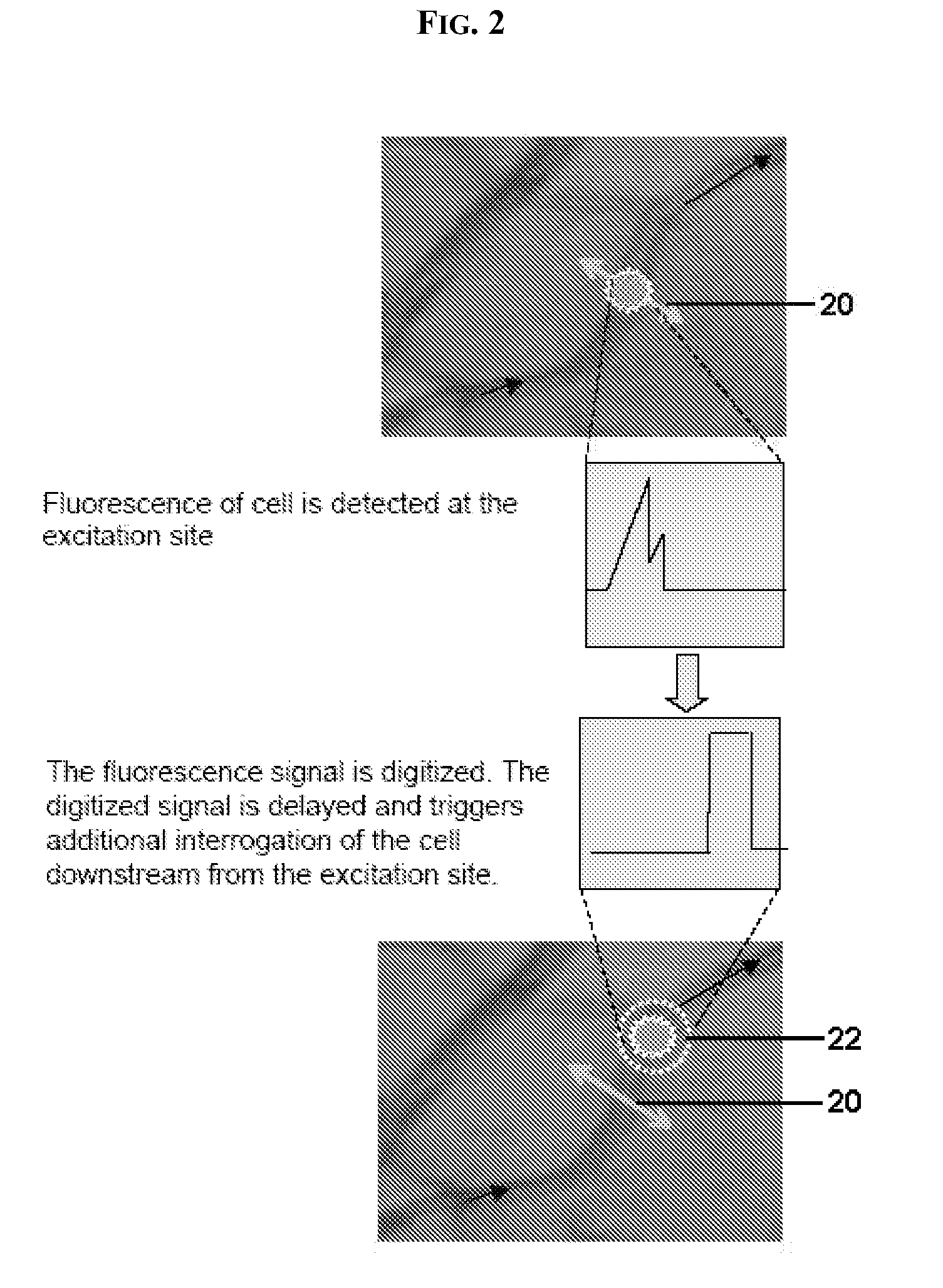
A two-photon in vivo flow cytometer (TIFC) similar to the exemplary embodiment shown above in FIG. 5 was used to detect both two-photon tryptophan fluorescence and one-photon DiD fluorescence from stationary DiD-labeled cells. As shown in FIG. 9A, the experimental TIFC system setup was similar to the embodiment shown in FIG. 5 except that a single multi-photon excitation source 74 forms the radiation source. The source 74 was used to excite both two-photon fluorescence of endogenous tryptophan and one-photon fluorescence of exogenous DiD. The source generates laser light with a wavelength of 590 nm and pulse width of 100 fs using the elements schematically shown in FIG. 9B. The source generally includes a femtosecond laser, an optical parametric oscillator, and a frequency doubler. In this experiment, the laser was a mode-locked Ti:sapphire laser (Spectra-Physics MaiTai-HP) that generates ultra-short pulses (100 fs) at a wavelength of 750 nm. This light is then injected into an opti...


PUM

PropertyMeasurementUnit
excitation wavelengthsaaaaaaaaaa
diameteraaaaaaaaaa

Abstract

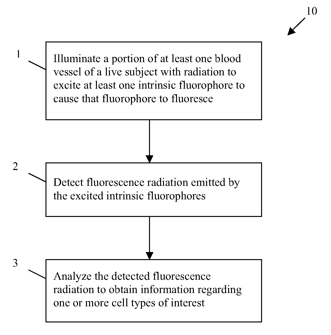
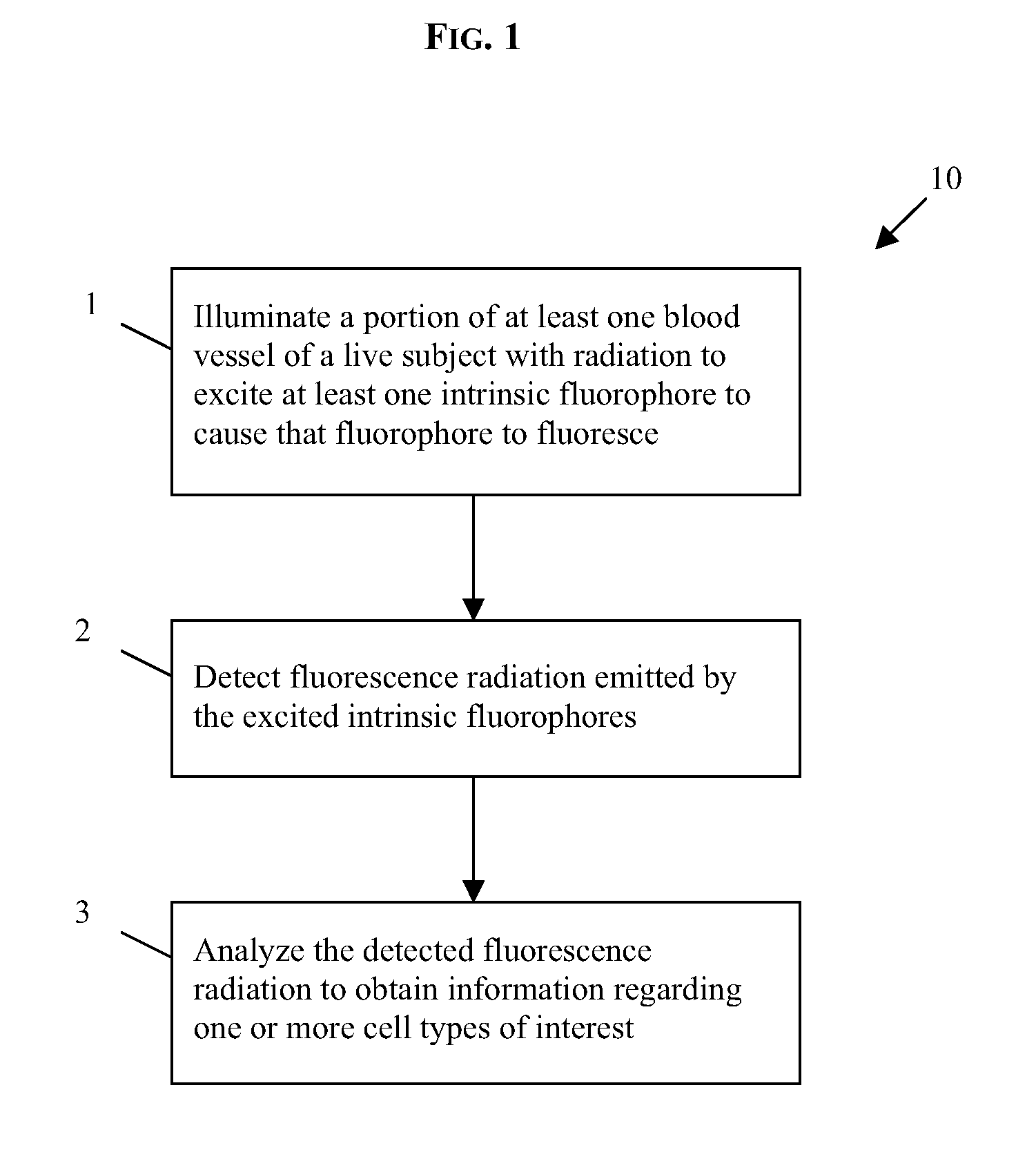
The present invention generally provides methods and systems for performing in vivo flow cytometry by using blood vessels as flow chambers through which flowing cells can be monitored in a live subject in vivo without the need for withdrawing a blood sample. In some embodiments, one or more blood vessels are illuminated with radiation so as to cause a multi-photon excitation of an exogenous fluorophore that was previously introduced into the subject to label one or more cell types of interest. In some other embodiments, rather than utilizing an exogenous fluorophore, endogenous (intrinsic) cellular fluorescence can be employed for in vivo flow cytometry. The emission of fluorescence radiation from such fluorophores in response to the excitation can be detected and analyzed to obtain information regarding a cell type of interest.

Description

BACKGROUNDThe present invention relates generally to methods and systems for performing flow cytometry, and more particularly to such methods and systems that allow performing in-vivo flow cytometry in a live subject without extracting blood.Many current techniques for detecting and quantifying various cell types circulating within a subject require extracting a blood sample from the subject followed by analysis of the extracted blood. For example, one such conventional method includes withdrawing a blood sample, fluorescently labeling specific cell populations ex vivo with antibody-targeted fluorescent markers, and passing those cells through a flow stream to be interrogated by a light source (typically a laser). Fluorescence and light scattering signals emitted, or reemitted, by the cells in response to the interrogating light can be employed to determine the types and the number of cells. Automated hematology analyzers can separate and count blood cell populations based on their ...

Claims


Application Information

Patent Timeline
Patent Type & Authority Applications(United States)
IPC IPC(8): A61K49/00G01J1/58
CPCG01N15/1434G01N33/56972G01N2015/008A61B5/0059G01N2015/1497G01N33/537Y10T436/13A61B5/0071A61B5/0068
Inventor LIN, CHARLES P.CARLSON, ALICIA L.ALT, CLEMENSBISS, DAVID P.PITSILLIDES, COSTAS M.LI, CHUNQIANG
Owner THE GENERAL HOSPITAL CORP
Why Eureka
Try Eureka