
Method for assaying nucleic acids by fluorescence

a nucleic acid and fluorescence technology, applied in the field of fluorescence assaying nucleic acids, can solve the problems of difficult comparison of methodologies, insensitiveness, and inability to allow the amount of circulating dna in patients to be measured

Inactive Publication Date: 2010-10-21
BIOCHEMIKON +1
View PDF2 Cites 48 Cited by
  • Summary
  • Abstract
  • Description
  • Claims
  • Application Information

AI Technical Summary

Benefits of technology

[0067]Apoptosis quantification within the frame of the present invention can proceed in kinetic condition, as experimentation goes along. Indeed, it is not necessary, for example, to stop cell culture to do the measure, since nucleic acids fragments eliminated in the external medium (e.g. culture medium) are measured. Remaining living cells allow continuation of the experimentation. Thus it is possible to repeat measurement a short time after, and so on, as often as necessary. In the same way it is possible to measure apoptosis occurring in humans or in animals, the measure medium will be here any type of biological sample.
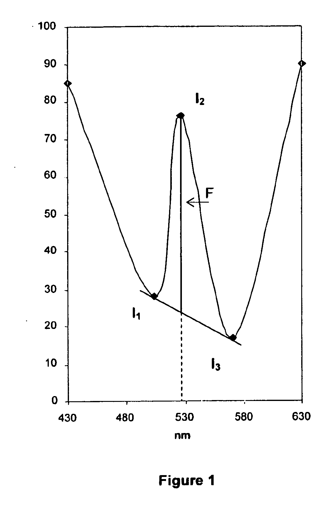
[0147]Advantageously, the method according to the invention allows the phenomenon of Rayleigh interference to be limited or prevented.
[0149]Although the use of just two wavelengths is less precise than the use of three wavelengths, it also allows the phenomenon of Rayleigh interference to be limited or prevented while at the same time being easy to carry out.Fluorimeter
[0153]The fluorimeter of the invention is preferably equipped with at least one means for supplying electric power, by mains or by battery (within a mobile use) and also at least one means for connecting to a computer system. If the fluorimeter is equipped with a plurality of connection means, these connection means will preferably be of several different standards. Within a mobile use, the fluorimeter is preferably as compact as possible, for example having the following dimensions: H<15 cm; L<25 cm; W<20 cm, and as light as possible so as to be able to be installed, without inconveniencing the user, for example on a laboratory benchtop, and to be able to be easily moved.
[0157]The various compartments of the tray (wells, reservoirs, vessel) are preferably connected by capillaries and the passage from one compartment to another is preferably caused by crushing, for example, of the buffer reservoir, the fluorophore reservoir and / or the standardisation reservoir. The passage of the buffer through the fluorophore reservoir allows the fluorophore to be diluted and the mixture as a whole to be driven toward the vessel. The sample, which may or may not have been diluted beforehand, can be deposited in the well in the tray before or after the tray is positioned in a fluorimeter. Preferably, a precise sample volume is deposited in the well, then an optimally determined amount of the sample is directed, by way of the standardisation reservoir, toward the vessel in order to be mixed therein with the fluorophore. The mixing in the vessel can then be carried out, for example by subjecting the vessel to pressure-vacuum or mechanic pressure. The tray according to the invention is advantageous because it allows extemporaneous mixing of the fluorophore in a concentrated solution and of the buffer in order to arrive at the final dilution which is optimal for fluorescence; it also allows homogenisation of the fluorophore and the sample and also, if appropriate, suitable dilution of the sample and measurement of the emitted fluorescence.

Problems solved by technology

A plurality of studies have attempted to approximate this threshold but it is quite difficult to compare them as methodologies, and the results obtained differ at many levels.
These process include, primarily, spectrophotometry [Greenstock et al., 1975]—a method used very commonly in all research laboratories and that has the benefit of being inexpensive but the drawback, which is a major one in clinical practice, of being extremely insensitive: it does not allow the amounts of circulating DNA in patients to be measured.
There are other more sensitive processes but they all have characteristics rendering them more or less difficult to use routinely, partly because they have a common step which is that of extracting the DNA from the biological environment prior to measuring:Radioimmunoassay processes [Leon et al., 1975] take a relatively long time (several hours per analysis series), have to be carried out in a series of measurements and not sporadically and, above all, require specialist structures and staff authorised to handle radioactive elements.Competitive PCR [Siebert et al., 1992; Yap et al., 1992], based on comparative display relative to known standards, is the addition of a plurality of processes in series providing detection sensitivity and specificity.
Nevertheless, it is not without drawbacks: compulsory extraction, relatively high handling costs, specific equipment which can not be used for large-scale routine analyses, and insufficient sensitivity.Quantitative fluorimetry [Greenstock et al., 1975].
However, the technical conditions under which it is used (microplate, volume reagent, measurement of fluorescence) prevent it from reaching the desired sensitivity limits and do not allow the origin of the detected DNA (human or non-human) to be identified.

Method used


Image

  • Method for assaying nucleic acids by fluorescence
  • Method for assaying nucleic acids by fluorescence
  • Method for assaying nucleic acids by fluorescence

Examples


example

Equipment

[0174]Plasmas or serums collected in herparinised EDTA tubes from centrifuged whole blood, then decanted; TBE 1× buffers (pH 8.5) (Tris 90 mM, boric acid 90 nM, EDTA 2 mM); polyethylene haemolysis tubes, acrylic vessels; synchronous fluorimeter (Shimadzu RF 535 reader, DR-3 data analyser); Picogreen® (dsDNA quantitation kit, Molecular Probes); standard DNA of known concentration for the standard range.

Method

A—Principle of Synchronous Fluorimetry

[0175]a) Difficulties Associated with Conventional Fluorimetry

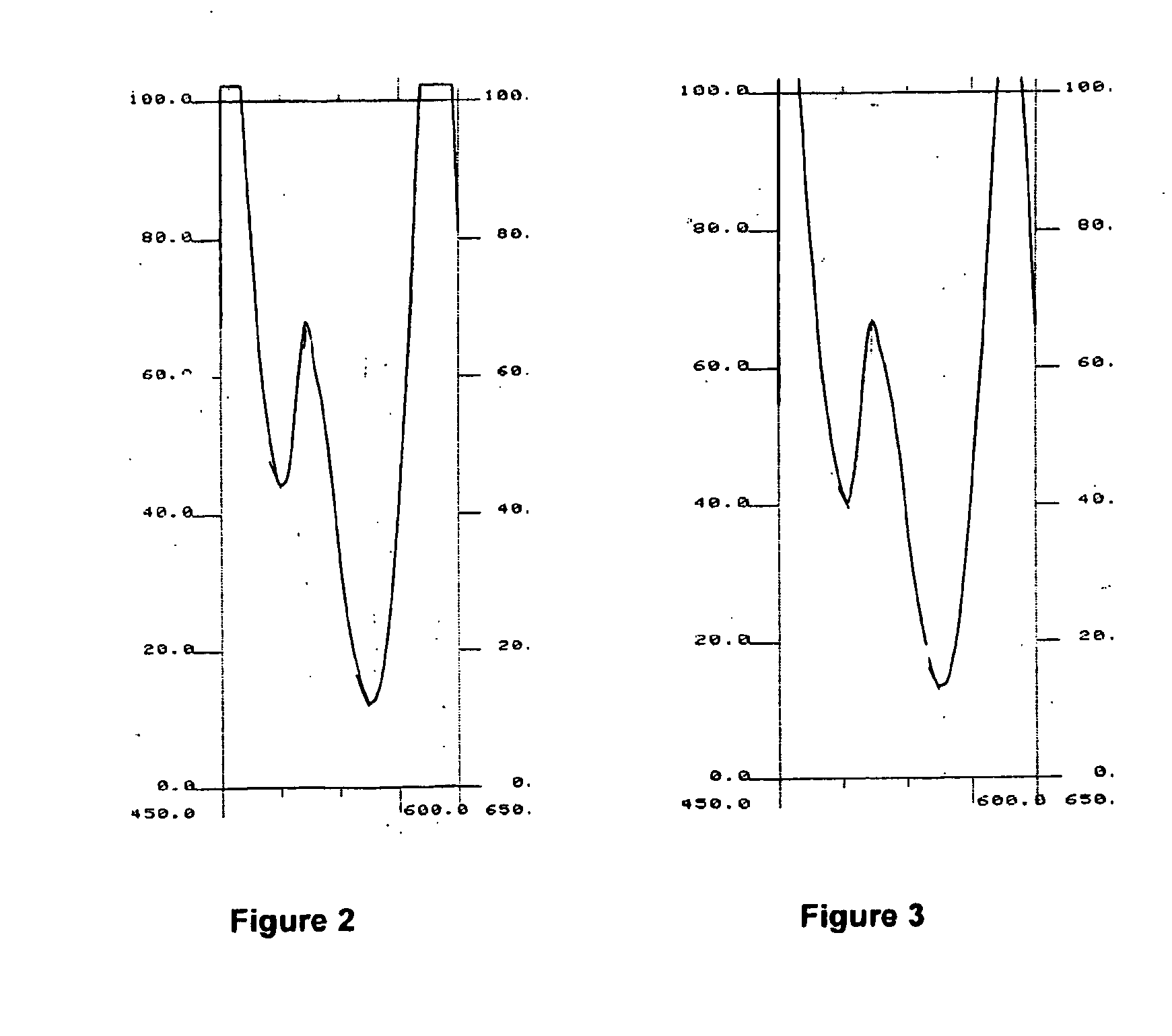
[0176]The main difficulty often associated with conventional fluorimetric detection is related to the Rayleigh scattering resulting from the proximity of the excitation and emission wavelength. This effect can be particularly intensive in the case of analysis of complex and proteinated media such as blood serum.

[0177]When determining a conventional fluorescence spectrum, one of the monochromators will sweep the spectrum while the second remains fixed. In order to produce a...


PUM

PropertyMeasurementUnit
fluorescence intensitiesaaaaaaaaaa
emission wavelengthsaaaaaaaaaa
excitation wavelengthsaaaaaaaaaa

Abstract

The invention relates to a method for determining the amount of nucleic acid present in a sample, wherein:—a fluorophore is added to the sample,—fluorescence intensities emitted by the fluorophore at least two emission wavelengths in response to light stimulations at least two excitation wavelengths respectively are measured, and—the amount of nucleic acid present in the sample is deduced from the measured fluorescence intensities.

Description

[0001]This application claims the benefit of prior U.S. provisional application 60 / 836,949 filed Aug. 11, 2006.[0002]The present invention relates to a fluorimetric method for determining the amount of nucleic acid present in a sample, to a fluorimeter suitable for carrying out said method and to a tray for fluorescence assay of a compound.[0003]The existence of free nucleic acid circulating in the blood has been known for a number of years [Mandel and Métais, 1947; Tan et al., 1966]. Since this time, it has been found using processes such as RIA (radioactive immunoassay) that patients suffering from cancers had a high level of circulating DNA relative to those suffering from benign tumors or relative to healthy subjects [Leon et al., 1977; Shapiro et al., 1983].[0004]However, these recent years, with the development of highly effective molecular biology processes such as real-time PCR, have seen a burgeoning interest in circulating DNA fragments with a view to use as a clinical bio...

Claims


Application Information

Patent Timeline
Patent Type & Authority Applications(United States)
IPC IPC(8): G01J1/58G01N23/00
CPCC12Q1/68C12Q1/6816G01J3/4406G01N21/6428G01N2021/6419G01N2021/6421G01N2021/6482C12Q2563/173C12Q2563/107C12Q2537/101C12Q2523/313
Inventor SACKO, MORYDELACOTTE, NICOLASBAH, MAMADOU SALIOUCONTI, MARC
Owner BIOCHEMIKON
Learn More