Dielectric spectroscopy assays for screening of ion channel ligands
- Summary
- Abstract
- Description
- Claims
- Application Information
AI Technical Summary
Benefits of technology
Problems solved by technology
Method used
Image
Examples
example 1
[0172]hERG cells used in the experiments are treated following the standard protocol: incubated at 37 degrees at 5% CO2 in DMEM media (with 10% fetal calf serum, penicillin streptomycin and 2 mM L-glutamine). When 80% confluent they are split and re-incubated. When the cells are needed for the experiment the flasks is treated with trypsin, incubated for 10 minutes. Trypsin is an enzyme that removes the cells from the flasks by cleaving. The cells are then gently diluted into DMEM media with 20% fetal calf serum (to annihilate the effects of trypsin) then span at 1000 g for 8 minutes. The pellet is gently and slowly resuspended in Tyrode's buffer for dielectric measurements.
example 2
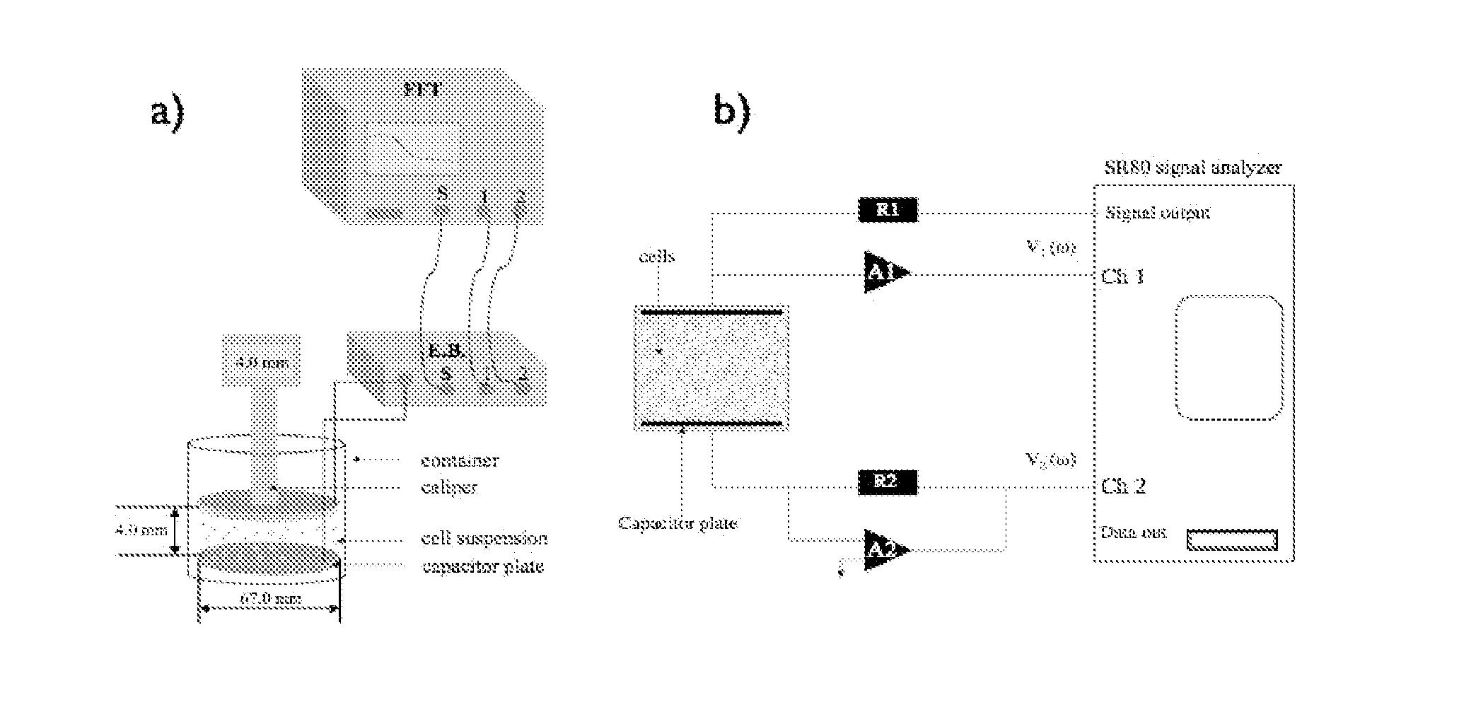
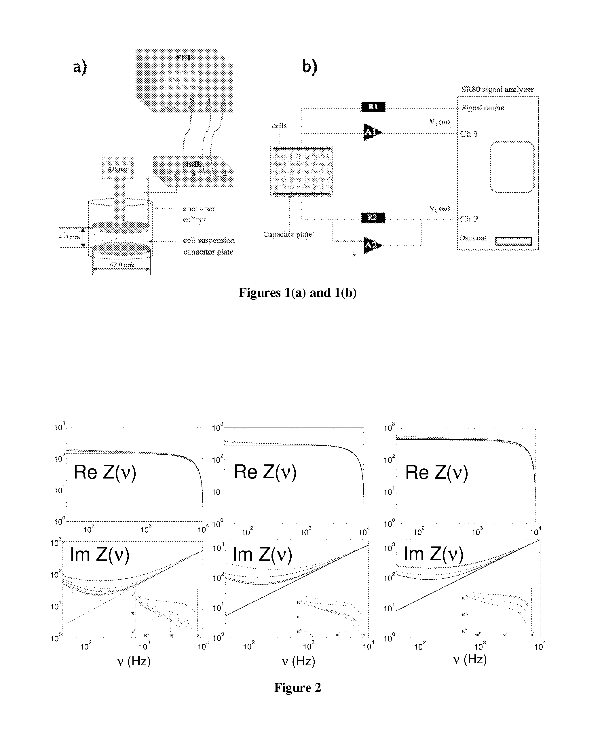
[0173]The solution to be measured is placed in a small, 30 ml glass beaker with 2 gold-plated flat circular plate electrodes as shown in FIG. 1. A Signal Analyzer SR80A provides a sinusoidal voltage at its signal output. It also digitizes the voltage at Channels 1 and 2 and takes the ratio of these two as function of frequency. The real and imaginary of this ratio is stored on a computer. The bottom electrode plate is held at ground potential through the negative input of the Amplifier A2. The output voltage from the Signal Analyzer is applied to the upper electrode, through resistor R1. As a result, the current I that flows through the cell suspension produces a voltage V1, equal to the product of I and impedance Z of the probe. The voltage V2 is equal to minus the product of I and resistance R2. Thus the transfer function, T, is directly related to the cell suspension impedance by: T=V1 / V2=Z / R2. The complex dielectric function is then extracted from Z. All the electronic component...
example 3
Activity of hERG Ion Channels in the Presence of Antiarrhythmic Agents and Antihistamines with Carditoxicity Effects
[0174]The hERG ion channel activity is studied on HEK-293 cell line with overexpresses hERG ion channels. Antiarrhythmic agents tested include quinidine, a class 1 agent, and propranolol, a class 2 agent. An antihistamine agent, astemizole, was also tested. For these compounds, the membrane potential variation was determined by dielectric spectroscopy and the IC50 curve is compiled. The results from the dielectric spectroscopy are then closely compared with patch clamping experiments done in parallel. The compounds analyzed herein were chosen to test a large range of potency. For example, 2 μLM quinidine is needed to block 50% of the hERG channels while only 1.3 nM astemizole is required to produce the same effect.
HEK-293 cells: Determining Drug Potency Using DS. Comparison with Traditional Methods
[0175]The cells used in the experiments were treated following the stand...
PUM
 Abstract
Description
Claims
Application Information
 - R&D
- Intellectual Property
- Life Sciences
- Materials
- Tech Scout
- Unparalleled Data Quality
- Higher Quality Content
- 60% Fewer Hallucinations
Browse by: Latest US Patents, China's latest patents, Technical Efficacy Thesaurus, Application Domain, Technology Topic, Popular Technical Reports.
© 2025 PatSnap. All rights reserved.Legal|Privacy policy|Modern Slavery Act Transparency Statement|Sitemap|About US| Contact US: help@patsnap.com


