
Method and apparatus for tissue equivalent solid state microdosimetry

a solid state microdosimetry and tissue technology, applied in the field of tissue equivalent solid state microdosimetry, can solve the problems of insufficient consideration of delta ray energy distribution and its relationship to spatial dose distribution, inability to predict the radiobiological effects of biological tissue for all types of radiation, and limited correlation of let approach with radiobiological effects. to achieve the effect of accurate observation

Active Publication Date: 2010-04-15
UNIV OF WOLLONGONG
View PDF4 Cites 28 Cited by
  • Summary
  • Abstract
  • Description
  • Claims
  • Application Information

AI Technical Summary

Benefits of technology

[0042]However, in other embodiments it is possible to provide submicron scale detectors (i.e. “nano-dosimetry”), with little noise from readout electronics.
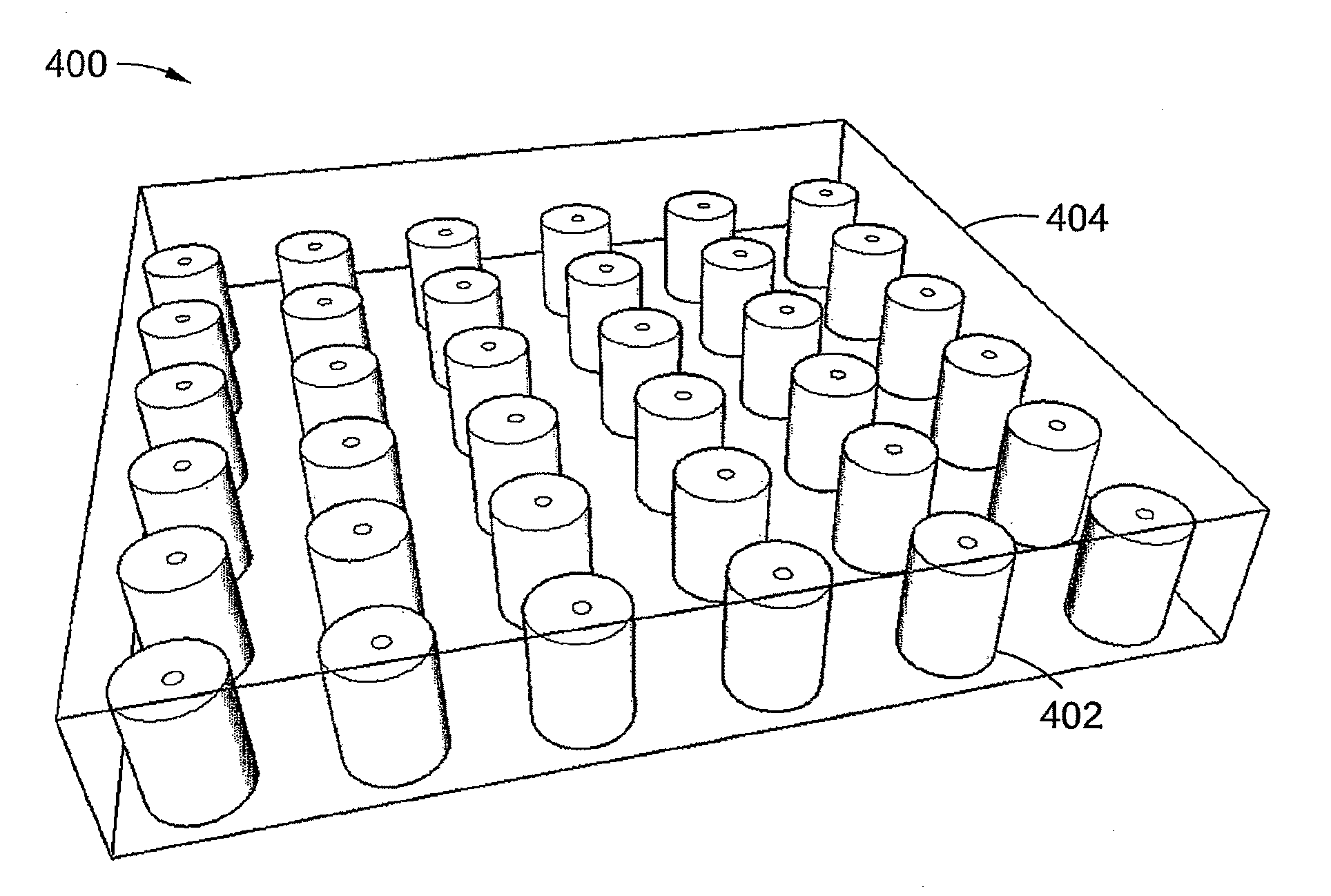
[0047]One advantage of this microdosimeter is its essentially 100% collection of charges produced by charged particles owing to the absence of lateral diffusion (which occurs with planar p-n junctions in existing arrangements). It allows accurate determinations of deposited energy event by event, and allows error reductions in the determination of dose equivalents.
[0049]In one particular embodiment, the detectors are connected such that each of the detectors is not connected to at least one of its immediately neighbouring detectors of the detectors. This allows the more accurate observation of the pattern of charge particle track produced by δ-electrons and the use of more sophisticated approaches in the determination of dose equivalent.

Problems solved by technology

J / kg, but cannot predict the radiobiological effects in biological tissue for all types of radiation.
It limits the correlation of the LET approach with radiobiological effects.
Firstly, delta ray energy distribution and its relationship to spatial dose distribution are not adequately considered.
These limitations in LET lead to the formulation of a set of measurable stochastic quantities that provide the fundamental basis for the field of microdosimetry.
1. TEPCs require a gas supply system that are inconvenient in many applications;
2. TEPCs are large, up to 1 to 2 cm in diameter, which limits their use where spatial resolution is required;
3. TEPCs require high voltage biases; up to 2 kV; and
4. TEPCs suffer from the “wall effect” and other physical size related effects, being much larger than the cell structures, leading to artefacts in microdosimetric spectra.
For example, MOSFET detectors (which have a very small SV of micron or submicron size) are able to measure absorbed doses with 1 micron spatial resolution, but cannot distinguish the energy deposited in the SV due to a particular event.
These detectors are not a suitable substitution for TEPCs, as do not have proper LET resolution and cannot be used in real time dosimetry.
Recombination of charge in the gate oxide is well known in MOSFET detectors, and reduces the utility of MOSFET detectors for dosimetry in proton and heavy ions fields (even in an active mode).
This microdosimeter is designed principally to distinguish the gamma and neutron components of a radiation field, but it can only with difficultly obtain dose equivalent using the weighting coefficient Q in arbitrary radiation fields as recommended by the ICRU.
Applications of such planar arrays of p-n junctions for regional microdosimetry are limited owing to uncertainty in the average chord, charge collection efficiency within the SV, overlayers and shape of the SV.
Increasing the total area of the p-n junction array leads to increases in the noise owing to an increase in capacitance that reduces the minimal LET detected by the microdosimeter.
Hence, interpretation of MCA spectra for conversion to dose equivalent is complex.
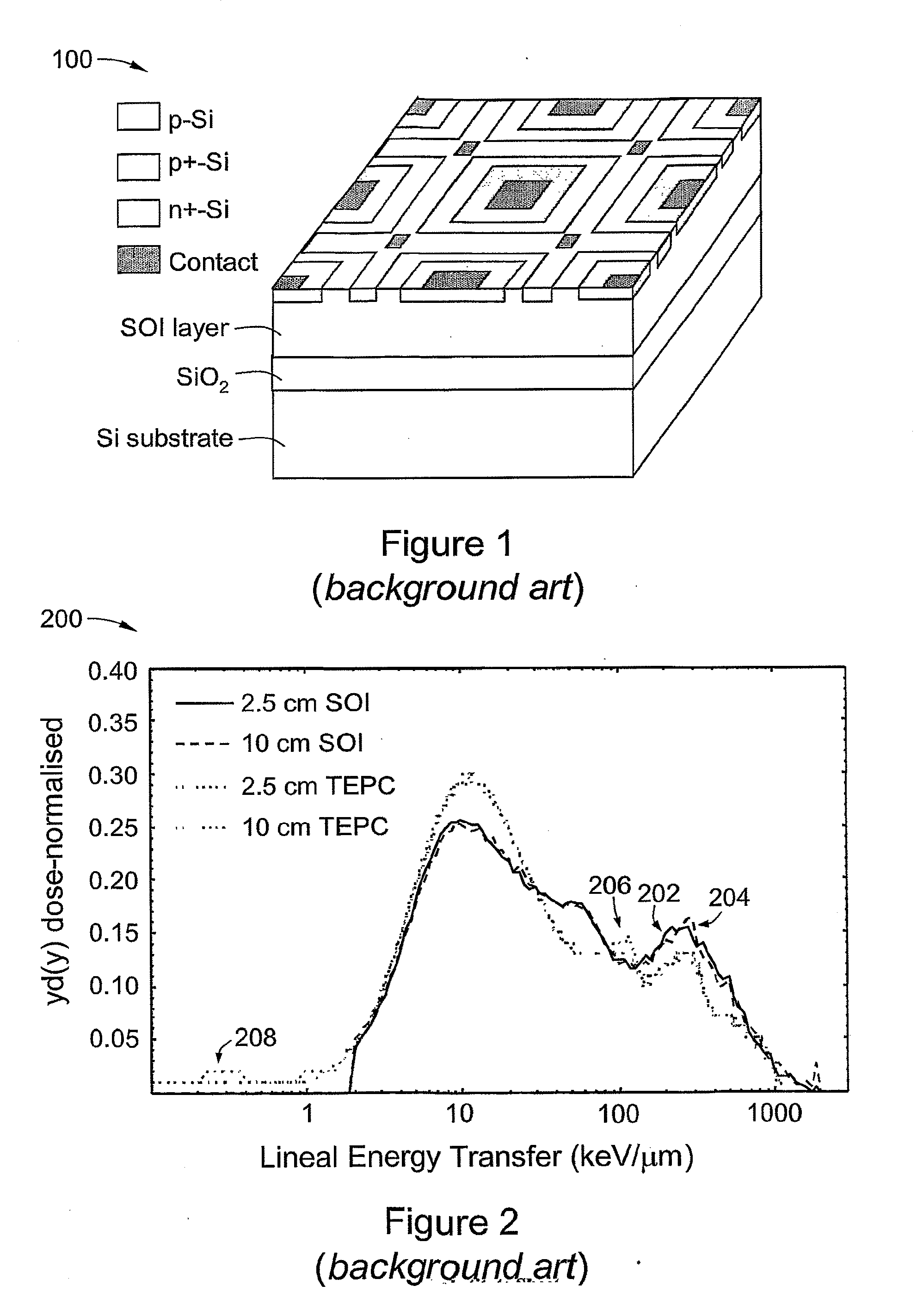
It is apparent from FIG. 2 that an SOI microdosimeter with an array of planar p-n junctions is essentially unable to measure events with LET (Lineal Energy Transfer) less than 1-2 keV / μm; this is due to detector noise.
Losses of charge in a planar P-N junction cell due to recombination prevents the accurate measurement of energy deposited in a cell from a single event, thus reducing the accuracy of equivalent dosimetry.
This is a disadvantage of the current design of solid state microdosimeters with planar p-n junction.
Also, reducing the size of the SV to reduce the charge deficit reduces the energy deposited in the SV, and this technique is also limited by the noise of the detector.
This produces an error in the determination of the cluster of deposited energy in a single SV.

Method used


Image

  • Method and apparatus for tissue equivalent solid state microdosimetry
  • Method and apparatus for tissue equivalent solid state microdosimetry
  • Method and apparatus for tissue equivalent solid state microdosimetry

Examples


example 1

[0116]Measurement of TE Microdosimetric Spectra on Pu—Be Gamma-Neutron Source.

[0117]The response of an SOI microdosimeter of the background art (cf. FIG. 1) in a mixed gamma / neutron field produced from a 238Pu—Be radioisotope neutron source was measured. The source was located in a general purpose neutron irradiation facility of floor area 7.5 m2; the facility was surrounded by walls 400 mm thick and 1.75 in high of borated paraffin and concrete. The source activity was 325 GBq with an estimated neutron emission rate of 2×10⁷ neutrons in 4π. The average energy of fast neutrons was approximately 4 MeV with a maximum energy of about 10 MeV. The dominant gamma component was a 4.4 MeV emission associated with the decay of the bound excited state of 12C* produced in the Be(α,n) reaction within the 238Pu—Be source. The 5 μm thick SOI microdosimeter with 4800 planar parallel p-n junctions was mounted at a distance of approximately 2.6 cm from the source.

[0118]FIG. 9 is an example of a set ...

example 2

[0119]Measurements were made of dose equivalent of neutron contamination in a patient phantom produced during proton therapy.

[0120]The interaction of 250 MeV protons with beam modifying devices and the patient produces fast neutrons that are deposit the-energy dose in the patient and may be the cause of secondary cancer. Estimates were made of the equivalent neutron dose within the phantom outside the proton field using solid state microdosimeter and the steps of the data analysis method of the above embodiment.

[0121]FIG. 10 is a set of plots 1000 of TE microdosimetric spectra obtained from the MCA spectra with a background art 10 μm SOI microdosimeter comparable to that of FIG. 1 in a water phantom at different depths in the water phantom along the proton Bragg peak. Proton energy was 190 MeV; the range was 26 cm. For clarity, the individual spectra—according to data collection position—are labelled as follows:

[0122]1002 x=2.2 cm

[0123]1004: x=20 cm

[0124]1006: x=23.2 cm

[0125]1008: x...


PUM


Abstract

A microdosimeter, comprising an array of three-dimensional p-n junction semiconductor detectors, each providing a sensitive volume-target and a tissue equivalent medium for generating secondary charged particles. The array is manufactured from a semiconductor on insulator wafer and the detectors are located to detect secondary charged particles generated in the tissue equivalent medium.

Description

RELATED APPLICATION [0001]This application is based on and claims the benefit of the filing date of AU application no. 2006907071 filed 19 Dec. 2006, the content of which is incorporated herein by reference in its entirety.FIELD OF THE INVENTION [0002]The invention relates to a method and apparatus for tissue equivalent solid state microdosimetry, of particular but by no means exclusive application in predicting the probability of cell damage in a radiation field and for predicting Single Event Upsets (SEUs) in microelectronics and dose equivalent measurement.BACKGROUND OF THE INVENTION[0003]Classical radiation absorbed dosimetry operates to determine the average energy deposited per unit mass,[0004]J / kg, but cannot predict the radiobiological effects in biological tissue for all types of radiation.[0005]Early attempts at understanding cellular radiation effects recognized that knowledge of the energy distribution at a scale comparable to the structures affected by irradiation (“end...

Claims


Application Information

Patent Timeline
Patent Type & Authority Applications(United States)
IPC IPC(8): G01T1/02G01T1/16
CPCH01L27/1446G01T1/026
Inventor ROZENFELD, ANATOLY
Owner UNIV OF WOLLONGONG
Learn More