
Promoters exhibiting endothelial cell specificity and methods of using same for regulation of angiogenesis

a technology of endothelial cells and promoters, which is applied in the field of promoters exhibiting endothelial cell specificity and methods of using same for regulation of angiogenesis, can solve the problems of difficult to suppress angiogenesis, limiting clinical use, bleeding and blindness,

Inactive Publication Date: 2010-04-01
VASCULAR BIOGENICS
View PDF80 Cites 17 Cited by
  • Summary
  • Abstract
  • Description
  • Claims
  • Application Information

AI Technical Summary

Benefits of technology

This approach enables precise regulation of angiogenesis in specific tissue regions, potentially leading to more effective treatments for diseases by enhancing the expression of angiogenic factors or inducing apoptosis in angiogenic cells, while minimizing systemic toxicity and improving therapeutic efficacy.

Problems solved by technology

In diabetes, new capillaries formed in the retina invade the vitreous humor, causing bleeding and blindness.
Until recently, the angiogenesis that occurs in diseases of ocular neovascularization, arthritis, skin diseases, and tumors, had been difficult to suppress therapeutically.
Unbalanced angiogenesis typifies various pathological conditions and often sustains progression of the pathological state.
However, for mass formation of long lasting functional blood vessel there is a need for repeated or long term delivery of the above described protein factors, thus limiting their use in clinical settings.
Furthermore, in addition to the high costs associated with the production of angiogenesis-regulating factors, efficient delivery of these factors requires the use of catheters to be placed in the coronary arteries, which further increases the expense and difficulty of treatment.
Though promising in pre-clinical models, to date systemic administration of all anti-angiogenic agents tested in clinical trials, have shown limited rate of success and considerable toxicities including thrombocytopenia, leukopenia and hemoptysis.
These results suggest that there may be limits to the use of current tumor anti-angiogenic agents as therapy for advanced malignancies.
Interestingly, poor results have also been obtained when anti-angiogenic therapy (e.g., heparin, heparin-peptide treatment) directed at smooth muscle cell proliferation has been practiced on myocardial ischemia in patients with coronary artery disease [Liu et al., Circulation, 79: 1374-1387 (1989); Goldman et al., Atherosclerosis, 65: 215-225 (1987); Wolinsky et al., JACC, 15 (2): 475-481 (1990)].
Various limitations associated with the use of such agents for the treatment of cardiovascular diseases included: (i) systemic toxicity creating intolerable level of risk for patients with cardiovascular diseases; (ii) interference with vascular wound healing following surgery; (iii) possible damage to surrounding endothelium and / or other medial smooth muscle cells.
Thus, these and other inherent obstacles associated with systemic administration of anti-angiogenic factors (i.e., manufacturing limitations based on in-vitro instability and high doses required; and peak kinetics of bolus administration attributing to sub-optimal effects) limit the effective use of angiogenic factors in treating neo-vascularization associated diseases.
However, a high mutation rate is not the only mechanism for cancer's genetic instability.
There is evidence of “apoptotic bodies” phagocytosed by tumor cells, resulting in aneuploidy and a further increase in genetic instability.
Although angiogenic endothelial cells involved in tumor progression proliferate rapidly, they differ from tumor cells by their genomic stability, and thus also in minimal drug resistance and low likelihood of the development of mutant clones.
However, since these agents are proteins and their administration therefore depends on frequent intravenous administration, their use poses serious manufacturing and maintenance difficulties.
Although, great efforts have been directed towards developing methods for gene therapy of cancer, cardiovascular and peripheral vascular diseases, there is still major obstacles to effective and specific gene delivery [for review see, Feldman A L.
However, these promoters showed only weak activity and did not allow for high levels of expression.
However, no expression was detected in the vascular beds of the spleen, lung, liver, kidney, heart, testes and aorta as well as in the thrombomodulin locus.
However, expression in adult was limited to the vessels of the lung and kidney and no expression was detected in the heart, brain, liver.
Thus, none of these sequences work uniformly in all endothelial cells of all developmental stages or in the adult animal.
To date, however, the toxicity of recombinant forms of endogenous anti-angiogenic agents has not been demonstrated although some synthetic anti-angiogenic agents have been associated with toxicity in preclinical models [Kong and Crystal (1998) J. Natl. Cancer Inst.
Angiostatin has also been used as a possible anti-angiogenic agent (Folkman et al, Cell 1997 Jan. 24; 88(2):277-85), however due to the redundancy of factors involved in regulation of angiogenesis in tumors, it is highly unlikely that angiostatin therapy alone would be effective.
However, inhibiting the formation of new blood vessels and / or partially destroying them may be insufficient in cancer pathologies where a dramatic anti angiogenic effect that destroys most or all existing angiogenic blood vessels and induce tumor necrosis is required.
However, this patent neither implies nor demonstrates that an endothelial-specific enhancer can be employed to increase the level of expression achieved with the PPE promoter while preserving endothelial specificity.
Further, this patent does not teach that the PPE-1 promoter is induced to higher levels of transcription under hypoxic conditions.
Moreover, since GCV toxicity is based on DNA synthesis, it affects mainly proliferating cells.
Nevertheless, results are disappointing, mostly limited in vivo by a low transduction percentage.
However, to date, clinical studies have demonstrated only limited results.

Method used


Image

  • Promoters exhibiting endothelial cell specificity and methods of using same for regulation of angiogenesis
  • Promoters exhibiting endothelial cell specificity and methods of using same for regulation of angiogenesis
  • Promoters exhibiting endothelial cell specificity and methods of using same for regulation of angiogenesis

Examples


example 1

In-Vitro Assay for Pro-Apoptotic Gene Activity in Endothelial Cells (BAEC) and 293 Cells

[0404]In cancer treatment, anti-angiogenic therapy targets the evolving vasculature which nourishes the growing tumor [Folkman J. N Engl J Med (1995) 333(26):1757-63]. As the research of apoptosis, or programmed cell death, has progressed, numerous genes that encode selective and efficient cell death regulators have been identified [Strasser et al. Annu Rev Biochem (2000) 69:217-45.].

[0405]The present study screened several pro-apoptotic genes in order to identify an agent most suitable for anti-angiogenic therapy. Several pro-apoptotic genes including MORT1 (FADD—Fas associated death domain protein, GenBank Accession number NM—003824), RIP (receptor-interacting-protein, GenBank Accession number U25995), CASH (c-FLIP, GenBank Accession number AF010127), MACH (caspase 8 GenBank Accession number X98172), CPP32 (caspase 3, GenBank Accession number U13737), caspase 9 (U60521) and Fas-chimera (Fas-c),...

example 2

Production of Recombinant Adenoviruses Encoding Fas-Chimera Under the Control of the Modified PPE-1 Promoter (PPE-1(3×)

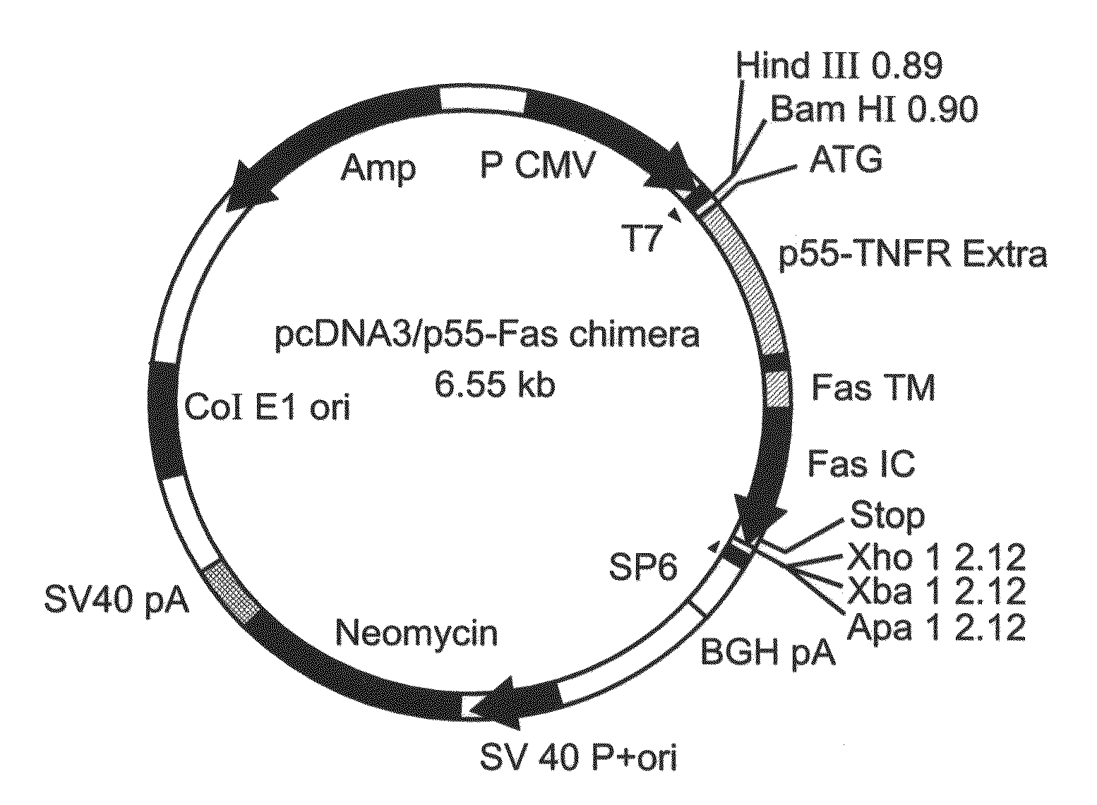
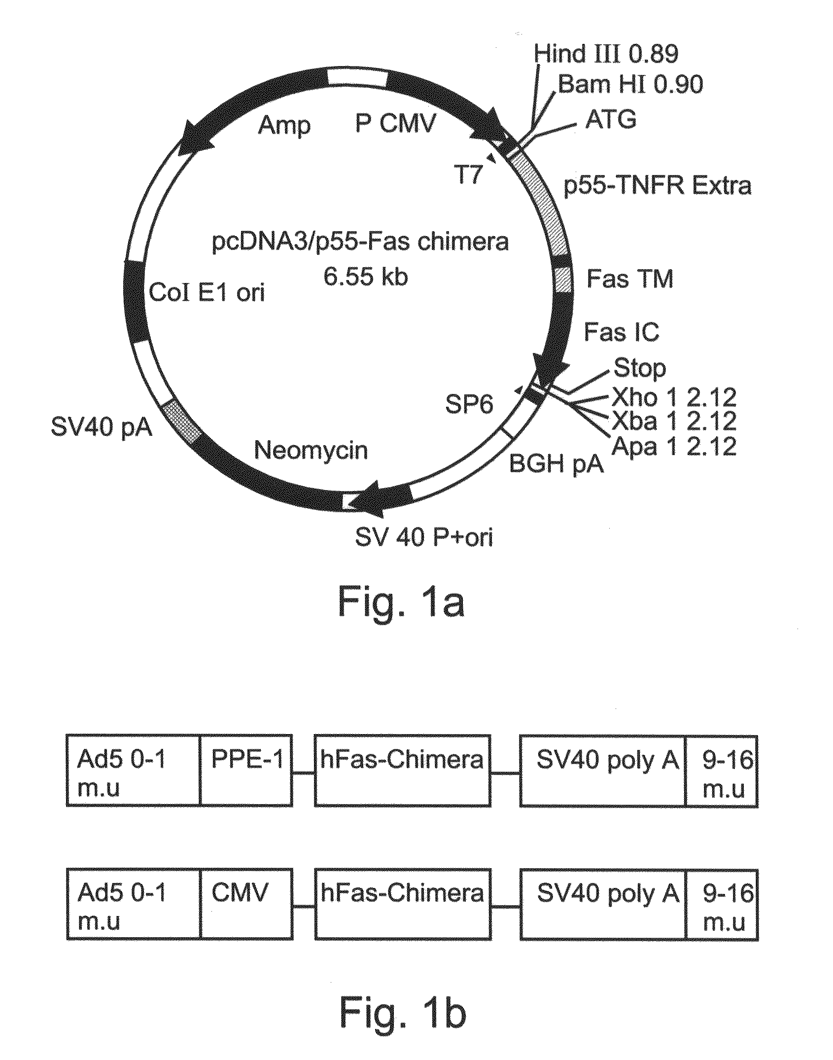
[0407]A cDNA encoding a full length Fas-chimera was subcloned into the plasmid pPACPPE1-3× containing the modified pre-proendothelin1 promoter (see FIG. 1b). Recombinant adenoviruses were produced by co-transfection of this plasmid with pJM17 plasmid into human embryonic kidney 293 cells. Successful viral cloning was verified via PCR amplification (FIG. 5a).

[0408]In order to determine the expression of Fas-c in the target cells, endothelial BAEC cells were transduced with the indicated titer of Ad-PPE-1(3×)-Fas-c. 72 h post transduction cells were lysed and cellular proteins resolved using a non-reducing SDS-PAGE gel. Western blot analysis was performed using anti-TNFR1 antibody (Sc-7895, Santa-Cruz Biotech). As demonstrated in FIG. 5b, a prominent band migrating at 45 kD was clearly evident and its expression was dose-dependent, suggesting correct folding and expre...

example 3

Ad-PPE-1 (3×)-Fas-c Expression Induces Apoptosis in Endothelial Cells

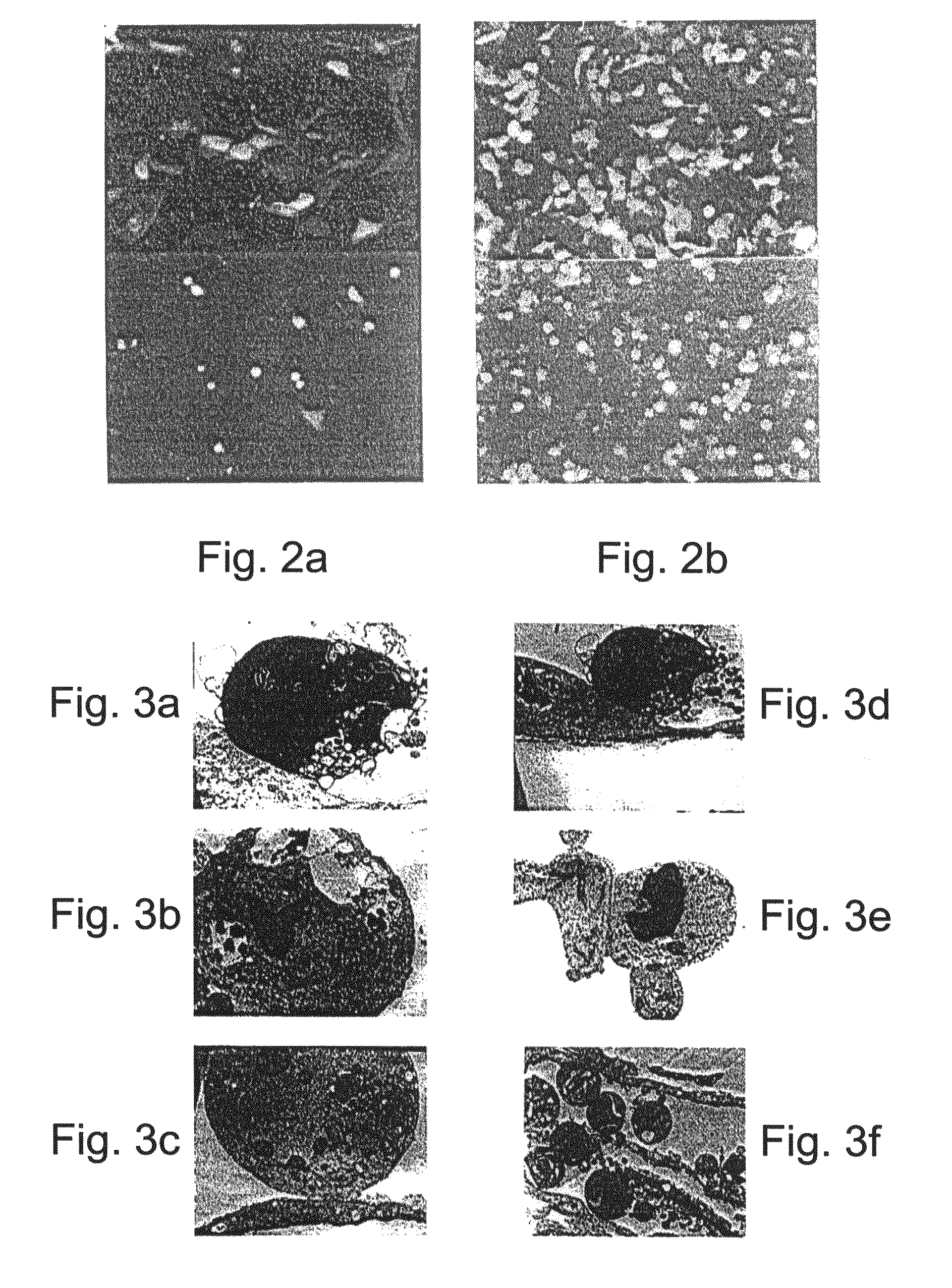
[0409]The ability of Ad-PPE-1(3×)-Fas chimera to induce apoptosis of endothelial cells was determined. As shown in FIGS. 6a-b, pre-proendothelin directed, adenovirus-mediated transduction of endothelial cells resulted in an evident and massive cell death; HUVEC and BAEC infected with Ad-PPE-1(3×)-Fas-c (103 MOI) had morphological features of adherent cells undergoing apoptosis including membrane blebbing, rounding and shrinking and detachment from the culture dish. In contrast, cells infected with control viruses at the same MOI, maintained normal appearance and growth rate. Cells transduced with 100 MOI presented only a minimal degree of cell death (data not shown).

[0410]Further assessment of the cytotoxic properties of Ad-PPE-1(3×)-Fas-c was effected by expressing this virus in cells expressing the reporter gene GFP under the control of the PPE-1 promoter. As is evident from FIGS. 6c-d, most of the transduced cel...


PUM

PropertyMeasurementUnit
thicknessaaaaaaaaaa
concentrationsaaaaaaaaaa

Abstract

Isolated polynucleotide sequences exhibiting endothelial cell specific promoter activity, novel cis regulatory elements and methods of use thereof enabling treatment of diseases characterized by aberrant neovascularization or cell growth are disclosed.

Description

[0001]This application is a Divisional of U.S. patent application Ser. No. 10 / 988,487, filed on Nov. 14, 2004, which is a Continuation-In-Part of U.S. patent application Ser. No. 10 / 135,447, filed May 1, 2002, now U.S. Pat. No. 7,067,649, issued on Jun. 27, 2006, which is a Continuation-In-Part of PCT / IL01 / 01059, filed Nov. 15, 2001, now expired, which claims the benefit of priority from U.S. Provisional Patent Application No. 60 / 248,582, filed Nov. 17, 2000, the specifications of which are hereby incorporated by reference.[0002]U.S. patent application Ser. No. 10 / 988,487, filed on Nov. 14, 2004 is also a Continuation-In-Part of U.S. patent application Ser. No. 10 / 490,746, filed Apr. 12, 2004, now U.S. Pat. No. 7,585,666, issued on Sep. 8, 2009, which is a National Phase Application of PCTIL02 / 00339, filed May 1, 2002, now expired, which claims the benefit of priority from U.S. Provisional Patent Application No. 60 / 330,118, filed Oct. 19, 2001, now expired, the specifications of whi...

Claims


Application Information

Patent Timeline
Patent Type & Authority Applications(United States)
IPC IPC(8): C07H21/04A61K38/17A61K48/00C07K14/475C07K14/515C07K14/575C12N15/85C12N15/861
CPCC12N2830/30C07K14/475C12N2840/203C07K14/52C07K14/49C12N15/85C12N2840/20C07K2319/70A61K48/0008C12N2800/108C07K14/57536C12N2830/15C12N2830/85C12N2830/008A61K48/0058C07K14/503A61K48/00C07K14/515C07K14/70578C12N2710/10343C12N15/86A61K38/00C12N2830/002A61P17/06A61P19/02A61P27/02A61P35/00A61P35/04A61P43/00A61P9/00A61P9/10A61P9/14C12N15/00C12N5/00C07H21/04
Inventor BREITBART, EYALBANGIO, LIVNATPELED, MICHAELGREENBERGER, SHOSHANAHARATS, DROR
Owner VASCULAR BIOGENICS
Learn More