
Pulsed cavitational ultrasound therapy

a cavitational ultrasound and ultrasound therapy technology, applied in the field of ultrasonic therapy, can solve the problems of inability to definitively establish the optimal treatment of small renal masses, internal adhesion formation and cosmetic problems, and the risk of infection, so as to enhance drug delivery and enhance uptake or transport

Inactive Publication Date: 2010-03-18
RGT UNIV OF MICHIGAN
View PDF106 Cites 55 Cited by
  • Summary
  • Abstract
  • Description
  • Claims
  • Application Information

AI Technical Summary

Benefits of technology

The present patent provides a non-invasive, non-thermal technology for inducing mechanical cavitation of tissue using pulsed, focused ultrasound energy. This technology can be used for controlled mechanical subdivision of soft tissue, which involves the formation of bubbles in the tissue that can be detected using ultrasound imaging. The bubbles can then be sustained for a period of time, resulting in the fractionation of the tissue. The technology can also be used for enhanced drug delivery by inducing mechanical disruption of tissue and allowing for the transport of drugs or other substances across cell membranes. The method is non-thermal, meaning it does not produce significant heating of the tissue, and can be easily monitored and adjusted in real-time during treatment. The technology can also be used to optimize the process of cavitation by monitoring and adjusting the bubble cloud dynamics. Overall, the technology provides a significant advantage over previous methods and can be used for various therapeutic purposes.

Problems solved by technology

Several negative effects can be associated with invasive therapies, including the risk of infection, internal adhesion formation and cosmetic issues related to skin surface scarring, and the need for pain management during and after the procedure.
However, the optimal treatment for small renal masses has yet to be definitively established and continues to evolve.
However, these methods are all invasive therapies.
These minimally invasive methods deliver energy via percutaneous probes to induce thermal effects that cause cellular injury and death in the targeted region.
However, inhomogeneous tissue heating / cooling, variable blood perfusion resulting in heat sink effects, and changing tissue characteristics during treatment, are factors that are difficult to predict or control and ultimately may limit these thermal ablative modalities.
Unfortunately, this technology may also be limited by the inability to precisely control the margin of thermal injury as well as the lengthy time required to closely pack hundreds of lesions necessary to ablate a clinically useful volume of tissue.
Furthermore, therapies that deliver therapeutic agents, including pharmaceutical compositions and various drugs, to a site in need to treatment can still be frustrated due to natural barriers in spite of local delivery.
Single injections and / or continuous administration via a cannula pump can deliver a therapeutic agent to a localized site, however, one or more barriers may still prevent optimal efficacy.
Ultrasound has been used to enhance drug uptake or delivery, although the mechanisms of observed effects, which are often modest at best, are poorly understood.
Many experiments or devices use acoustic parameters that are arrived at by trial and error approaches with no rational basis for optimization.
The primary reason for avoiding cavitation is that it is very unpredictable due to significant variations in cavitation thresholds which are usually depend on the quantity and quality of small gas bubbles and other cavitation nuclei in different tissues.
This makes it impossible to obtain reliable results with predictable dose-effect relationships, and make it very difficult to predict the degree of enhanced transport of deliverable substances.
Moreover, prior art methods of assisted drug delivery do not allow easy assessment or feedback of when the process is operating effectively, and often do not provide any feedback which can be used to optimize the process.
However, use of ultrasound based therapies has been problematic due the phenomenon of acoustic cavitation.
Specifically, when an acoustic field is propagated into a fluid, the stress induced by the negative pressure produced can cause the liquid to rupture, forming a void in the fluid which will contain vapor and / or gas.
However, previous invasive methods of tissue removal or ablation, bulk tissue fractionization, and delivery of therapeutic agents, and even noninvasive methods, do not allow easy assessment or feedback of when the process is operating effectively, and often do not provide any feedback which can be used to optimize the process.

Method used


Image

  • Pulsed cavitational ultrasound therapy
  • Pulsed cavitational ultrasound therapy
  • Pulsed cavitational ultrasound therapy

Examples


example 2

Optical and Acoustic Feedback and Monitoring of Bubble Cloud Dynamics

[0174]Optical Detection: The optical attenuation method detects light absorption and scattering by the bubbles when a bubble cloud is created. A laser beam is projected through the ultrasound focus in front of the tissue and the light intensity is monitored continuously by a photodetector. Optical attenuation detection is capable of monitoring real-time bubble cloud dynamics without interference from the tissue or disturbing the ultrasound field, yet simple and of low cost. The temporal resolution of the optical attenuation method depends on the response time of the photo-detector. It can easily reach nanoseconds or better with very reasonable cost equipment. This enables almost continuous monitoring of the bubble cloud compared to the time scale of acoustic therapy pulse (on the order of μs and above). Using this detection scheme, we expect to gain much fundamental knowledge of the temporal dynamics of the bubble ...

example 3

Selection of Parameters to Detect Initiation of Variable Acoustic Backscatter

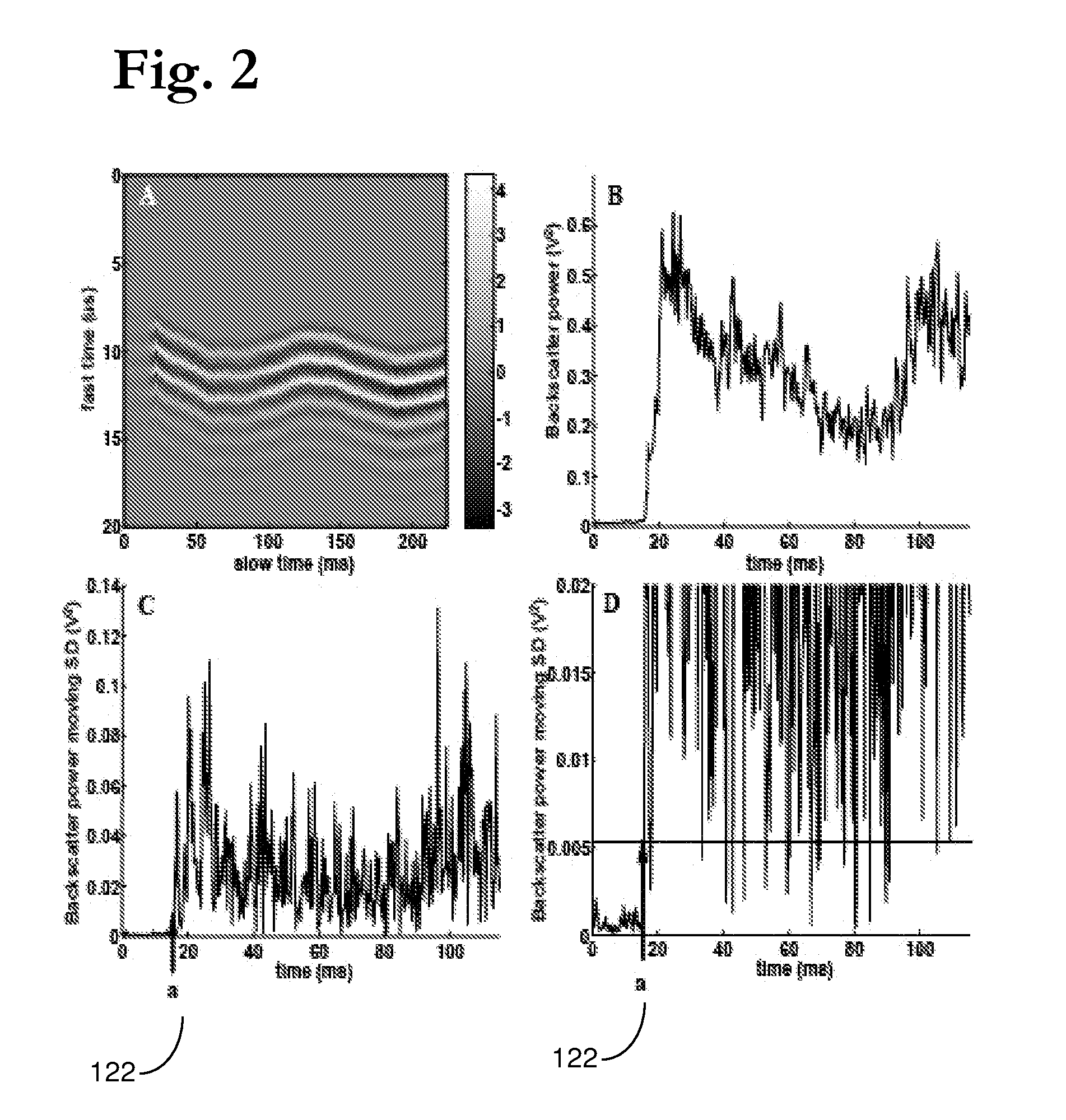
[0199]To identify points of initiation and extinction based on variability in the backscatter signal, we applied a common technique from the area of statistical quality control of industrial processes, the Shewhart chart [G. B. Wetherill and D. W. Brown, Statistical Process Control Theory and practice: Chapman and Hall, 1991]. Depending on the data, different Shewhart charts are used to identify changes in a time series process. For our particular situation, we used the s-chart, where the sample standard deviations (SD) of the backscatter power at point i in the time series is used as the measure of variability. Because only a single measurement of the backscatter power was made at each time point in a given experiment, the SD at a single point can not be directly estimated. For such “one-at-a-time” data, a moving SD approach is employed to estimate the acoustic backscatter variability at certain time point...

example 4

Ablation of Kidney Tissue

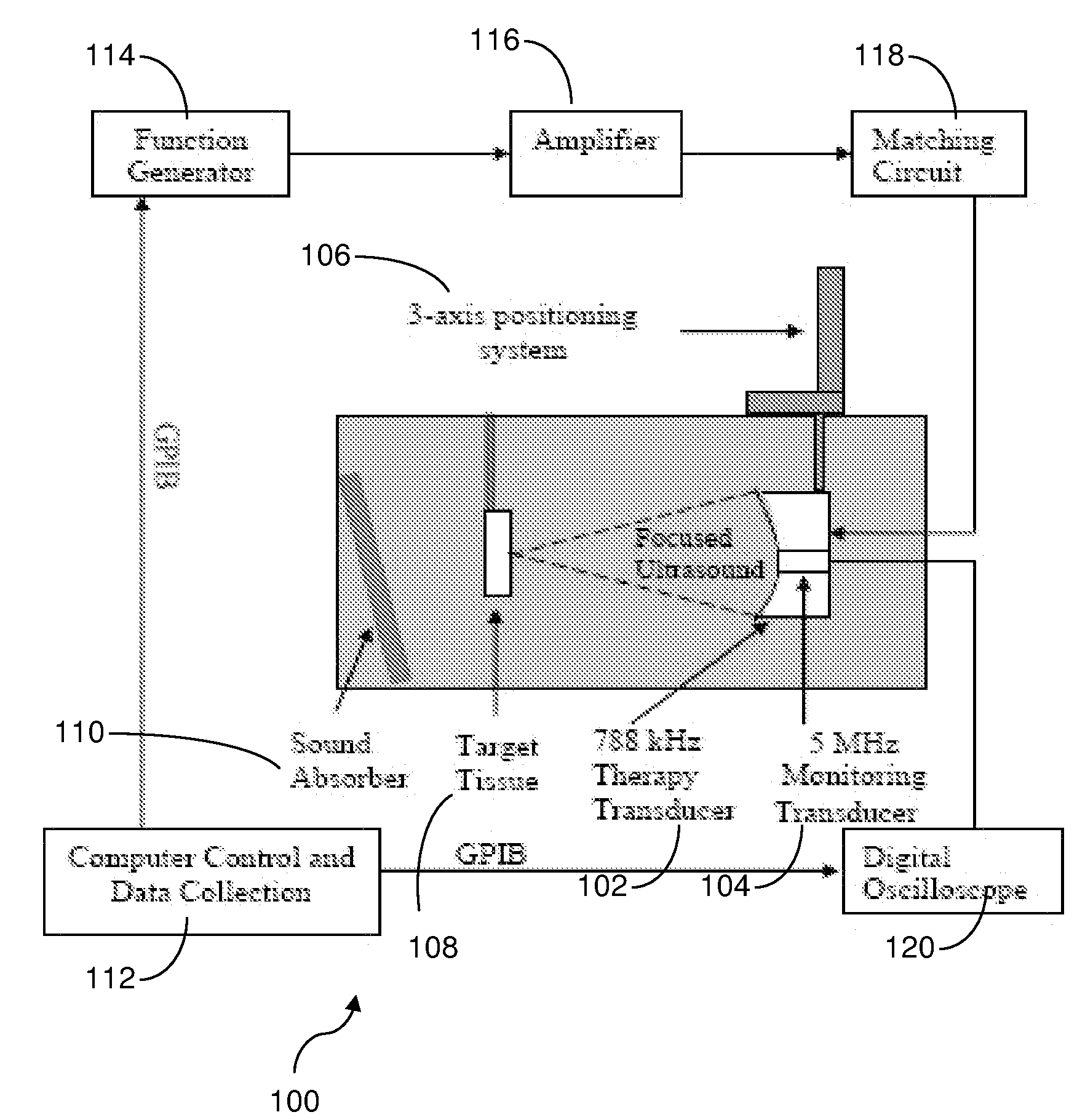
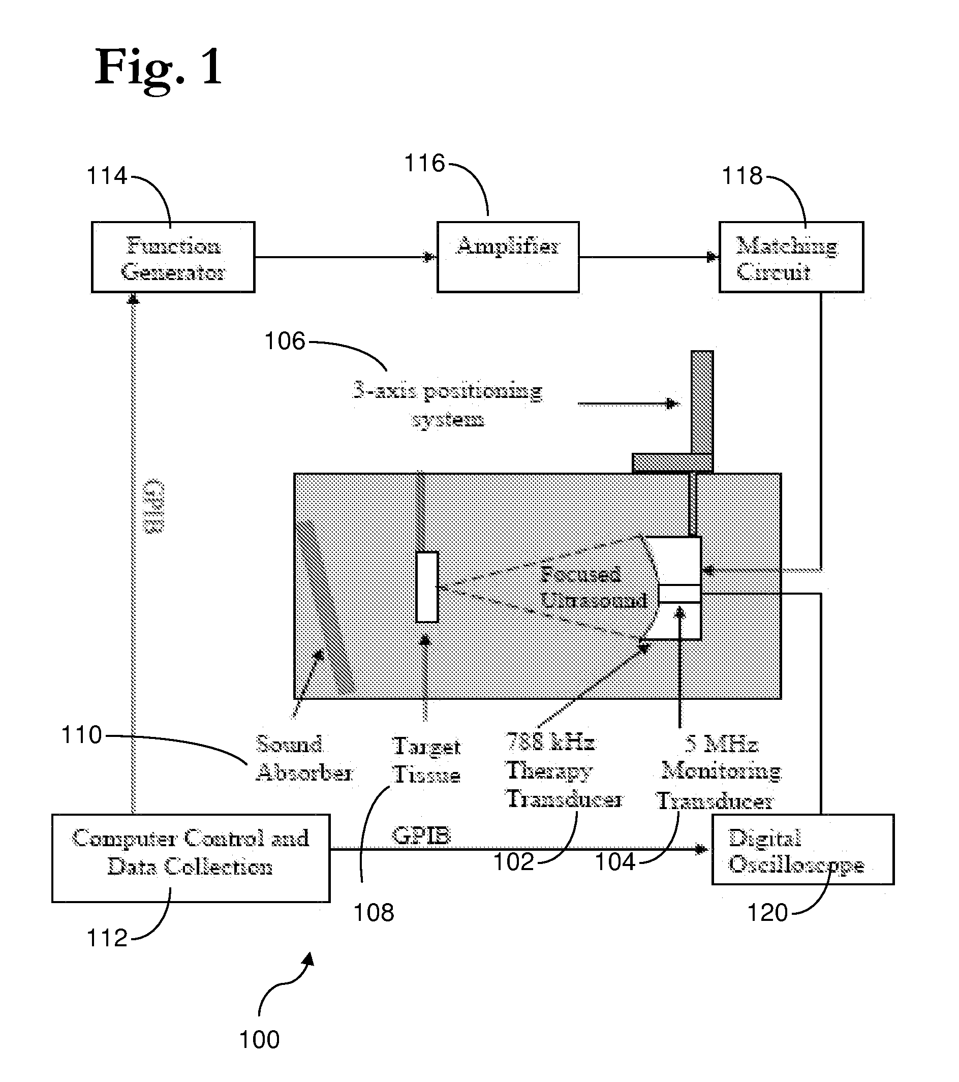
[0223]Ultrasound Apparatus: The therapeutic ultrasound unit, shown in FIG. 16, consisted of a large, high power annular 18 element piezo-composite phased transducer array (750 kHz, 145 mm diameter, 100 mm focal length) [Imasonic, Besançon, FR]. A commercial diagnostic 2.5 MHz imaging probe (General Electric Medical Systems, Milwaukee, Wis.) was coaxially aligned through the central hole of the phased-array and operated in a 1.8 MHz octave mode (harmonic imaging) with a nominally 30 Hz frame rate. The imaging probe was offset from the back of the therapeutic transducer by 40 mm resulting in an imaging distance of 60 mm. This provided sufficient image quality without substantially blocking the path of the therapeutic transducer. The transducer system was mounted on a brass frame tilted 20 degrees from vertical (to reduce reverberations from the animal skin surface) and placed at the bottom of a tank filled with degassed water. The focal pressure field could no...


PUM


Abstract

Therapy methods using pulsed cavitational ultrasound therapy can include the subprocesses of initiation, maintenance, therapy, and feedback of the histotripsy process, which involves the creation and maintenance of ensembles of microbubbles and the use of feedback in order to optimize the process based on observed spatial-temporal bubble cloud dynamics. The methods provide for the subdivision or erosion of tissue, liquification of tissue, and the enhanced delivery of therapeutic agents. Various feedback mechanisms allow variation of ultrasound parameters and provide control over the pulsed cavitational process, permitting the process to be tuned for a number of applications. Such applications can include specific tissue erosion, bulk tissue homogenization, and delivery of therapeutic agents across barriers.

Description

CROSS-REFERENCE TO RELATED APPLICATIONS[0001]This application is a continuation of U.S. application Ser. No. 11 / 523,201 filed on Sep. 19, 2006, which claims the benefit of U.S. provisional patent application No. 60 / 786,322, filed Mar. 27, 2006, U.S. provisional patent application No. 60 / 719,703, filed Sep. 22, 2005, and U.S. provisional patent application No. 60 / 753,376, filed Dec. 22, 2005. The disclosures of the above applications are incorporated herein by reference.GOVERNMENT RIGHTS[0002]Portions of this invention were made with government support under Contract Nos. RR14450, R01-HL077629-01A1, and RO1 DK42290, all awarded by the National Institutes of Health. The U.S. Government has certain rights in the invention.INTRODUCTION[0003]The present teachings relate to ultrasound therapy and, more particularly, relate to methods and apparatus for the controlled use of cavitation during ultrasound procedures.[0004]Treatment relating to tissue defects, various medical conditions, and d...

Claims


Application Information

Patent Timeline
Patent Type & Authority Applications(United States)
IPC IPC(8): A61N7/00
CPCA61B8/00A61B17/22004A61B8/485A61B2017/22089A61M37/0092A61B2017/22088
Inventor CAIN, CHARLES A.FOWLKES, J. BRIANXU, ZHENHALL, TIMOTHY L.
Owner RGT UNIV OF MICHIGAN
Learn More