Multicolor bioluminescent visualization probe set, or single-molecule-format multicolor bioluminescent visualization probe

a bioluminescent and probe technology, applied in the field of multicolor bioluminescent probes or single molecule-format multicolor bioluminescent probes, can solve the problems of difficult detection of target proteins/lipids, life phenomena, and high cost of facilities, and achieve high sample throughput, rapid and accurate detection, and high screening speed

Inactive Publication Date: 2009-05-14
NAT INST OF ADVANCED IND SCI & TECH
View PDF2 Cites 14 Cited by
  • Summary
  • Abstract
  • Description
  • Claims
  • Application Information

AI Technical Summary

Benefits of technology

[0026]The present invention provides novel means for detecting the presence/absence of multiple bioactivities (pharmacological activity, toxicity, carcinogenicity etc.) of a target-specific ligand including, for example, drugs, toxins, and carcinogens as a multicolor and two-dimensional spectrum simply, rapidly and accurately. The present invention also provides a basic technique for screening unknown antagonist/agonist for a wide range of “ligand recognition proteins” by using living cell lines transformed to express a multicolor bioluminescent probe set or a single-molecule-format multicolor bioluminescent probe via an expression vector therefore.
[0027]Consequently, multiple...

Problems solved by technology

However, inside cells, (i) uncountable numbers of proteins are present; (ii) internal structures thereof are partitioned into such as organelles and complicated; and (iii) the molecular mechanisms are precisely regulated by unknown complicated and sophisticated control systems.
In addition, (iv) detection of target proteins/lipids, which play key roles in life phenomena, is very difficult because they have no specific absorption spectra and generally exist in a small amount in the cells.
For example, the FRET method and the reporter gene assay which utilize fluorescence as analytical signals have a problem of increase in autofluorescence (background) and hence it requires expensive facility such as sophisticated filtering system for detection of fluorescent signals.
They also require an external light source, which makes an in vivo analysis very difficult.
On the other hand, the protein complementation and the protein splicing which use luminescence as analytical signals have such a problem of the necessity to introduce external substrates (Kim, S. B.; Ozawa, T.; Watanabe, S.; Umezawa, Y.
102, 9802-9807), as described above, enables rapid detection of a ligand and it also enables an agonist and antagonist to be detected individually; however, background luminescence is disadvantageously high, and high-resolution fluorescence microscope and sophisticated filter apparatus, as well as an expert technician are required for measuring energy transfer between two chromophores at high accuracy.
Additionally, it requires external li...

Method used


Image

  • Multicolor bioluminescent visualization probe set, or single-molecule-format multicolor bioluminescent visualization probe
  • Multicolor bioluminescent visualization probe set, or single-molecule-format multicolor bioluminescent visualization probe
  • Multicolor bioluminescent visualization probe set, or single-molecule-format multicolor bioluminescent visualization probe

Examples


example 1

Construction of Plasmid Encoding Single-Molecule-Format Bioluminescent Probe Set

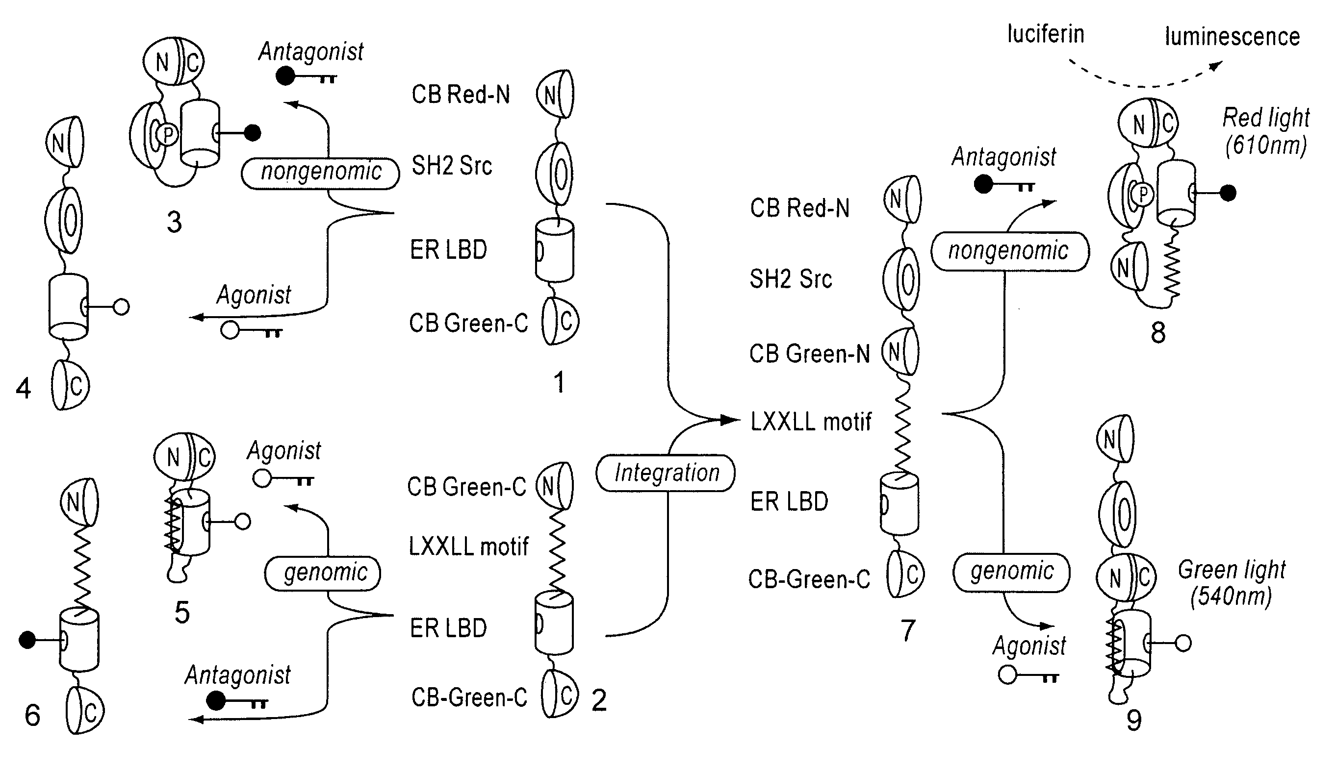
(1-1) Construction of Nucleic Acids Encoding Single-Molecule-Format Bioluminescent Probe Emitting Red Light by Phosphorylation-Dependent Protein-Protein Interaction

[0078]cDNAs of ER LBD (305-550 AA) and phosphorylation-recognition domain of Src (SH2) were amplified by PCR, and specific restriction sites were introduced at both ends. Recombinant DNAs in which cDNAs encoding ER LBD and Src SH2 were connected to each other were produced by ligation. Subsequently, the cDNAs encodes full CB Red were fragmented at five different points roughly localizing a sequence region encoding hydrophilic amino acids around 4 / 5 from the beginning of the cDNAs. As a result, five sets of cDNA fragments of CB Red (CB Red-N and CB Red-C) were created. Between the N- and C-terminal fragments (CB Red-N and CB Red-C, respectively) of these five sets, the connected components, Src SH2-ER-LBD, were inserted. Consequently, DNA const...

example 2

Gene Tansfection of Plasmid Expressing Single-Molecule-Format Bioluminescent Probe into Living Cells

[0081]COS-7 cells derived from African Green Monkey's kidney were cultured in Dulbecco-modified Eagle's medium (DMEM; Sigma) supplemented with 10% steroid-deficient fetal bovine serum (FBS) and 1% penicillin-streptomycin (P / S), and incubated in a 12-well plate in a cell incubator at 37° C. and 5% CO2. Using TransIT-LT1 (mirus), commercially available gene transfection kit pSimer-G, pSimer-R, or pSimer-RG series plasmids were transfected into COS-7 cells in the 12-well plate. The COS-7 cells containing respective plasmids were incubated in a cell incubator for 16 hours, allowed to sufficiently express respective bioluminescent probes, and used in the following experiments.

example 3

Determination of Optimal Dissection Site in CBLuc-Red for Red Luminescent Probe Used for Measuring Protein Phosphorylation (pSimer-R Series)

[0082]As shown in the above (Example 2), plasmids of pSimer-R1 through —R5 were transfected into COS-7 cells respectively, incubated for 16 hours, and allowed to sufficiently express the bioluminescent probes. Intensity of luminescence emitted by 20 minutes-stimulation was measured by luminometer, provided OHT (4-hydroxytamoxifen), which is a commercially available anticancer drug, is present and absent. As a result, it was observed that cells carrying pSimer-R2 showed the strongest luminescence intencsity in the presence of OHT (FIG. 3).

[0083]This result demonstrates that the optimal dissection point of CBLuc-Red is that of pSimer-R2 (between 412 and 413 AA).

[0084]Further, measurement of the light spectrum from cells carrying pSimer-R2 with all wavelengths showed that the wavelength of the luminescence was about 610 nm (FIG. 4).


PUM

PropertyMeasurementUnit
Ratioaaaaaaaaaa
Wavelengthaaaaaaaaaa
Luminescenceaaaaaaaaaa

Abstract

The present invention provides ligand detection means capable of exhibiting two-dimensional information (wavelength and intensity of luminescent signal) responding to multiple signals triggered by a ligand via a target protein, while taking advantage of the merit of the single-molecule-format bioluminescent probe.
The present invention could provide a luminescent probe set which is comprised of a fusion protein comprising a ligand recognition protein and a molecular recognition domain to which the ligand recognition protein bind upon conformational change, wherein the fusion protein is sandwiched between split Lighting Enzyme fragments, the probe set can emit multiple luminescence by utilizing Lighting Enzymes emitting lights with multiple wavelength, wherein these multiple components can be tandemly arranged by using C-terminal fragment of the Lighting Enzyme.
By using a living cell line transfected with gene of multicolor luminescent probe set or single-molecule-format multicolor bioluminescent probe, it becomes possible to distinguish and detect bioactivity level of a target ligand in a complex context of the living cell two-dimensionally (wavelength versus intensity) in multi colors, and to quantitatively evaluate multiple effects (anticancer and carcinogenesis actions, agonist and antagonist) of a ligand represented by a drug at once by two-dimensional information of different colors in short time.

Description

TECHNICAL FIELD[0001]The present invention relates to a set of multicolor bioluminescent probes or a single-molecule-format bioluminescent probe capable of quickly visualizing two distinctive activities of a ligand that is specific to a target protein.BACKGROUND ART[0002]In the natural world, abundant nature and organisms coexist, and maintain their life phenomena based on common molecular mechanisms and principles such as DNA expression. Many researches in medical, pharmaceutical and natural scientific fields aim at finding out life phenomena of living organisms. To understand such life phenomena and ensure safe life environments, it is essential to develop effective novel pharmaceuticals while excluding potential risk factors. For achieving this, it is necessary to biologically analyze various molecular phenomena occurring in a cell line which are the basic units of living organisms (for example, protein-protein interaction, phosphorylation, molecular trafficking, transcription / tr...

Claims


Application Information

Patent Timeline
IPC IPC(8): C12Q1/66C12Q1/48C07H21/00C12N15/63C12N5/00
CPCC07K14/4702C07K14/721C07K2319/00C07K2319/61C07K2319/70C07K2319/72G01N33/542C12Q1/485G01N33/5008G01N33/5011G01N33/533G01N33/535C12N9/0069
Inventor KIM, SUNG BAETAO, HIROAKIUMEZAWA, YOSHIO
Owner NAT INST OF ADVANCED IND SCI & TECH
Why Eureka
Try Eureka