
Ultra Low Strength Electric Field Network-Mediated Ex Vivo Gene, Protein and Drug Delivery in Cells

a low-power electric field and ex vivo gene technology, applied in specific use bioreactors/fermenters, biomass after-treatment, biochemistry apparatus and processes, etc., can solve the problems of misleading and inaccurate reference to the present bioelectric application, and achieve low strength, minimize or even avoid the effect of reducing the number of induced ion exchanges

Inactive Publication Date: 2008-07-31
RGT UNIV OF CALIFORNIA
View PDF7 Cites 3 Cited by
  • Summary
  • Abstract
  • Description
  • Claims
  • Application Information

AI Technical Summary

Benefits of technology

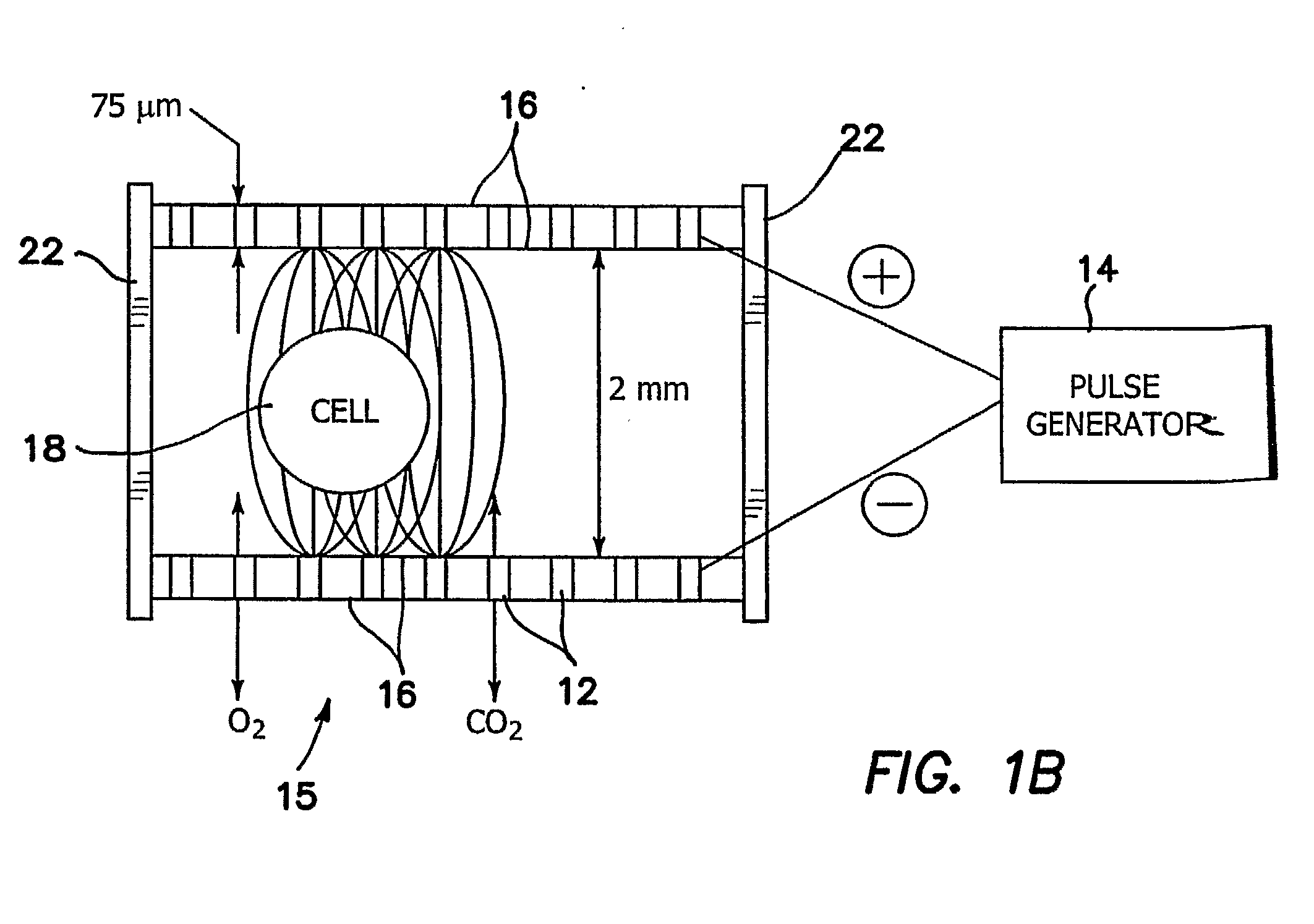
[0011]An LSEFN chamber is used which is shaped and sized to intimately contain the cells, cell clusters, or tissues in a transfusion chamber between opposing gas permeable membrane encapsulated electrode arrays across which low voltage LSEFN pulses are applied. The gas or gases introduced into the culture fluid through the gas permeable membrane can be chosen to optimize the specific metabolism and health required by the cell, cell clusters, or tissues. The high percentage of cell death, which is typical of prior art electroporation, is minimized or even avoided in the present application by the synergistic combination of low strength electric field network and optimal culture and gas environment for the cell, cell clusters, or tissues.
[0012]The illustrated invention is thus characterized by and has the advantages of: low voltage electro-permeabilization which results in less damage to the cell; dynamic electro-permeabilization, i.e. cells are moving in a static field, and rotating in a constant rate of buffer flow, therefore a constant temperature is maintained and heat damage to the cells is avoided thus allowing a long term LSEFN treatment as compared to the prior art; use of an electric array of very small electrodes to minimize heat and to use diffusing electric fields for providing a more nearly uniform average LSEFN exposure and transfusion into the cells; and a non-cuvette system which uses long exposure cell in a compact chamber to transfuse a large number of cells and to reintroduce them at a single time for large batch processing.
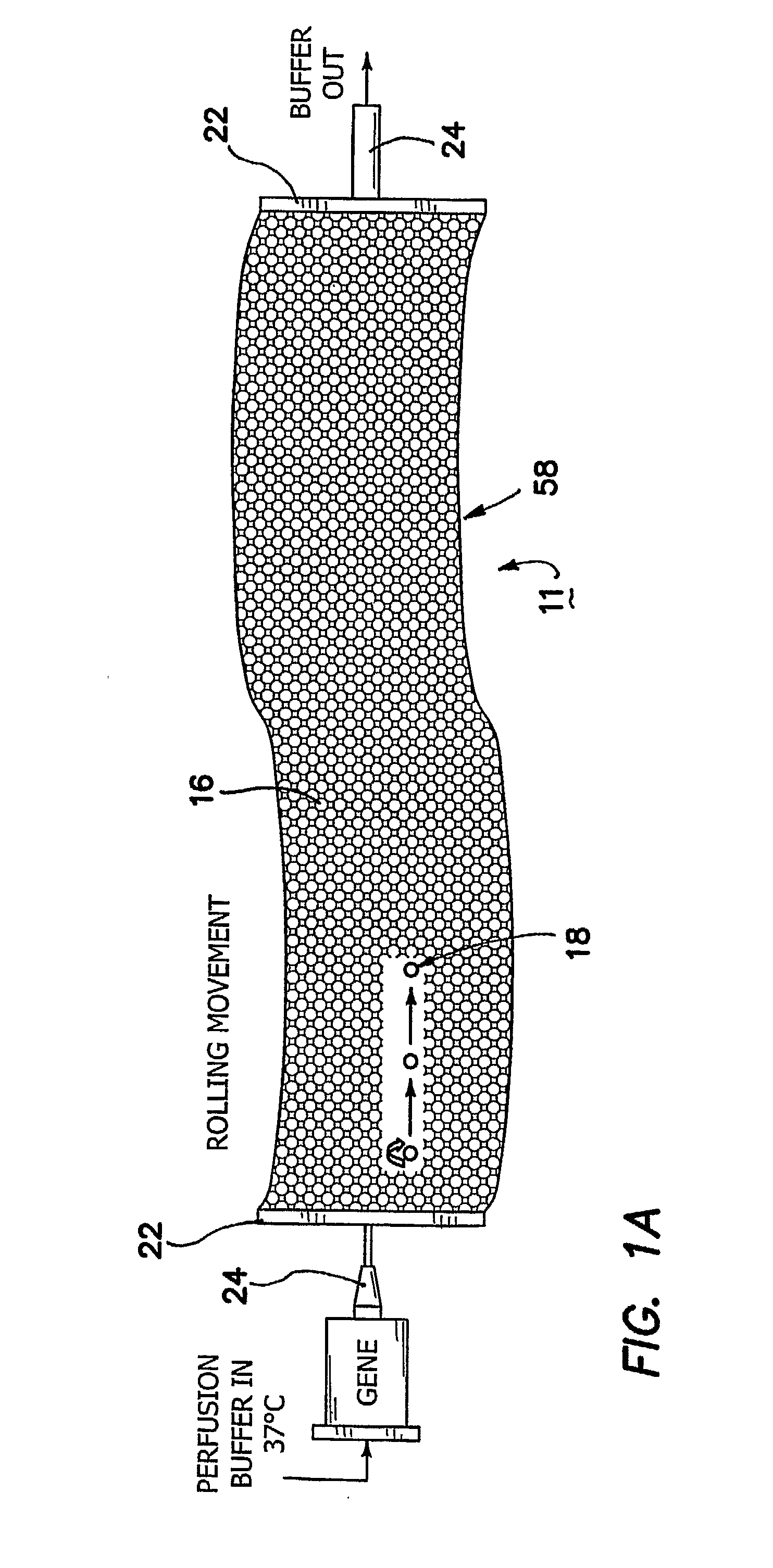
[0015]The method further comprises the step of flowing a culture fluid to bathe the cells, cell clusters, or tissues during application of ex vivo an LSEFN electric field and during systemically transfusing the gene, protein or drug materials. The fluid may be used to culture the cells, cell clusters, or tissues. The flowing culture fluid easily mixes with the drug, protein or gene and increases the chance of the drug, protein or gene interaction with the cell membrane.
[0017]A flowing fluid maintains the temperature of the fluid substantially constant to avoid heat damage to the cells, cell clusters, or tissues in the LSEFN electric field.
[0021]The method is easily modified to mass production or batch production of a large number of cells, cell clusters, or tissues during a single exposure time interval over an extended exposure path along which the cells, cell clusters, or tissues are moved, whereby mass production of mediated cells, cell clusters, or tissues are produced.

Problems solved by technology

Hence, to refer to the present bioelectric application as electroporation is misleading and inaccurate.

Method used


Image

  • Ultra Low Strength Electric Field Network-Mediated Ex Vivo Gene, Protein and Drug Delivery in Cells
  • Ultra Low Strength Electric Field Network-Mediated Ex Vivo Gene, Protein and Drug Delivery in Cells
  • Ultra Low Strength Electric Field Network-Mediated Ex Vivo Gene, Protein and Drug Delivery in Cells

Examples


second embodiment

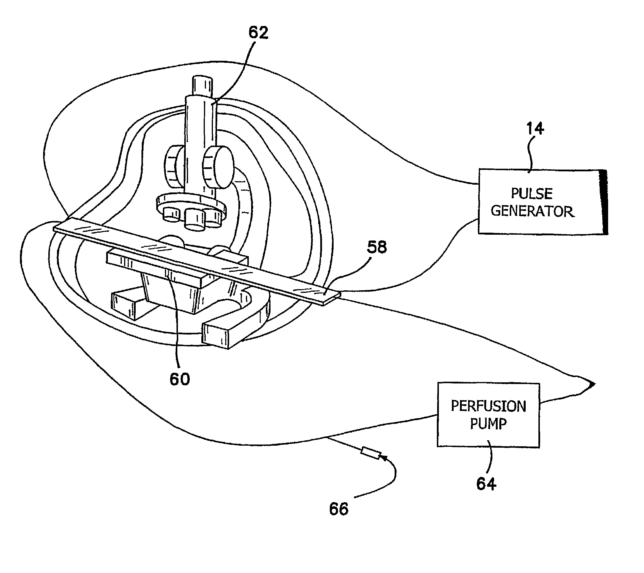
[0050]In FIGS. 3 and 4 we provide an apparatus 11 for ultra low strength electric-field mediated ex vivo gene, protein and drug delivery in cells, and clusters, such as islets. A cylindrically shaped or tubular culture chamber 26 is provided with an electrode array 12 similar to that shown in FIGS. 1a, and 2b and is used for applying the electric field from generator 14 to the cell clusters 28. As shown best in the cross-section view of FIG. 3b, chamber 26 is provided with encapsulated electrodes 12 between concentric membranes 16, the electrodes 12b being connected to the negative terminal 40 and electrodes 12a being connected to the positive terminal 42. Electrodes 12a and 12b may be provided in any geometric arrangement desired, but the preferred embodiment is shown in FIG. 3b where the negative electrodes 12b and positive electrodes 12a are geometrically alternated to maximize the fringing field which extends into chamber 26 and hence to cluster 28. The number of electrodes is s...

first embodiment

[0051]FIG. 4 shows that the cylindrical chamber 26, which is unrolled or shown in a linear shape in FIG. 3a, can be helically coiled to form a more three-dimensionally compact system. In this chamber 26 as best seen in the perpendicular cross-sectional view of FIG. 3b, the electrode arrays 12 are closer to the cell clusters 28 than in the first embodiment because the chamber dimensions more nearly approximate the size of the cell clusters themselves. Thus, the LSEFN voltage can be further reduced. For example of field of about 1 V / cm can be employed across chamber 26, which is again included within the definition of ultra low field strength.

third embodiment

[0052]In FIGS. 5 and 6 we provide another apparatus 11 for ultra low strength electric-field mediated ex vivo gene, protein and drug delivery in tissue 32. In this embodiment, the chamber size and shape can be modified to match the various shapes of cultured tissue, or bioengineered scaffolds 32. Two opposing electrode arrays 12 in sealed membranes 16 are provided on the both side of the culture tissue 32 in chamber 30 in a manner similar to FIGS. 1-4. However, the size and shape of frame 22 and the chamber 30 defined within it is customized to the particular shape of the tissue being treated. For example, a section of skin graft tissue or cornea may be the treated tissue in which case frame 22 and the chamber 30 will be contoured to match the section of skin graft tissue, so that low field LSEFN can be effectively performed on the section during perfusion.

[0053]Consider now how the invention is used to provide gene, protein and drug therapy for isolated cells. Any biological cells,...


PUM

PropertyMeasurementUnit
Temperatureaaaaaaaaaa
Volumeaaaaaaaaaa
Strengthaaaaaaaaaa

Abstract

Ex vivo gene, protein or drug delivery to macroscopic quantities of various types of cells, cell clusters, or tissues using ultra low strength LSEFN strategies is disclosed in which the bioengineered cells and tissues are then systemically transfused, delivered or implanted into the various organs or tissue for the treatment of diseases. An LSEFN chamber is used which is shaped and sized to intimately contain the cells, cell clusters, or tissues in a transfusion chamber between opposing membrane encapsulated electrode arrays across which LSEFN pulses are applied.

Description

[0001]The present application is related to U.S. Provisional Patent Application Ser. No. 60 / 663,562, filed on Mar. 19, 2005, which is incorporated herein by reference and to which priority is claimed pursuant to 35 USC 119.BACKGROUND OF THE INVENTION[0002]1. Field of the Invention[0003]The invention is in the field of methodologies to facilitate the ex vivo gene, protein or drug delivery in large quantity to various types of cells or cell clusters, such as islets, or various of tissues using an ultra low strength electric field network.[0004]2. Description of the Prior Art[0005]Electroporation is a technique involving the application of short duration, high intensity electric field pulses to cells or tissue. The electrical stimulus causes membrane destabilization and the subsequent formation of nanometer-sized pores. In this permeabilized state, the membrane can allow passage of DNA, enzymes, antibodies and other macromolecules into the cell. Electroporation holds potential not only...

Claims


Application Information

Patent Timeline
IPC IPC(8): C12N15/01C12M1/42C12Q1/68C12Q1/02
CPCC12N15/87C12M35/02
Inventor SEN, LUYICUI, GUANGEN
Owner RGT UNIV OF CALIFORNIA
Learn More