
Optical Method and Device for the Detection and Enumeration of Microorganisms

a microorganism and optical method technology, applied in the direction of microorganisms, bioreactors/fermenters, biomass after-treatment, etc., can solve the problems of insufficient enumeration tests, slow sensor speed, limited device determination,

Active Publication Date: 2008-07-24
NEOGEN CORP
View PDF11 Cites 31 Cited by
  • Summary
  • Abstract
  • Description
  • Claims
  • Application Information

AI Technical Summary

Problems solved by technology

Consequently, these devices are limited to the determination of presence or absence of microorganisms in the tested samples which is adequate for clinical and sterility tests.
Due to the solid nature of the sensor, the diffusion rate of the metabolites to the sensor is quite slow and may not be consistent for duplicate samples and, consequently, is inadequate for enumeration tests.
The disadvantage of this device is that the semi-liquid phase disintegrates in higher temperatures and, therefore, the device cannot be thermally sterilized.
Consequently, it cannot be used for clinical and sterility tests.
Another disadvantage of this device is that the agar occasionally gets dislodged during transportation.

Method used


Image

  • Optical Method and Device for the Detection and Enumeration of Microorganisms
  • Optical Method and Device for the Detection and Enumeration of Microorganisms
  • Optical Method and Device for the Detection and Enumeration of Microorganisms

Examples


example 1

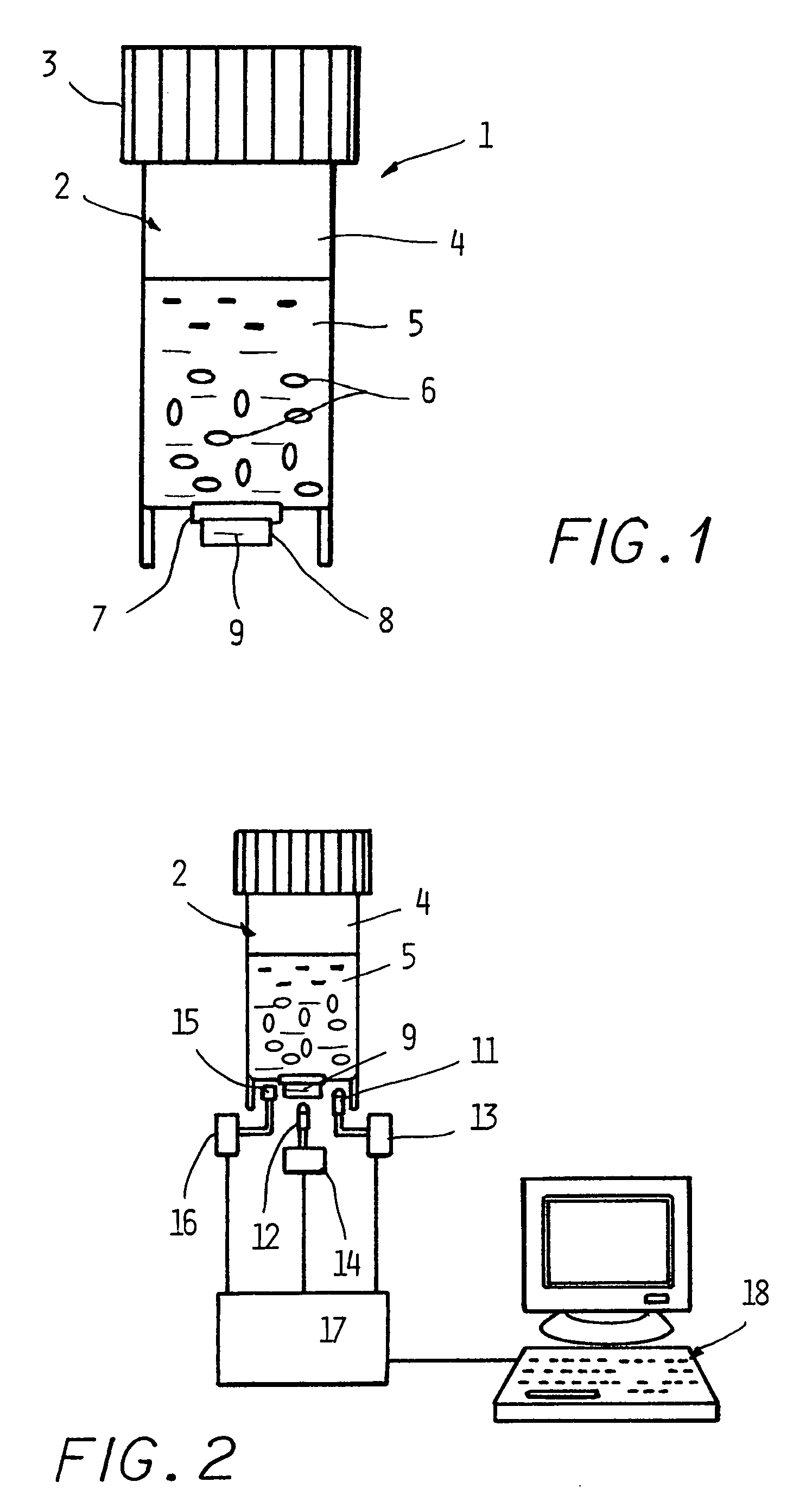
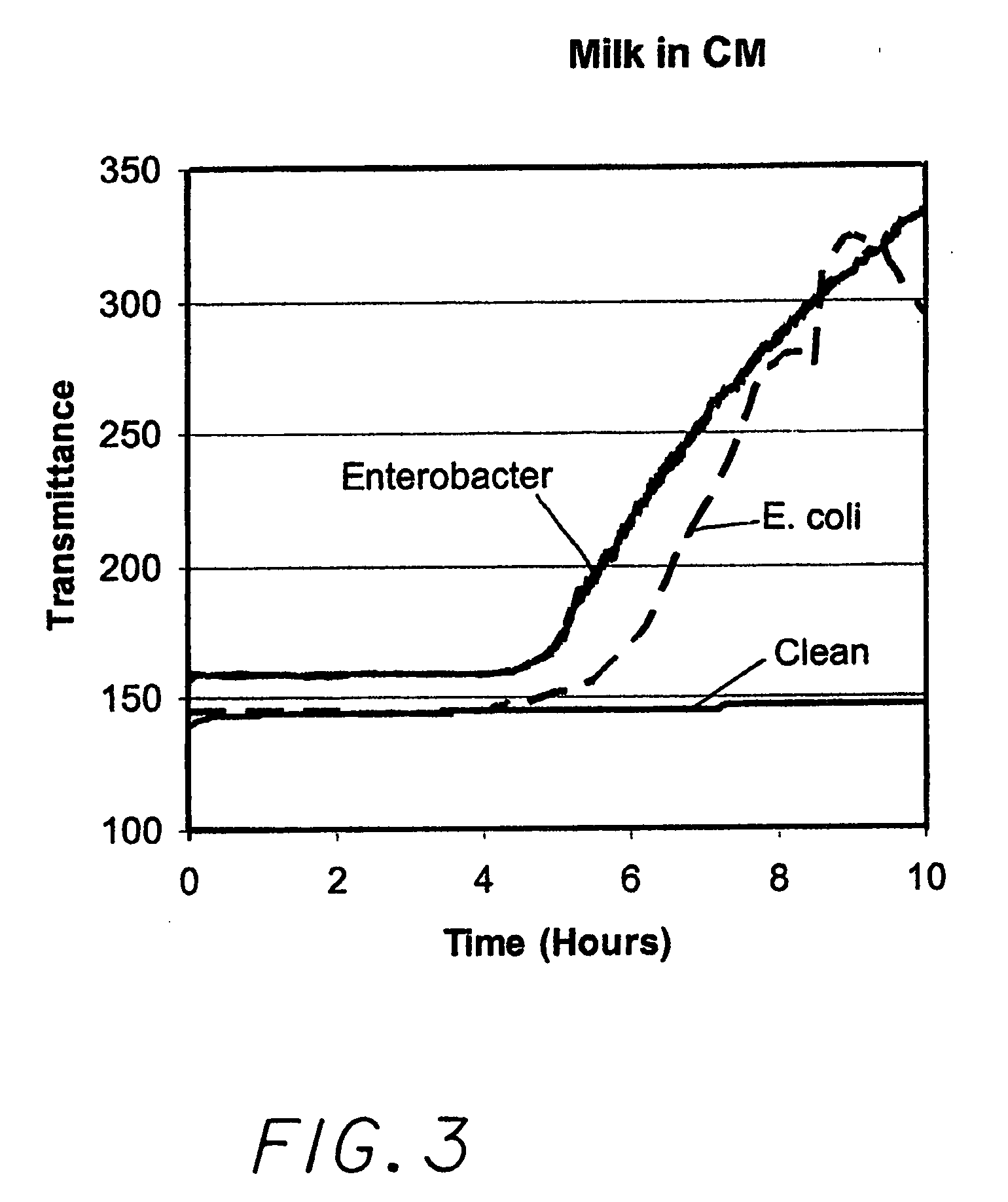
[0018]Three containers were prepared with a barrier layer made of hydrophilic Porex disks (material #4897) and pre-filled with 9 ml of mixture of coliform media (BioMérieux CM) that also contains an adequate amount of Bromocresol Purple. Bromocresol Purple changes its color from purple to yellow due to fermentation of lactose resulting in the reduction of pH. Three samples of 1 ml of pasteurized milk were prepared, one inoculated with E. coli, another with Enterobacter aerogenes, and the third remained un-inoculated. Each 1 ml sample was pipetted into one of the three containers and placed into the fixture assembly inside an incubator set at 35° C. Data corresponding to the transmittance value picked up by the photodiode 15, due to activation of the light source 11, was collected and stored every 10 minutes. The collected data for each of the three containers is shown in FIG. 3. It is evident that the clean sample did not change the pH in the container during the duration of the tes...

example 2

[0019]Similar tests were carried out with three different samples of ground beef. The growth media is mPCB (Difco, Becton Dickenson and Company, Sparks, Md., USA), which is a non-selective medium for total aerobic count, mixed with 20 mg. / liter of Bromocresol Purple. Each container contained 9 ml of mixture of media and indicator. Each of the meat samples was diluted 1:10 in Butterfield's phosphate buffer, 2 ml of each sample was added into each container, and the containers were placed in an incubator and monitored over 15 hours. The meat sample, shown as Meat A in FIG. 4, was fresh and relatively clean from microorganisms. The sample Meat C resulted from letting the same meat of Meat A stay at room temperature for 8 hours, and Meat B stayed at room temperature overnight. As expected, Meat B resulted in short Detection Time of approx. 5 hours, Meat C detected in 9.5 hours, and the “clean” sample did not detect during the hours duration of the test. In addition, it can be seen that ...


PUM


Abstract

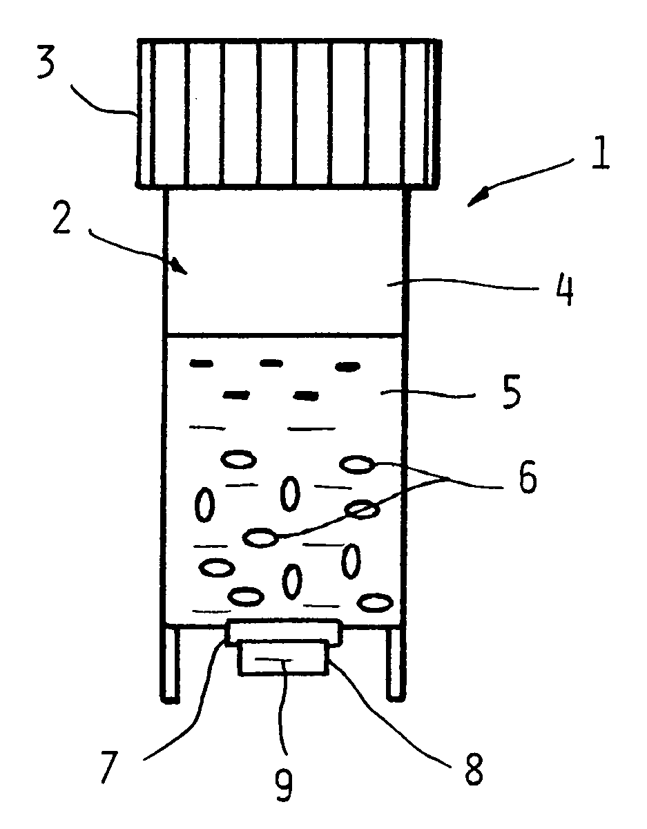
A new device and method for detecting the presence of living microorganisms in test samples are described. The device comprises a container with at least one section transparent to light, a growth zone located in said container containing a mixture of growth media capable of supporting growth of the microorganisms, and at least one indicator substrate that changes its optical properties due to growth of the microorganisms. A detection zone is located in the container adjacent to the transparent section, and a barrier layer comprising porous solid material separates the two zones, allowing diffusion of molecules and ions of metabolic by-products of the organisms, while preventing microorganisms and particulate matter of the test sample from penetrating into the detection zone.

Description

CROSS REFERENCE TO RELATED APPLICATIONS[0001]This patent application claims the benefit of provisional patent application No. 60 / 857,917 filed Nov. 10, 2006.BACKGROUND AND PRIOR ART[0002]The presence and enumeration of microorganisms in industrial samples have been traditionally determined by growing the microorganisms on the surface of agar in Petri dishes and counting the colonies. In the last two decades, other tests have been practiced for clinical and industrial specimens. These methods are based on culturing the specimens in liquid media and monitoring the metabolites generated during the growth of the micro-organisms. Several systems, such as the Bactometer (bioMerieux, Hazelwood, Mo., USA), BacTrak (Sy-Lab, Neupurkersdort, Austria), Malthus Systems (Lab M, Crawley, UK) and the RABIT (Bioscience International, Bethesda, Md., USA), are based on monitoring the electrical properties of the media measured via two metallic electrodes immersed in the growth media. The conductivity ...

Claims


Application Information

Patent Timeline
IPC IPC(8): C12Q1/20C12M1/34
CPCC12M23/34C12Q1/06C12Q1/04C12M41/36
Inventor EDEN, GIDEONEDEN, RUTH
Owner NEOGEN CORP
Learn More