Nanoparticle radiosensitizers

a radiosensitizer and nanoparticle technology, applied in the field of nanomaterials, can solve the problems of difficult clinical implementation, anti-enhancement and other side effects, damage to dna, cellular membranes or other targets, etc., and achieve the effects of enhancing specificity, enhancing x-ray absorption, and enhancing the specificity of both detection and treatmen

Inactive Publication Date: 2008-01-03
RGT UNIV OF CALIFORNIA
View PDF5 Cites 48 Cited by
  • Summary
  • Abstract
  • Description
  • Claims
  • Application Information

AI Technical Summary

Benefits of technology

[0009] The present invention meets this need by providing a new type of radiosensitizer that utilizes nanomaterials targeted to cellular structures and molecules such as DNA. This method is called Nanostructure Enhanced X-ray Therapy (NEXT). In this method, nanomaterials of heavy elements such as gold are used to enhance x-ray absorption; and chemically targeted delivery of the nanomaterials to the target sites enhances specificity. In addition, x-ray beams such as those used in Computed Tomography or Computerized Axial Tomography (CT or CAT scan) and multiple collimated x-ray beams used in Gamma Knife technology can be used to further enhance the specificity of both detection and treatment of disease.
[0010] There are several distinct advantages to this methodology. First, heavy elements such as gold absorb more high energy x-rays, thus increasing the linear energy transfer (LET) from x-rays to the target. Second, although nanostructures such as a nanoparticle absorbs the same amount of x-rays as the same number of individual atoms, the former helps to localize the toxicity of the x-rays to the nanoparticle site by emitting an intense burst of Auger electrons and other secondary electrons from a single nanoparticle. These low energy electrons are highly localized at the targeted sites, thus producing high concentrations at these sites. This means the effective LET is even higher for gold nanoparticles than gold atoms spread over a much larger volume in solution. Third, if gold nanoparticles are used, the x-ray energy to be used would be above 81 keV (the K absorption edge of gold), leading to less absorption by light elements in the body and thus deeper penetration. Fourth, smaller nanoparticles, patterned aggregates of small nanoparticles, or other materials such as gold nanorods or nanobeads with a higher percentage of surface atoms offer the Auger or secondary electrons an easier escape with minimal residual positive charges on each individual nanoparticles. Fifth, gold is one of the most benign elements to the body, although gadolinium, gold, lanthanum (La), cerium (Ce), praseodymium (Pr), neodymium (Nd), promethium (Pm), samarium (Sm), europium (Eu), gadolinium (Gd), terbium (Tb), dysprosium (Dy), holmium (Ho), erbium (Er), thulium (Tm), ytterbium (Yb), lutetium (Lu) iodine, tungsten, rhenium, osmium, iridium, platinum, and bismuth may also be utilized. Lastly, nanoparticles have been modified chemically and used in many cases to connect to DNA segments, so it is possible to extend this method to tackle specific sites (Shenhar et al., 2003, Alivisatos et al, 1996, McDevitt et al., 2000, Zanchet et al., 2002, Sandstrom et al., 2003).
[0014] The complex nanostructures such as strings of nanoparticles at one location can be used to increase double strand breaks (DSBs), which are much more difficult to repair than single strand breaks. Again, it is important to chemically conjugate a small number of nanoparticles to the target to achieve enhanced damage.

Problems solved by technology

Second, although nanostructures such as a nanoparticle absorbs the same amount of x-rays as the same number of individual atoms, the former helps to localize the toxicity of the x-rays to the nanoparticle site by emitting an intense burst of Auger electrons and other secondary electrons from a single nanoparticle.
These electrons and especially the radicals generated by them will cause damage to DNA, cellular membranes, or other targets.
Typically, the lower limit of the weight percentage of the nanostructures is 1%, which makes it difficult to implement clinically.
In most cases, such implementation can even lead to anti-enhancement and other side effects because of the presence of a large amount of nanostructures and chemical ligands that can scavenge the electrons and radicals generated in water.

Method used


Image

  • Nanoparticle radiosensitizers
  • Nanoparticle radiosensitizers
  • Nanoparticle radiosensitizers

Examples


example 1

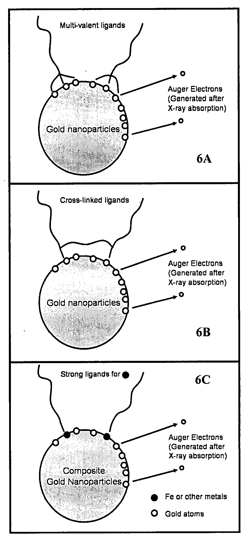
The Use of Gold Nanoparticles to Enhance X-Ray Absorption

[0094] Materials and Methods

[0095] Synthesis of TMAnAuNP:

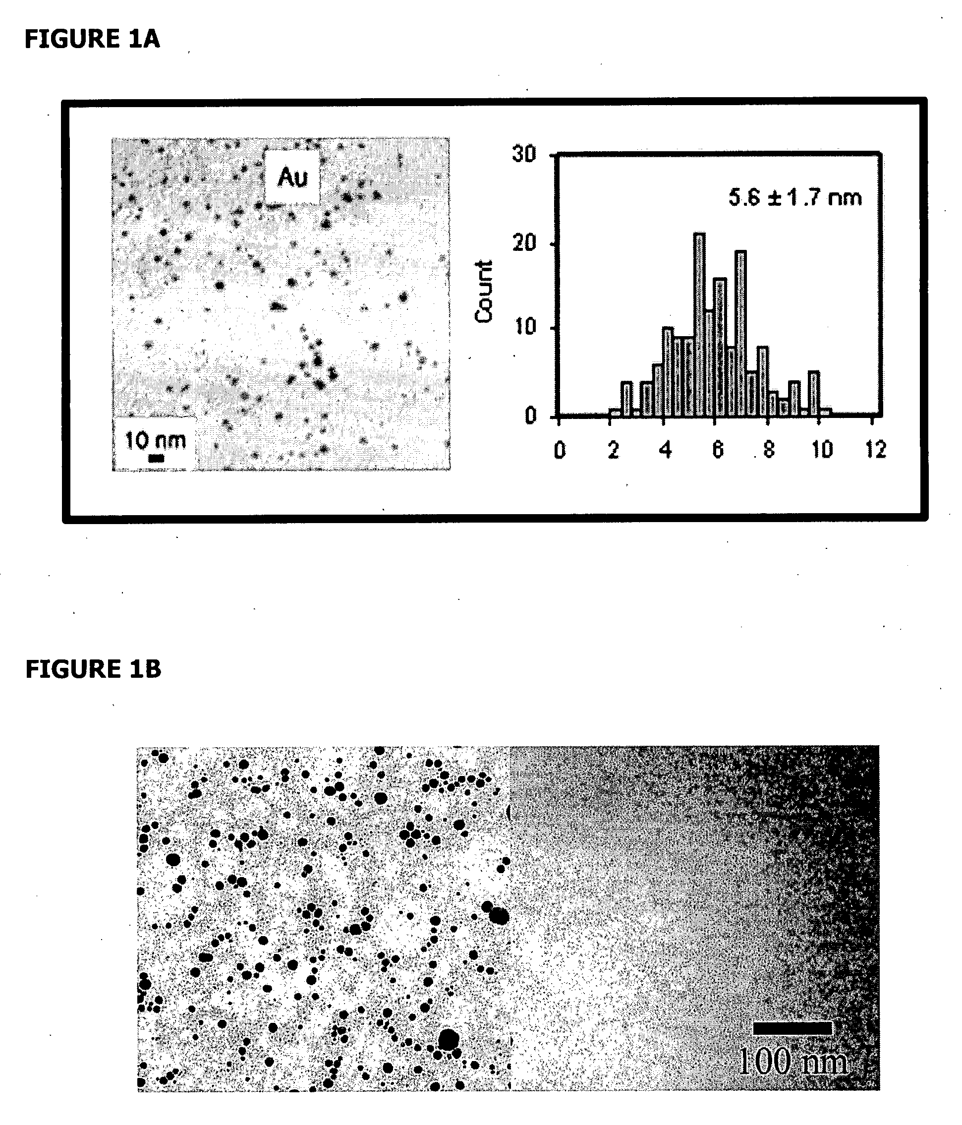
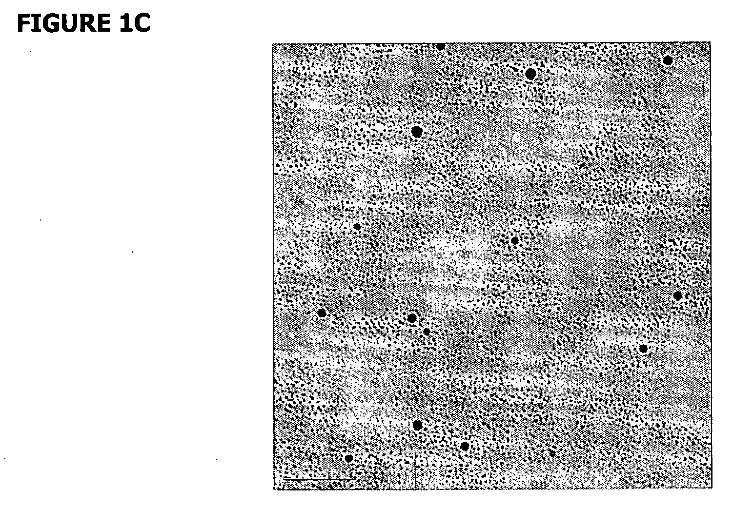
[0096] Dodecanethiol (C12) functionalized gold nanoparticles (AuNP), NNN trimethyl(11-mercaptoundecyl) ammonium chloride ligands, and trimethylammonium (TMA) C12 functionalized gold nanoparticles (TMAnAuNP, n denotes the number of TMA ligands on a nanoparticle) were synthesized using the available procedures (Tien et al., 1997, Brust et al., 1994, McIntosh et al., 2001). Different reaction times (24-hour or 51-hour) were used in the ligand exchange reactions to make TMAnAuNP, which yielded different n values. NMR and UV-VIS were used to verify the reaction intermediates and products.

Gel Electrophoresis

[0097] 1.2% and 0.8% Agarose E-gels (Invitrogen) and Agarose gels prepared in the lab were used to detect supercoiled DNA, gold nanoparticles, and their complexes. The running conditions were between 50 or 60 V for 45 min (E-gels) or 3 hours (self-poured gels). Gels w...

example 2

Use of Gold Nanoparticles for Local Enhancement

[0114] Gold nanoparticles conjugated to scDNA using an ethidium-based intercalating ligand were used. In this experiment, 3-nm gold nanoparticles covered with a mixture of ethidium thiol ligands (<10 per nanoparticle) and charge-neutral surfactants were prepared. The charge-neutral ligands were used to avoid any DNA aggregation. Such a small amount of ethidium in the samples (<150 nM) did not result in any detectable change to the scDNA. Tris buffer was added to control the diffusion distance of hydroxyl radicals in water (Hodgkins et al., 1996). The ratio of nanoparticles to scDNA was ˜10. FIG. 12A shows a transmission electron microscope (TEM) image of nanoparticle-conjugated scDNA in which all the nanoparticles are conjugated to scDNA. FIG. 12B shows the results of DNA damage as a function of buffer concentration, which directly controlled the diffusion distance of OH radicals. At low buffer concentrations the enhancement was nearly...

example 3

Use of Gold Nanotubes for Remote Enhancement

[0115] Remote enhancement may be demonstrated using gold nanotubules (Qu et al, 2006). In this demonstration, scDNA was mixed with a matrix of ˜20 mg ligand-free gold nanotubules in 20 μl of water, as shown in FIG. 13A. The ratio of the weight of these gold nanotubules to water in the scDNA samples was ˜1:1. Because the shell thickness of the gold layer was about 40 to 70 nm, only high energy photoelectrons could escape the nanostructures. The gaps between these nanotubules were of the order of several hundred nanometers and scDNA could easily move through the matrix. This creates a mismatch between the penetration distance (many microns) of high energy photoelectrons and the distance between the nanotubules, which leads to the re-absorption of those photoelectrons by gold nanotubules. A factor 2 reduction in enhancement was found with the gold nanotubule case because many low energy electrons cannot escape the nanotubules. The re-absorpt...


PUM

PropertyMeasurementUnit
Fractionaaaaaaaaaa
Fractionaaaaaaaaaa
Lengthaaaaaaaaaa

Abstract

Herein is described Nanostructure Enhanced X-ray Therapy (NEXT), which uses nanomaterials as radiosensitizers to enhance electromagnetic radiation absorption in specific cells or tissues. The nanomaterial radiosensitizers emit Auger electrons and generate radicals in response to electromagnetic radiation, which can cause localized damage to DNA or other cellular structures such as membranes. The nanomaterial radiosensitizers contain moieties for specific targeting to molecules or structures in a cell or tissue, and can be functionalized for increased stability and solubility. The nanomaterial radiosensitizers can also be used as detection agents to help in early diagnosis of disease. Together with known techniques such as Computed Tomography or Computerized Axial Tomography (CT or CAT scan), these nanomaterial radiosensitizers could allow early diagnosis and treatment of diseases such as cancer and HIV.

Description

RELATED APPLICATIONS [0001] This application is a Continuation-in-Part of International Application number PCT / US2005 / 034949, filed Sep. 27, 2005, which claims the benefit under 35 USC § 119(e) of U.S. Provisional application No. 60 / 614,137, filed Sep. 28, 2004.GOVERNMENT RIGHTS [0002] This invention was made with U.S. Government support from the National Science Foundation grant number 0135132. The U.S. Government may have certain rights in this invention.FIELD OF THE INVENTION [0003] Described herein are compositions, devices and methods for use as radiosensitizers, particularly the use of nanomaterials as radiosensitizers for therapy and diagnosis of diseases such as cancer and HIV. BACKGROUND OF THE INVENTION [0004] The toxicity of x-rays to biological species has been the cornerstone of cancer treatment for decades (Moss et al., 2003 and Hall et al., 1973). However, x-rays alone are an ineffective modality because they lack the selectivity toward killing malignant cells while s...

Claims


Application Information

Patent Timeline
IPC IPC(8): A61K49/04A61K33/24A61P31/18A61P35/00A61K39/395
CPCA61K41/0085A61K41/0038A61P31/18A61P35/00
Inventor GUO, TING
Owner RGT UNIV OF CALIFORNIA
Why Eureka
Try Eureka