Vascular graft sterilization and decellularization
- Summary
- Abstract
- Description
- Claims
- Application Information
AI Technical Summary
Benefits of technology
Problems solved by technology
Method used
Image
Examples
example 1
Processing Parameters
Cell Removal:
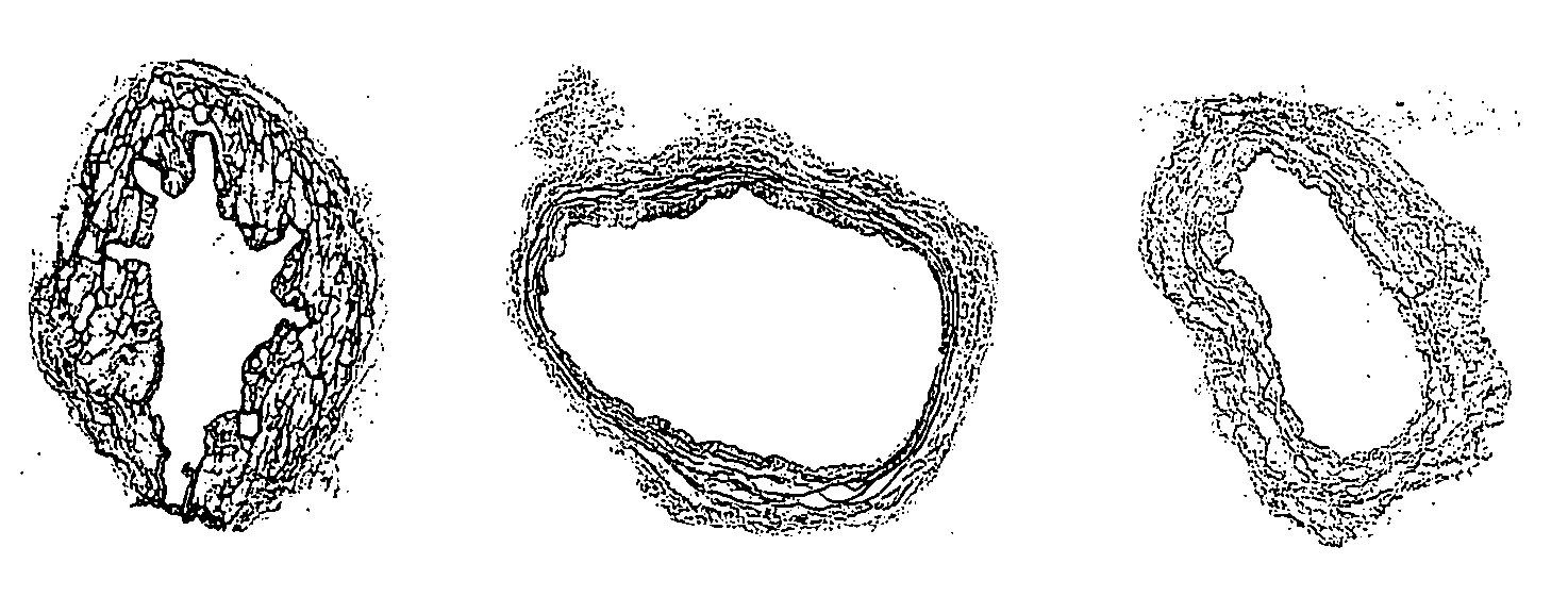
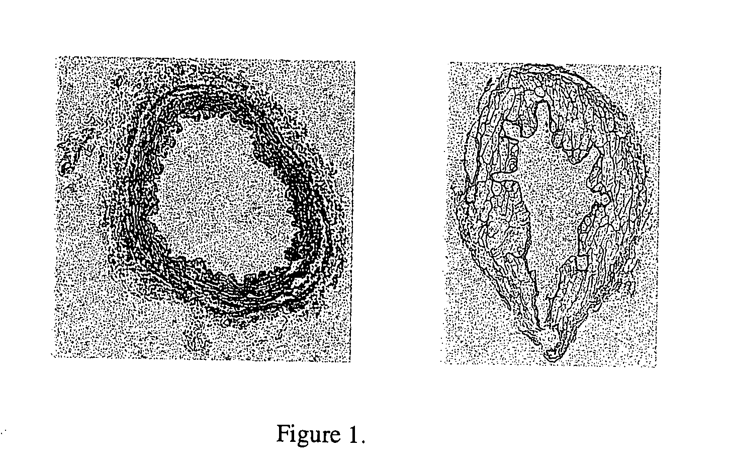
[0194] The effectiveness of cell and cellular debris removal was evaluated using two detergent and two enzyme solutions at various concentrations (Table 2). The detergents used in this study were Triton-X 100 at 0.25% (v / v) and 1.0% (v / v) and sodium deoxycholate (Cholate) at 0.25% (w / v) and 1.0% (w / v). Due to viscosity issues with sodium deoxycholate at low temperatures, tests were repeated at 0.5% (w / v). One treatment group combined Triton-X 100 with sodium deoxycholate. Trypsin alone at 0.05% (w / v) or 1.0% (w / v) and in combination with 0.02% (w / v) EDTA were the enzyme solutions used in the decellularization process. EDTA was also combined with Triton for a comparison since the Trypsin samples appeared grossly degraded after treatment.
[0195] Samples of human saphenous veins 10 mm in length were placed in each of the test solution at 37° C. or 48° C. for both 6 and 24 hours while continuously shaking. Samples were then rinsed in PBS at room tem...
example 2
Reduction Curves (Survivor Curves) for Spores
[0208] In order to determine the reduction in spores after processing with cleaning agents, an effective and consistent method was developed. Initially, a 107 spores / ml titre of B. stearothermophilus was used to enumerate the remaining population after treatment on both TSA media plates and sheep's blood agar (BA) plates. It was determined that residual hydrogen peroxide inhibited the growth at concentrations of 0.6% or higher for TSA plates and 6% or higher for blood plates (FIG. 12). It is believed that the catalase contained in the blood agar plates inactivated the hydrogen peroxide. A similar investigation was performed on PAA, but no inhibition was observed within the processing concentrations (FIG. 13). Even though the blood agar plates allowed for dilutions as low as 1:10, the titre used was too low to detect more than a 3 log reduction in the process limiting the data collection for the survivor curves. Therefore, the titre used...
example 3
Derivation of Governing Equations
[0224] The vein wall is divided into five layers: endothelial cell monolayer, intima, internal elastic lamina (IEL), media and adventitia. Since the endothelial layer acts as an active barrier to transport it is likely to offer more resistance to flow than bulk tissue (Kenyon, 1979). The study mentioned above on rabbit aortic tissue found that removal of the endothelial cells increased the macromolecular uptake at both 70 and 160 mm Hg of applied pressure (Meyer, 1996). The resistance due to this layer will not be a factor in this study since the grafts will be deendothelialized. The layer in contact with the luminal fluid will then be the intima. The intima itself provides very little resistance to flow and it has been shown that it provides no support to matrix stress while consolidation occurs at the interior wall at the border between the intima and rigid IEL (Kenyon, 1979). When it is combined with the IEL there is a greater pressure drop (ΔP / ...
PUM
 Abstract
Description
Claims
Application Information
 - R&D
- Intellectual Property
- Life Sciences
- Materials
- Tech Scout
- Unparalleled Data Quality
- Higher Quality Content
- 60% Fewer Hallucinations
Browse by: Latest US Patents, China's latest patents, Technical Efficacy Thesaurus, Application Domain, Technology Topic, Popular Technical Reports.
© 2025 PatSnap. All rights reserved.Legal|Privacy policy|Modern Slavery Act Transparency Statement|Sitemap|About US| Contact US: help@patsnap.com


