Implantable medical articles having laminin coatings and methods of use
a technology of medical articles and laminin coatings, applied in the field of implantable medical articles, can solve the problems of device failure in vivo, foreign body reaction, inflammation, etc., and achieve the effect of improving the function of the article and promoting the formation of blood vessels
- Summary
- Abstract
- Description
- Claims
- Application Information
AI Technical Summary
Benefits of technology
Problems solved by technology
Method used
Image

Examples
example 1
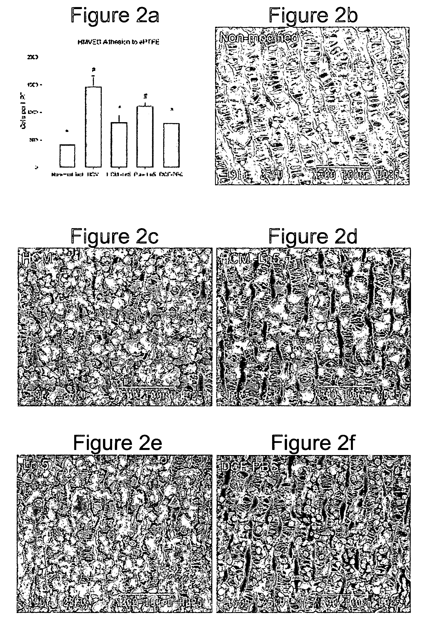
[0145] The HaCaT and II-4 cell lines (Dr. Norbert Fusenig (German Cancer Research Center) were maintained in culture medium (Dulbecco's Modified Eagle's Medium with high glucose, 10% fetal bovine serum, 2 mM L-glutamine, and 5 mM HEPES buffer). Cells at 70% confluence were rinsed with di-cation free phosphate buffered saline (DCF-PBS), pH 7.4, and placed in serum free medium for 48 hrs prior to collection of conditioned medium. Collected conditioned medium was centrifuged at 750 g for 5 min to remove debris prior to coating procedure.
[0146] Human microvessel endothelial cells (HMVEC) were isolated from human liposuction fat as previously described in Williams et al. (Williams, S. K., Wang, T. F., Castrillo, R. & Jarrell, B. E. Liposuction-derived human fat used for vascular graft sodding contains endothelial cells and not mesothelial cells as the major cell type. J Vasc Surg 19, 916-923 (1994)). Cells were maintained in culture medium (Medium 199, 10% fetal b...
example 2
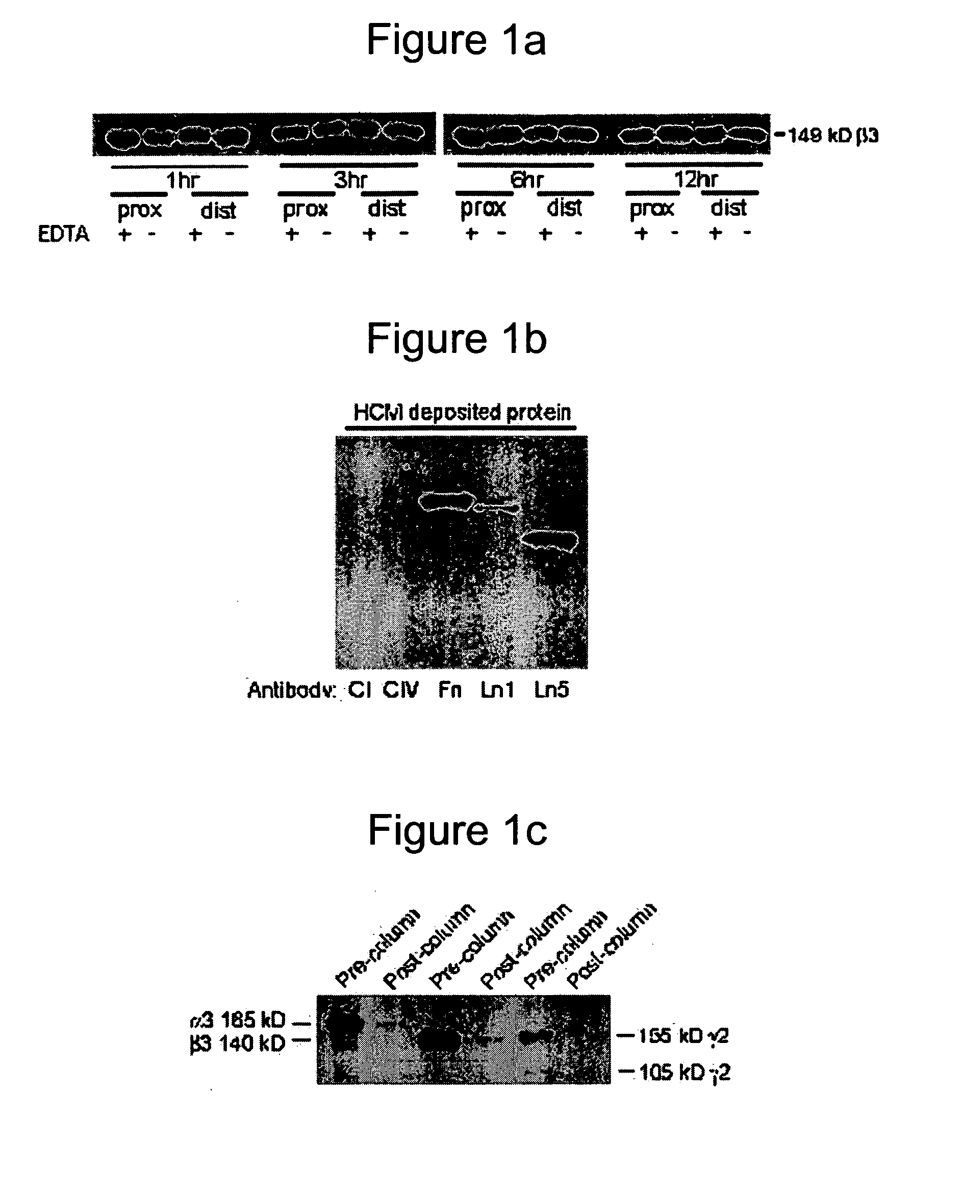
[0159] A heterobifunctional polyacrylamide reagent (HBPR, made as described in Example 9—U.S. Pat. No. 5,858,653) that contains amine-reactive and photo-reactive groups was used to immobilize extracellular matrix proteins onto ePTFE vascular graft (4 mm straight, C. R. Bard, Impra Corporation, Tempe, Ariz.). Matrix proteins were obtained from the following sources: bovine collagen-I (Kensey Nash), human collagen-IV (BD Biosciences), human fibronectin (BD Biosciences), mouse laminin-I (BD Biosciences), and human laminin-V (University of Arizona). Asceptic technique was used during all handling of the grafts and reagents. Grafts were cut to a 3.2 cm length. Female luer fittings (Small Parts, Inc.) were secured to each end of the graft with surgical suture. Grafts were denucleated (removing trapped air from the interstices of the graft) by soaking in isopropyl alcohol (IPA) for 20 minutes and then placing the graft in degassed Dulbecco's cation-free phosp...
example 3
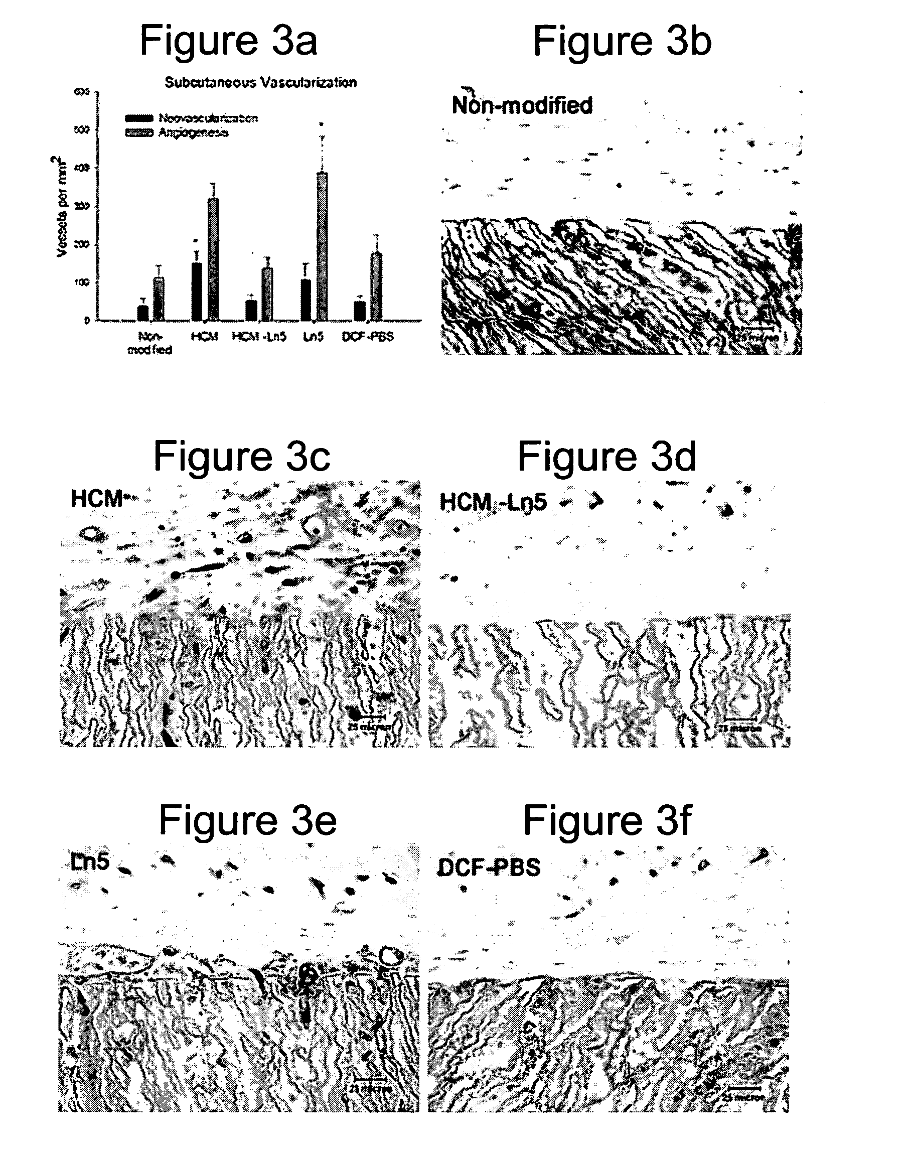
Rat Implant
[0164] An in vivo study evaluated the wound healing and inflammation associated with ePTFE discs coated with the reagent and protein coatings. ePTFE Discs (4 mm diameter size, (4 mm straight, C. R. Bard, Impra Corporation, Tempe, Ariz. A photoactivatable copolymer (HBPR) was prepared as described in Example 9 of U.S. Pat. No. 5,858,653. The following samples were evaluated: uncoated ePTFE, HBPR alone, HBPR Collagen-I, HBPR Laminin-I, HBPR Laminin-V, HBPR Collagen-I / Laminin-I, and HBPR Collagen-I / Laminin-V, Photo Collagen I and Photo Laminin 1. The laminin and collagen samples were obtained from the sources described in Example 2. Photo collagen 1 and Photo laminin 1 were made by the procedures described in Example 1 of U.S. Pat. No. 5,744,515, except that collagen 1 or laminin 1 was substituted were specifically made for this example. The coating procedure for HBPR and the protein samples is described in Example 2 except that the Collagen I / Laminin V example was prepare...
PUM
| Property | Measurement | Unit |
|---|---|---|
| Thickness | aaaaa | aaaaa |
| Thickness | aaaaa | aaaaa |
| Atomic weight | aaaaa | aaaaa |
Abstract
Description
Claims
Application Information
 - R&D
- Intellectual Property
- Life Sciences
- Materials
- Tech Scout
- Unparalleled Data Quality
- Higher Quality Content
- 60% Fewer Hallucinations
Browse by: Latest US Patents, China's latest patents, Technical Efficacy Thesaurus, Application Domain, Technology Topic, Popular Technical Reports.
© 2025 PatSnap. All rights reserved.Legal|Privacy policy|Modern Slavery Act Transparency Statement|Sitemap|About US| Contact US: help@patsnap.com