
Method and device for identifying micro organisms

a microorganism and identification method technology, applied in the field of methods and devices for identifying microorganisms, can solve the problems of slow and cumbersome species-specific identification and calculation of microorganisms from mixed microorganism samples, slow and cumbersome, and relatively rapid decrease of intensity

Inactive Publication Date: 2006-07-13
CYFLO
View PDF6 Cites 6 Cited by
  • Summary
  • Abstract
  • Description
  • Claims
  • Application Information

AI Technical Summary

Benefits of technology

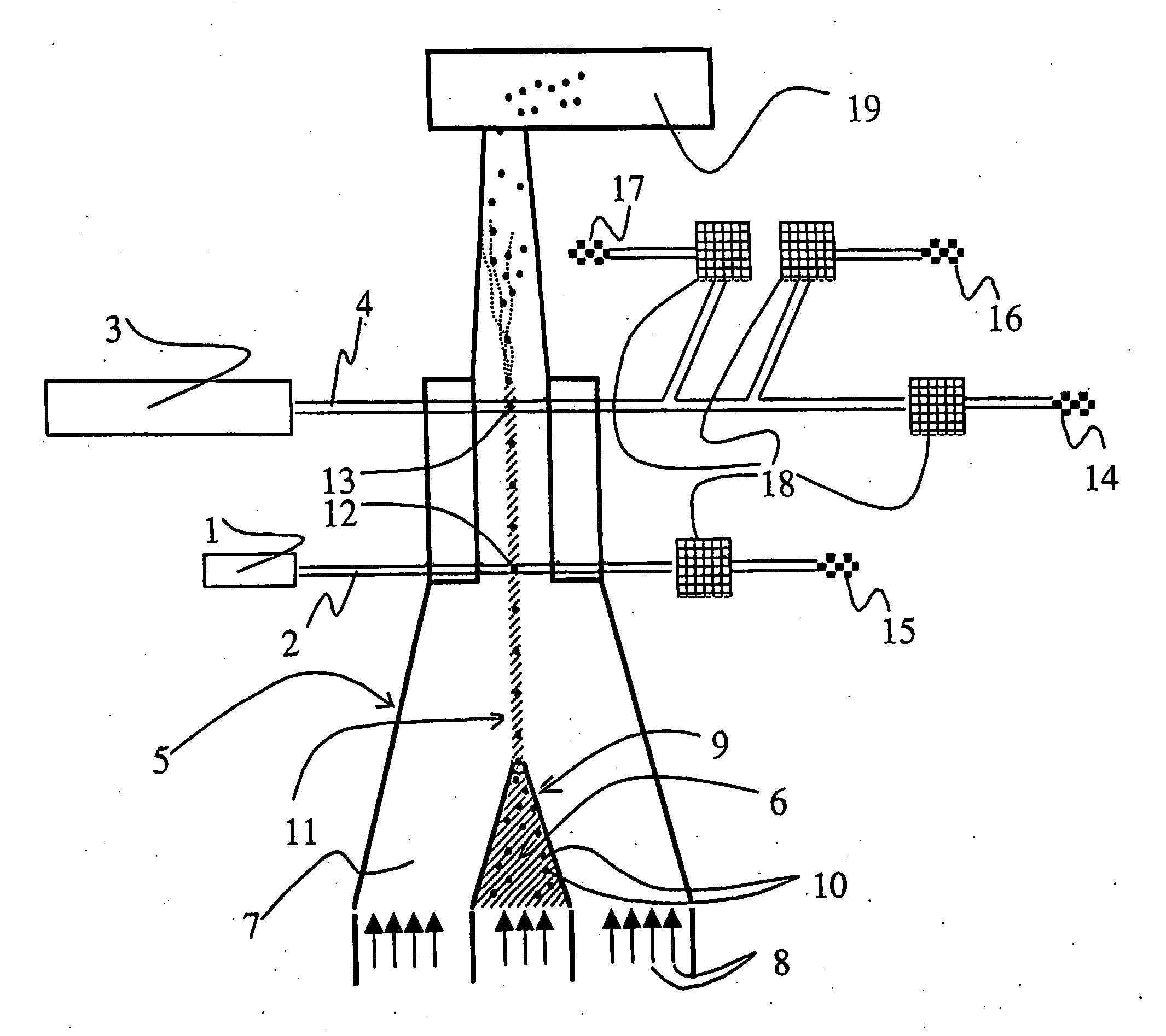
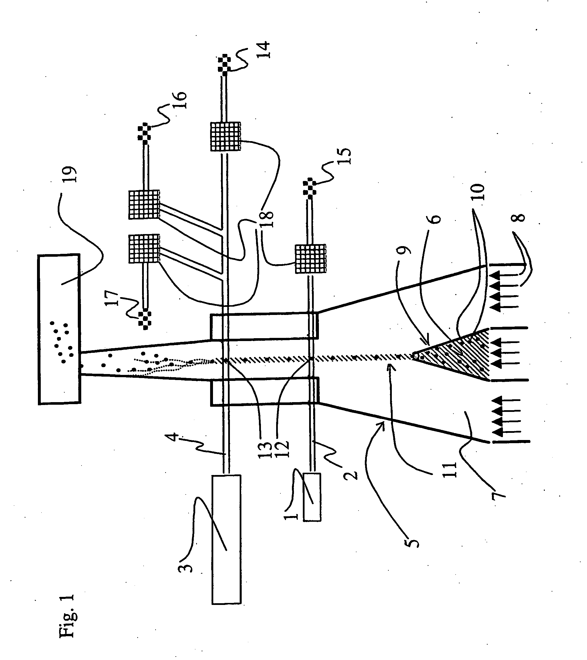
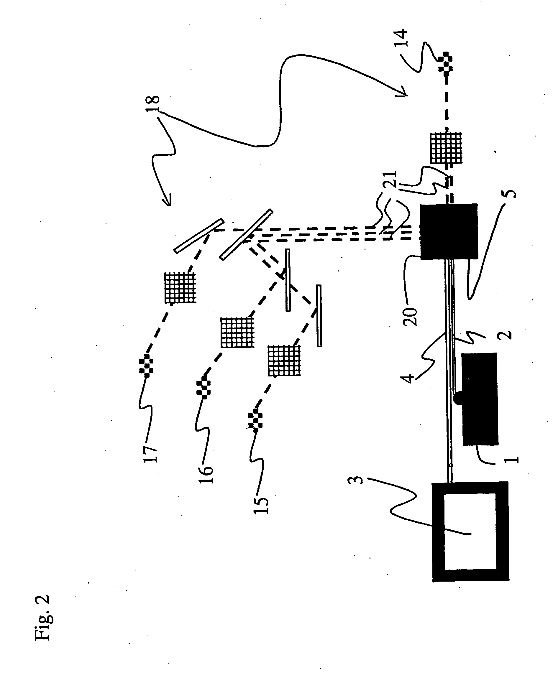
[0020] a) binding to a structure individualising least one micro organism species or group and enabling the identification a first fluorescent agent which absorbs light in a first wavelength area,
[0038] A considerable advantage by the method and device of this patent application is gained in that it enables a dependable, simultaneous distinguishing of all the three populations: the target micro organism population, the population formed by the rest of the micro organisms in the sample and the background population. This makes the analysis of the samples faster and makes the species-specific identification and calculation of micro organisms contained in mixed bacterial organism samples more dependable than before and enables a fast clarification of the concentration of micro organisms in a sample.
[0039] In the method according the invention, the hybridised probes can really be proven to be in the micro organisms and not e.g. in the particles of the background population, since the hybridised particles can be detected as being DNA stained in the same analysis and dot diagram. By using (e.g. by means of a hybridisation probe) as the bound fluorochrome, a fluorochrome sufficiently absorbing and emitting the light of the red wavelength area, and as the fluorochrome (e.g. a DNA colour) bound to all the micro organisms being examined, a fluorochrome sufficiently absorbing and emitting the light of the orange or a shorter wavelength area, there will be no hindering energy transfer between the fluorochromes. If the fluorochromes were used in such a manner that to the hybridisation probe, a fluorochrome absorbing and emitting the light of the shorter wavelength area would be attached and as the DNA colour, a fluorochrome absorbing and emitting the light of the longer wavelength area would be used, there could be an energy transfer between the fluorochromes hindering the distinguishing of the target micro organisms from the rest of the micro organisms in the sample.
[0040] As a method being both fast, automatic, and capable of being automated, the analysis of the micro organisms hybridised with the FISH technique according to the invention is a considerably better method than the microscopy-FISH for the species-specific examination and calculation of complicated mixed bacterial micro organism samples. The device according to the invention enables one to dependably identify even thousands of particles per second. In a unit of time, the number of identified micro organisms is thus multiple as compared to microscopy. The information given by a device correctly enabled is unambiguous, which reduces the error caused by human factors. The method according to the invention also enables one to count the number of the micro organisms contained in the sample more accurately and faster than by other methods.
[0044] According to one embodiment of the invention, at step d) of the method, in the sample to be subjected to flow there are in addition also micro particles, which are distinguished by means of their scattering properties and / or fluorescence properties. In addition, in the method and device in accordance with the invention there is a possibility of using a feeding device portioning out a standard amount of sample, a flow meter or some other device known to a person skilled in the art by means of which it is possible to measure the amount of the analysed sample. In this way, it is possible to determine the concentration of the micro organisms and micro organism species to be analysed in the sample. To calculate the accurate number of the micro organism cells contained in the sample to be analysed, the concentration of micro organisms and the portion of the target micro organisms, it is thus possible to use e.g. fluorescing micro particles or a feeding device portioning out a standard amount of sample.

Problems solved by technology

The species-specific identification and calculation of micro organisms from a mixed micro organism sample is slow and cumbersome with the methods used at present.
The disadvantages of the use include a relatively rapid decreasing of intensity (photobleaching), which renders difficult the calculation of the bacteria in the microscopy-FISH method.
In addition, the pH sensitiveness of the intensity of the light emitted by the fluorescaine makes it difficult to use it in many applications, and slows down the production of reagents.
Fluorescaine also has a wide emission spectrum, which makes it difficult to use it in applications utilising several fluorochromes.
Disadvantages of the microscopy-FISH method involve slowness and interpretative nature of results due to the non-specific hybridization.
This causes difficulty of interpretation in the microscopy-FISH.
Due to these reasons, the repeatability of the results obtained by the ‘microscopy-FISH method often’ remains unsatisfactory.
Using this method, the analyse velocity can be improved a little, but the analysing of the sample is nevertheless rather slow.
As in a manual microscopy-FISH, the problem with the automated microscopy-FISH is the determination of the luminance limit to be identified and the distinguishing of the non-specifically hybridized bacteria from the hybridized target bacteria.
Instead, flow cytometric analysis methods of prokaryotic cells i.e. bacteria have not spread into wide use.
The level of technique of flow cytometry equipment and the level of know-how of flow cytometry have been an obstacle to becoming general of bacteriological analysis and calculation methods based on flow cytometry, the level not allowing a dependable analysis of prokaryotic cells considerably smaller than the eukaryotic cells.
The methods known at present are not suitable for routine use and they cannot be used to calculate the micro organism concentrations of mixed micro organism samples.
118-126, and U.S. Pat. No. 6,225,046 of D. Vail, and patent EP0347039 of L. Terstappen. The methods based on the use of antibodies, have, however, not enabled a dependable species-specific examination of mixed bacterial samples, since antibodies are not bacterium species-specific.
A considerable difference between the FISH applications based on microscopy and flow cytometry is the dissimilarity of the light sources used for the exciting of the fluorescent agents such as fluorochromes in the sample.
The use of such fluorochrome combinations is general in the analysis of eukaryotic cell samples, but no fluorochrome combinations suitable for the FISH technique are known (Handbook of Fluorescent Probes and Research Products, Molecular Probes).
In practice this has meant that using the flow cytometry-FISH it has not been possible to distinguish and calculate the target population hybridized with the probe and DNA stained from solely a DNA stained population containing the other micro organisms of the sample as well as from the background population formed by the particles of non micro organism origin in the same analysis.
It has not been possible to calculate the number of micro organism cells contained in the sample and the portion of the hybridized target micro organisms in the same analysis.
The exciting and emission wavelength spectra of the fluorochromes of the probes are so far from each other that the exciting of the fluorochromes with just one laser is not successful, instead one must use two lasers having different wavelengths, the beams of which hit the particles of the sample at different times. In this method, both lasers must be used to distinguish the target population from the rest of the bacteria of the sample.
In the same manner, both axes of the dot diagram are used to distinguish the target population from the rest of the bacteria of the sample, and it is not possible to distinguish the total bacterial population from the background population at the same time.
Although one has used in the method very strong and expensive water-cooled lasers having the power of hundreds of milliwatts, the intensity of the fluorochromes used remains weak, and the population cannot be satisfactorily distinguished from each other in one analysis.
This weakens the dependability of the method.
Also in this method, the low intensity of the fluorescence of the fluorochromes used in the probes does not make it possible to distinguish the target bacteria i.e. the bacteria to be analysed from the rest of the bacteria contained in the sample.
The probe's fluorochrome used to distinguish the target bacteria from the rest of the bacteria in the sample uses its emission energy to excite the DNA colour, and the fluorescence of the target bacteria is not sufficient for their dependable distinguishing from the rest of the bacteria in the sample.
The target bacterial population and the population formed by the rest of the bacteria in the sample are overlapping in the dot diagram, and it is not possible to calculate the number of bacterial cells and the portion of the target bacteria from the total number of bacteria.
Thus, these methods are not applicable for the calculation of concentrations of bacteria contained in complicated mixed bacterial samples such as faeces, as well as for the specific and dependable identification and calculation of separate bacterial species.
As a results of this, the flow cytometric analyses of mixed bacteria have been unreliable, and the microscopy-FISH is still the only method to be reckoned for the species-specific identification and calculation of the bacteria contained in mixed bacterial samples.

Method used


Image

  • Method and device for identifying micro organisms
  • Method and device for identifying micro organisms
  • Method and device for identifying micro organisms

Examples


example

[0076] The method and device in accordance with the invention were used to examine the bacteria contained in human faecal samples by hybridising them using the 16S rRNA technique and the DNA staining (as is disclosed in publication Quantitative fluorescence in situ hybridization of Bifidobacterium spp. with genus-specific 16S rRNA targeted probes and its application in fecal samples; P. S. Langendijk et al., Applied and Environmental Microbiology, 1995, vol. 61, p. 3069-3075). As the probe, a bifidobacterium-specific probe was used that had been labelled with the Cy5 label (manufacturer Eurogentec) of the red wavelength area, which Cy5 label has an absorption maximum of about 643 nm and an emission maximum of about 667 nm and which can thus be identified by the FL4 detector. As the DNA colour, the SYTOX™ Orange colour of the orange wavelength area was used, the absorption maximum of which is about 547 nm and the emission maximum about 570 nm and which was identified by the FL2 detec...


PUM

PropertyMeasurementUnit
Electrical conductanceaaaaaaaaaa
Nanoscale particle sizeaaaaaaaaaa

Abstract

The invention relates to a method and device for identifying at least one micro organism and / or micro organism species and for measuring the portion of at least one micro organism and / or micro organism species from a sample. The method includes the use of two different fluorescent agents and the excitation with light in two different wavelengths. The sample is subjected to a flow. Furthermore, the invention relates to the use of the aforementioned method and device for identifying micro organisms and for measuring their portions.

Description

[0001] The invention relates to a method and device for identifying one or more micro organisms and / or micro organism species, and for measuring the portion of at least one micro organism and / or micro organism species from a sample, as well as the use of the aforementioned method and the aforementioned device. PRIOR ART [0002] The species-specific identification and calculation of micro organisms from a mixed micro organism sample is slow and cumbersome with the methods used at present. A mixed micro organism sample is herein used to mean a sample containing several micro organisms and micro organism species. Typical examples of mixed micro organism samples include faeces and waste water. For example, human faeces has been found to contain 300 to 400 different bacterial species, the bacterial density in the sample being of the order of 1011 bacterial cells per gram of the sample (Human fecal flora: the normal flora 20 Japanese-Hawaiians; W. E. C. Moore and L. V. Holdeman, Applied Mi...

Claims


Application Information

Patent Timeline
IPC IPC(8): G01J3/30G01N21/64C12Q1/68C12Q1/689G01N15/14G01N33/58
CPCC12Q1/689G01N15/147G01N15/1475G01N33/582G01N2015/1438G01N2015/1477G01N15/1433
Inventor KORKEAMAKI, MIKAVAAHTOVUO, JUSSI
Owner CYFLO
Learn More