
Assays for trichomonal and other hydrolases

a technology of trichomonal hydrolase and hydrolase, which is applied in the field of hydrolases, can solve the problems of inability to detect approximately half of the infected women, labor-intensive methods, and inconvenience of vaginal discharge, and achieve the effects of improving selectivity toward trichomonal hydrolase activity, convenient use, and convenient wetting

Inactive Publication Date: 2006-06-15
QUIDEL
View PDF5 Cites 0 Cited by
  • Summary
  • Abstract
  • Description
  • Claims
  • Application Information

AI Technical Summary

Benefits of technology

[0013] One discovery is that trichomonads release a particular hydrolase into vaginal fluid that actively hydrolyzes a narrowly defined class of substrates at a low pH without interference from other hydrolases that are unrelated to trichomoniasis and that are also present, or frequently present, in vaginal fluid. A diagnosis of trichomoniasis can thus be made by assaying vaginal fluid for hydrolytic activity against one or more members of this class of substrates at low pH. Hydrolysis of the substrate is readily converted to a signal that is either machine-readable or visually detectable.
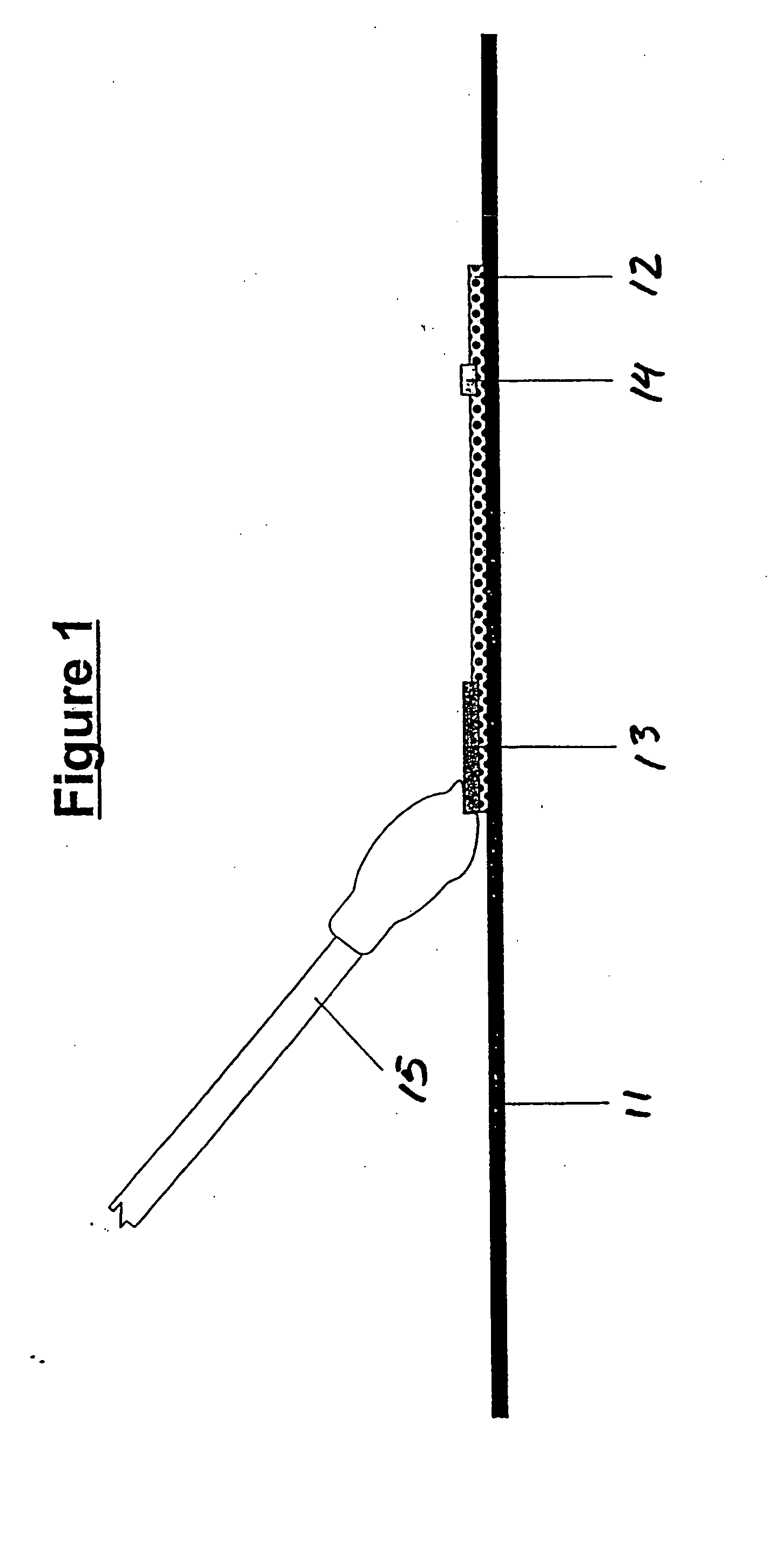
[0014] This discovery can be implemented in various diagnostic methods and test materials. One example is the use of an implement that is capable of retaining liquid together with a solid support on whose surface are deposited the assay components, in distinct regions if necessary, depending on the particular assay methodology. The implement can be applied to the surface and the assay result can be read directly on or in the implement. The implement can for example be a swab, dropper, pipette, or other liquid transfer device, and the deposited assay components can include a substrate and an indicator. Swabs are particularly convenient implements since a swab can easily be wetted with the sample, then rubbed on the operative surface of the solid support, causing the assay components deposited on the surface to adhere to the swab. This allows the user to perform the assay determination by simply noting whether a detectable change, preferably a color change, has occurred on the swab.
[0015] A further discovery is that trichomonal hydrolase activity, without being restricted to low pH, can be separated from other sources of hydrolase activity in a specimen of bodily fluid by size exclusion. Thus, by extracting from a specimen of bodily fluid, particularly vaginal fluid, a fraction that is devoid of particulate matter above a certain size threshold, one can detect the presence or absence of trichomoniasis in the specimen by determining whether hydrolytic cleavage of the substrate has occurred. Selectivity toward trichomonal hydrolase activity can be improved further by combining size exclusion with the use of a narrowly defined class of hydrolase inhibitors. Thus, in preferred embodiments of this discovery, a sample of vaginal fluid is processed to extract a fraction that is devoid of particulate matter above a certain size threshold and, in the presence of one or more of these inhibitors, the extracted fraction is brought into contact with an appropriate substrate to detect hydrolase activity. Restricting the substrate to a narrowly defined class provides even greater selectivity for trichomonal hydrolase activity.
[0016] Related to the discovery addressed in the preceding paragraph is the discovery of a test device that includes a migration path through which a sample of bodily fluid that is applied to the device travels by capillary force. The device contains porous material along the migration path to filter out particulate matter above a selected size threshold. The device is useful for the detection of any soluble enzyme that is not adhered to cells or other particulate matter in the bodily fluid, and bodily fluids on which the device can be used include vaginal fluid, urine, blood, saliva, and various others. The device also contains a substrate that is acted upon by the enzyme of interest and an indicator that produces a signal as a result of action of the enzyme upon of the substrate. In assays for trichomoniasis, for example, the substrate will be one that is hydrolyzed by trichomonal hydrolase activity. An alternative to the substrate-indicator combination is a substrate that includes a chromogen that undergoes a color change as a direct result of the action of the enzyme of interest. For enzymes that cause cleavage of the substrate, the chromogen may undergo a color change upon release of the chromogen from the remainder of the substrate. Hydrolases, including trichomonal hydrolases, are examples of such enzymes. Regardless of whether a substrate-indicator combination is used or a substrate is used that includes a chromogen whose color changes upon action of the enzyme, the substrate, the indicator if one is present, or both substrate and indicator are positioned far enough along the migration path that the visual signal is attributable only to hydrolytic activity in the filtered liquid. In preferred such devices, one of the class of inhibitors referred to above is also included to further improve the selectivity of the assay.

Problems solved by technology

Trichomoniasis in women may cause discomfort or a foul smelling vaginal discharge, and can be associated with adverse clinical sequelae.
This is a labor-intensive method which requires a microscope and a skilled technician, and fails to detect approximately half of the infected women (Baron et al., Laboratory Diagnosis of Female Genital Tract Infections, Cumitech 17A, American Society for Microbiology (1993)).
Due to the labor involved, the expense of culture media and supplies, the training required, and the delay of up to a week to detect trichomonal growth, few clinics or laboratories routinely use culture to diagnose trichomoniasis.
This test is sensitive but, like culture, not rapid enough to produce the results during the patient's visit to the clinic.
Due to the cost of the DNA-based test kits and the training and laboratory equipment required to perform these tests, this methodology is not widely used in clinics or in medical practice in general, particularly in the parts of the world where the need for a trichomoniasis test is greatest.
Thus, to date there is no rapid, accurate, cost-effective and simple method or test device for point-of-care diagnosis of trichomoniasis.
Unfortunately, the laboratory-based hydrolase detection methods used in the studies reported by these authors required equipment, skill and training and were labor-intensive and slow, so that the results were obtained only after several hours.
Both methods require a high level of expertise and costly equipment and take many hours to complete, and are therefore unsuitable for point-of-care testing.
Unfortunately, this procedure still entails the unwieldy polyacrylamide gel electrophoresis step prior to testing for hydrolase activity with the fluorogenic substrates.
Unfortunately, the methods used in this study are impractical for a point-of-care clinical diagnostic test, since gel electrophoresis is slow and cumbersome and observation of fluorescence requires instrumentation or a darkroom and ultraviolet illuminator.
The difficulty is that vaginal fluid contains many different hydrolases secreted by a variety of sources, including bacteria, which are present at extremely high levels, white blood cells, vaginal epithelial cells, and others.

Method used


Image

  • Assays for trichomonal and other hydrolases
  • Assays for trichomonal and other hydrolases
  • Assays for trichomonal and other hydrolases

Examples


experiment 1

[0083] This experiment examines the hydrolysis of the substrates CBZ-Arg-Arg-MNA (ZRR-MNA) and D-Val-Leu-Lys-MNA (VLK-MNA) by trichomonal hydrolases over the pH range 2.8 to 9.2 and examines the impact of vaginal fluid on hydrolase activity at each pH.

Materials

[0084] 200 mM buffer solutions: [0085] Glycyl-glycine (Gly-gly), pH 2.8 [0086] Sodium acetate, pH 5.1 [0087] 2-(N-Morpholino)propane-sulfonic acid (MOPS), pH 7.0 [0088] N-2-Hydroxyethylpiperazine-N′-2-ethanesulfonic acid (HEPES), pH 7.5 [0089] Tris(hydroxymethyl)aminomethane (TRIS), pH 8.0 [0090] Sodium borate, pH 9.5 [0091] Pooled normal vaginal fluid supernatant (NVS), prepared as follows: vaginal fluid specimens collected on Dacron swabs from normal, uninfected women were centrifuged to extract the fluid from the swabs, and the pooled fluid was frozen until use. The thawed fluid was centrifuged to pellet the particulate matter, the particulate materials and the supernatant were separated and each was diluted 1:10 in acet...

experiment 2

[0101] This experiment examines the hydrolysis of the substrates CBZ-Arg-Arg-MNA (ZRR-MNA), BOC-Leu-Arg-Arg-AMC (BLRR-AMC), BOC-Leu-Lys-Arg-AMC (BLKR-AMC), and CBZ-Lys-Lys-Arg-AMC (ZKKR-AMC) by trichomonal hydrolases and vaginal fluid hydrolases.

Materials

[0102] Buffer solutions consisting of 200 mM sodium acetate, pH 5, or 200 M Tris-(hydroxymethyl)aminomethane (TRIS), pH 8 [0103] 100 mM CBZ-Arg-Arg-MNA acetate (ZRR-MNA), in ethanol [0104] 100 mM BOC-Leu-Arg-Arg-AMC (BLRR-AMC) in dimethylformamide [0105] 100 mM BOC-Leu-Lys-Arg-AMC (BLKR-AMC) in dimethylformamide [0106] 100 mM CBZ-Lys-Lys-Arg-MNA (ZKKR-AMC), in water [0107] 6.3 mM L-cysteine hydrochloride in water [0108] pooled normal vaginal fluid supernatant (NVS) prepared by collecting vaginal fluid specimens on Dacron swabs from normal, uninfected women, centrifuging to extract the undiluted fluid from the swabs and to pellet the particulate matter, and pooling the supernatants [0109] whole normal vaginal fluid (NVF) from a si...

experiment 3

[0118] This experiment demonstrates that most of the interfering hydrolytic activity present in vaginal fluid collected from normal, uninfected women is present in the particulate matter and can be largely removed by centrifugation.

Materials

[0119] Swabs (two per donor) containing vaginal fluid specimens collected from normal, uninfected women at intervals of 1-2 days apart [0120] Substrate solutions consisting of 8 mM CBZ-Arg-Arg-MNA acetate (ZRR-MNA), 0.8 mM L-cysteine, and either 100 mM sodium acetate, pH 5, or 100 mM Tris(hydroxymethyl)aminomethane (TRIS), pH 8 [0121] Indicator solution consisting of 0.4 mM para-dimethylamino cinnamaldehyde, 1.4% w / v sodium dodecyl sulfate, and 100 mM citric acid (pH 4)

Procedure

[0122] Pairs of vaginal fluid specimens were obtained from donors on three separate days—Day 1, Day 3, and Day 4—and tested on the day of collection. The vaginal fluid was extracted from each swab by centrifugation. The vaginal fluid supernatant from the first swab f...


PUM

PropertyMeasurementUnit
diameteraaaaaaaaaa
diameteraaaaaaaaaa
diameteraaaaaaaaaa

Abstract

The release by trichomonads of a hydrolase that hydrolyzes a narrowly defined class of substrates at a low pH without interference from hydrolases that are unrelated to trichomoniasis is the basis for a selective diagnostic assay for trichomoniasis that measures hydrolysis of any of these substrates by vaginal fluid at a low pH. Selective assays for trichomoniasis are also obtained by removing particulate matter from a sample of vaginal fluid to extract a fraction devoid of particles greater than a selected size, and where desired, combining the extracted fraction with any of certain specified hydrolase inhibitors, then testing the fraction for enzymatic hydrolase activity. These qualities of trichomoniasis are the basis for a series of diagnostic tests and test devices that produce results that are detectable by visual and other means with a high degree of accuracy.

Description

BACKGROUND OF THE INVENTION [0001] 1. Field of the Invention [0002] This invention resides in the field of hydrolases (i.e., hydrolytic enzymes), and in particular, methods and devices for the detection of hydrolase activity in a sample or specimen and the diagnosis of disease based on the detection of hydrolase activity. A specific area of interest of this invention is the detection of trichomoniasis in female subjects by assaying for the presence of enzymatically active trichomonal hydrolases in a vaginal fluid specimen. [0003] All literature cited in this specification, including patents, technical articles and books, are incorporated by reference in their entirety. [0004] 2. Description of the Prior Art [0005] Trichomoniasis is a clinically important Sexually Transmitted Disease (STD) caused by the protozoan Trichomonas vaginalis. In 1955, the World Heath Organization estimated approximately 170 million new cases of trichomoniasis would arise annually among adults worldwide, wit...

Claims


Application Information

Patent Timeline
Patent Type & Authority Applications(United States)
IPC IPC(8): C12Q1/04C12M1/34C12Q1/34G01N33/58
CPCC12Q1/04C12Q1/34G01N33/581G01N2333/44G01N2800/26G01N2800/36
Inventor LAWRENCE, PAUL J.HUGHES, MARK A.CHAUDHURI, AULENAANDREASEN, TERRENCE J.
Owner QUIDEL
Learn More