
Hydrogels for biomolecule analysis and corresponding method to analyze biomolecules

a biomolecule and gel technology, applied in the field of hydrogels for biomolecule analysis and corresponding methods to analyze biomolecules, can solve the problems of difficult scaling-up to yield large microarrays suitable for analyzing an entire proteome, and the analysis that rely solely on expression profiles does not provide a complete picture of cellular control mechanisms. , to achieve the effect of eliminating the need for purifying complex samples

Inactive Publication Date: 2006-06-08
WISCONSIN ALUMNI RES FOUND
View PDF2 Cites 14 Cited by
  • Summary
  • Abstract
  • Description
  • Claims
  • Application Information

AI Technical Summary

Benefits of technology

[0009] A primary advantage of the present invention is that it eliminates the need to purify complex samples.
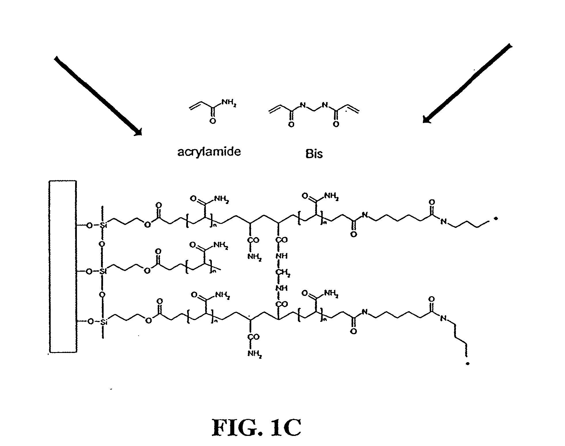
[0010] Thus, one embodiment of the invention is directed to a method of attaching a peptide, protein, and / or protein-containing complex to a surface. The method comprises providing a surface having immobilized thereon at least one domain comprising a first polymerizable ethylene-containing moiety. Also provided is a peptide, protein, or protein-containing complex modified to include a second polymerizable ethylene-containing moiety. Then, the functionalized peptide, protein, or protein-containing complex is polymerized with the domain, such that the peptide, protein, or protein-containing complex is immobilized upon and / or within the domain.
[0011] Another embodiment of the invention is directed to a method of assaying the presence and / or activity of a peptide, protein, and / or protein-containing complex using the composition of matter resulting from the method recited in the immediately preceding paragraph. Here, the method comprises providing a surface having immobilized thereon at least one domain comprising a first polymerizable ethylene-containing moiety; and providing a peptide, protein, or protein-containing complex modified to include a second polymerizable ethylene-containing moiety. The functionalized peptide, protein, or protein-containing complex is then polymerized with the domain, such that the peptide, protein, or protein-containing complex is: (i) immobilized upon and / or within the domain, and (ii) remains accessible to participate in chemical and enzymatic reactions, thereby yielding a reactive surface. Then, the reactive surface is contacted with a reagent mixture to be assayed under pre-defined reaction conditions. The reactive surface is then examined to detect whether the functionalized peptide, protein, or protein-containing complex within the reactive surface reacted with the reagent mixture.
[0012] In yet another embodiment, the invention is directed to a method of attaching a peptide, protein, and / or protein-containing complex to a surface. The method comprises providing a surface having immobilized thereon at least one domain comprising an acrylic acid- or acrylamide-based gel. Also provided is an acrylic- or acrylamide-functionalized peptide, protein, or protein-containing complex. The functionalized peptide, protein, or protein-containing complex is then co-polymerized with the domain such that the peptide, protein, or protein-containing complex is immobilized upon and / or within the domain.
[0013] In the preferred embodiment, the functionalized peptide, protein, or protein-containing complex is immobilized upon and / or within the domain such that it remains accessible to participate in chemical and enzymatic reactions.
[0014] The peptide, protein, or protein-containing complex can be any such entity, without limitation, including single-subunit and multi-subunit proteins, metal-containing proteins, proteins containing metallic or non-metallic prosthetic groups, etc. In a particularly preferred embodiment, the peptide, protein, or protein complex is an enzyme or fragment thereof, an enzyme substrate or fragment thereof; or a fusion protein or fragment thereof.

Problems solved by technology

In short, protein analyses that rely solely on expression profiles do not provide a complete picture of cellular control mechanisms.
While numerous methods for creating protein microarrays have been reported, a specific surface attachment strategy has yet to be widely accepted or adopted because each of the conventional approaches suffers from one drawback or another.
These transformations are easily performed on a proof-of-concept scale, but are quite difficult to scale-up to yield large microarrays suitable to analyze an entire proteome.
In addition, the close proximity of the attached proteins to the microarray surface can sterically block potential active sites or result in protein denaturation [9].
All of these prior art approaches, however, suffer from various shortcomings.

Method used


Image

  • Hydrogels for biomolecule analysis and corresponding method to analyze biomolecules
  • Hydrogels for biomolecule analysis and corresponding method to analyze biomolecules
  • Hydrogels for biomolecule analysis and corresponding method to analyze biomolecules

Examples


example 1

Preparation of Glass Surface

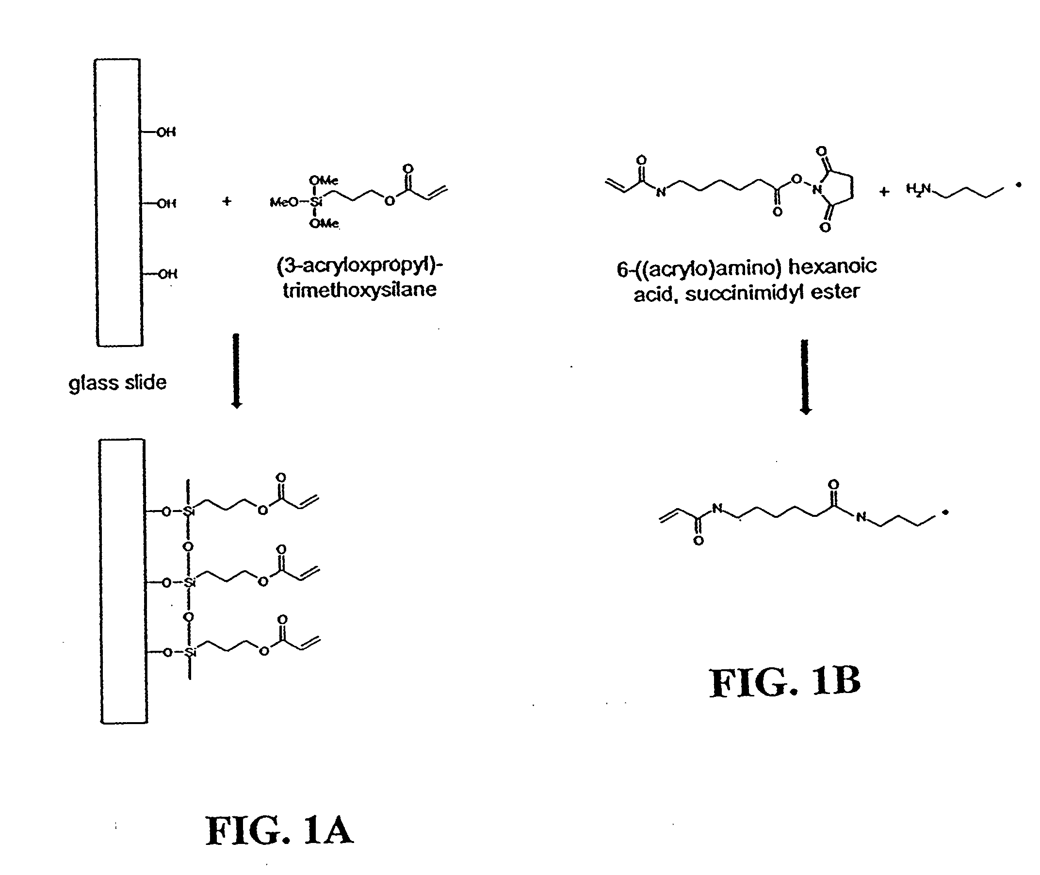
[0058] Glass microscope slides (Fisher Scientific, Hanover Park, Ill.) were cleaned in a 70 / 30 (v / v) mixture of H2SO4 / 30% H2O2 at 95° C. for 30 minutes. The slides were thoroughly washed with Milli-Q water (Millipore, Billerica, Mass.) and dried overnight at 140° C. To deposit the silane coupling agent on the surface, a 95 / 5 (v / v) solution of ethanol / Milli-Q water was prepared and brought to pH 5.0 with acetic acid. To the ethanol / water solution was added (3-acryloxypropyl)-trimethoxysilane (Gelest, Tullytown, Pa.) with stirring until a final concentration of 2% was reached. Clean glass slides were immersed in this solution for two minutes, rinsed by briefly dipping into ethanol, and dried overnight at room conditions. The slides were then stored in a desiccator at room temperature until needed.

example 2

Preparation of Fusion Proteins

[0059] The DNA sequence encoding a mutant green fluorescent protein (GFP-S65T) from Aequorea Victoria was amplified by PCR with primers containing BamH1 restriction sites flanking the GFP gene and cloned into the BamH1 restriction site of the pGEX-4T-1 vector (Amersham Biosciences, Piscataway, N.J.). This plasmid was transformed into E. Coli DH5α cells and the correct insertion of the GFP gene was confirmed by the isolation of colonies capable of producing the fluorescent fusion product. (Vectors containing the GFP-S65T insert are also available commercially from BD Biosciences, San Jose, Calif.; see also GenBank Accession No. U36202.)

[0060] For production of the GST-GFP fusion protein, the cells were grown to mid-log phase at 37° C. in 2× YT medium (16 g tryptone, 10 g yeast extract, 5 g NaCl, pH 6.6 in 1 (one) liter of water). The culture was then cooled to 30° C. and protein expression was induced by addition of isopropyl-β-D-thiogalactopyranoside ...

example 3

Labeling of Fusion Proteins

[0062] Purified fusion proteins were transferred to 100 mM sodium bicarbonate, pH 8.3 buffer and brought to a concentration of approximately 3 mg / ml through centrifugal concentration. Protein concentration was measured with a Pierce BCA protein assay kit (Pierce, Rockford, Ill.). Fusion proteins were then labeled with 6-((acryloyl)amino) hexanoic acid, succinimidyl ester (i.e., succinimidyl-6-[(acryloyl)amino]hexanoate) (Molecular Probes, Eugene, Oreg., catalog no. A20770) according to the manufacture's directions. Briefly, 100 μl of a 10 mg / ml solution of 6-((acryloyl)amino) hexanoic acid, succinimidyl ester in DMSO was slowly added to 1 ml of the fusion protein. The reaction was allowed to proceed for one hour at room conditions with maximum stirring. The reaction was then quenched by adding 100 μl of freshly prepared 1.5 M hydroxylamine, pH 8.5. The quenching reaction was allowed to proceed for one hour at room conditions. The acrylic-labeled fusion pr...


PUM

PropertyMeasurementUnit
diameteraaaaaaaaaa
fluorescence exposure timeaaaaaaaaaa

Abstract

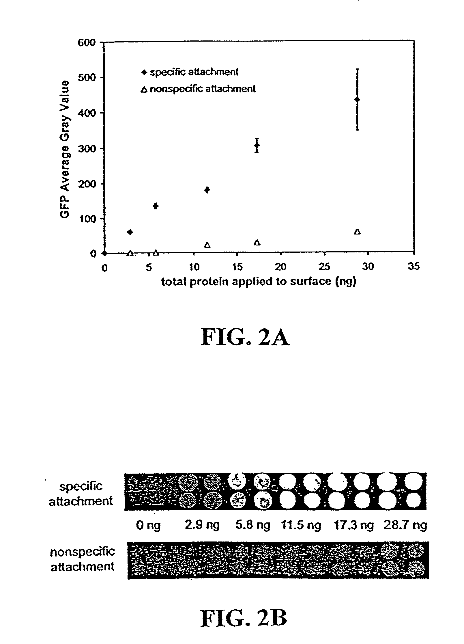
Disclosed are a polyacrylamide-based method of fabricating surface-bound peptide and protein arrays, the arrays themselves, and a method of using the arrays to detect biomolecules and to measure their concentration, binding affinity, and kinetics. Peptides, proteins, fusion proteins, protein complexes, nucleic acids, and the like, are labeled with an acrylic moiety and attached to acrylic-functionalized glass surfaces through a copolymerization with acrylic monomer. The specific attachment of GST-green fluorescent protein (GFP) fusion protein was more than 7-fold greater than the nonspecific attachment of non-acrylic labeled GST-GFP. Surface-attached GST-GFP (0.32 ng / mm2) was detectable by direct measurement of GFP fluorescence and this lower detection limit was reduced to 0.080 ng / mm2 using indirect antibody-based detection. The polyacrylamide-based surface attachment strategy was also used to measure the kinetics of substrate phosphorylation by the kinase c-Src. The surface attachment strategy is applicable to the proteomics field and addresses denaturation and dehydration problems associated with protein microarray development.

Description

CROSS-REFERENCE TO RELATED APPLICATIONS [0001] This is a continuation-in-part of application Ser. No. 11 / 066,136, filed Feb. 24, 2005, which claims priority to provisional application Ser. No. 60 / 547,198, filed Feb. 24, 2004, both of which are incorporated herein.FEDERAL FUNDING STATEMENT [0002] The invention was made with United States government support awarded by the following agencies: NIH CA103235. The United States has certain rights in this invention.INTRODUCTION [0003] Data from DNA microarrays has been used to elucidate the control mechanisms of cells and organisms based on gene expression profiles [1-3]. However, most cellular processes are the direct result of protein-protein interactions. Thus, a thorough understanding of the regulation of cellular processes must include a parallel analysis of protein activity to supplement the gene expression analysis. Protein activity depends on mRNA stability and translation rates, protein stability and degradation rates, post-transla...

Claims


Application Information

Patent Timeline
Patent Type & Authority Applications(United States)
IPC IPC(8): G01N33/53C12Q1/37G01N33/00
CPCG01N33/6842G01N33/6851H01J49/04
Inventor BRUEGGEMEIER, SHAWNKRON, STEPHENPALECEK, SEANPARKER, LAURIEKENT, STEPHEN
Owner WISCONSIN ALUMNI RES FOUND
Learn More