
Novel screens to identify agents that modulate retinal blood vessel function and pericyte function and diagnostic and therapeutic application therefor

a technology of retinal blood vessel and agent, applied in the field of drug screening, can solve the problems of affecting the function of the retinal vasculature, affecting the permeability of the retinal vasculature, and affecting the function of the pericyte, so as to reduce the contractile

Inactive Publication Date: 2005-10-20
MARTIN DONALD KEITH +1
View PDF18 Cites 4 Cited by
  • Summary
  • Abstract
  • Description
  • Claims
  • Application Information

AI Technical Summary

Benefits of technology

[0011] By “protagonise contraction” is meant that the compound enhances the contraction of the blood vessel or pericyte, causing them to contract more fully or to a greater extent, or for a longer duration or more rapidly.
[0012] By “antagonise contraction” is meant that the compound reduces the extent of contraction of the blood vessel or pericyte, thereby causing the blood vessel or pericyte to relax, to relax more fully, or that the duration of any contraction is reduced or that the period of contraction is shortened or the relaxation phase of the cell is enhanced or prolonged.
[0016] As used herein, the term “contractile state” shall be taken to mean the extent to which a blood vessel or pericyte is contracted. Accordingly, with regard to a pericyte, a reduced contractile state is one wherein the pericyte is more turgid or expanded or relaxed, or alternatively less contracted or slower to contract or relaxed for a longer duration. Conversely, an enhanced or increased contractile state is one wherein the pericyte is more contracted or constricted or contracts more rapidly or remains contracts for a longer period.
[0020] In one embodiment, such compounds are capable of enlarging the retinal blood vessels and thereby increase blood flow to the retina. In an alternate embodiment, such compounds are capable of restricting the retinal blood vessels and thereby decreasing blood flow to the retina. Further, the inventors have developed a diagnostic test based on the potential for the retinal blood vessels including small capillary blood vessels to have an impaired function in controlling blood flow in the eye. The effects of the agonists can be observed using recognised means according to a skilled person in the art. In a particularly preferred embodiment the diagnostic test enables a practitioner to assess the risk of a subject developing glaucoma, corneal angiogenesis, retinopathy of prematurity, or diabetic retinopathy.

Problems solved by technology

For example, impaired blood flow at and around the optic nerve head may be a major cause of complications associated with glaucoma.
In diabetic retinopathy, blood vessels (arterioles and capillaries) throughout the retina become damaged or blocked resulting in a lack of blood supply to small areas of the retina.
Diabetes can thereby lead to changes in the permeability of the retinal vasculature.
In more advanced cases of diabetic retinopathy, retinal neovascularisation can lead to leakage of blood into the retina, and retinal detachment, with consequent loss of vision.
If diabetic eye disease is left untreated, it can lead to serious visual impairment or blindness.
Unfortunately, there is currently no easily performed test for susceptibility of retinal vessels to develop permeability defects.

Method used


Image

  • Novel screens to identify agents that modulate retinal blood vessel function and pericyte function and diagnostic and therapeutic application therefor
  • Novel screens to identify agents that modulate retinal blood vessel function and pericyte function and diagnostic and therapeutic application therefor
  • Novel screens to identify agents that modulate retinal blood vessel function and pericyte function and diagnostic and therapeutic application therefor

Examples


example 1

Preparation of Silicone Rubber Substrate

[0213] A small volume of dimethylpolysiloxane (Sigma Chemical Co.) of either 60,000 cps (DMPS-60M) or 12,500 cps (DMPS-12M) viscosity was applied to 12 mm diameter glass cover slips. In some cases an intermediate viscosity dimethylpolysiloxane (30,000 cps) was prepared by blending 54% by weight of 60,000 cps dimethylpolysiloxane with 46% by weight of 12,500 cps dimethylpolysiloxane. The coated cover-slip was heated for 2 seconds using a Bunsen burner to induce cross-linking of the surface of the dimethylpolysiloxane and formation of a thin silicone rubber sheet bonded to the cover slip. After preparation, the cover slips were placed in 24-well tissue culture dishes and sterilised by UV irradiation overnight.

example 2

Growth of Pericytes on Silicone Rubber Substrate

[0214] Pericytes from primary cultures (first passage) were plated on the cover slips in DMEM supplemented with 10% FCS. The experiments were performed 48 h later, when almost all the cells were spontaneously in a contracted state as manifest by wrinkles in the rubber sheet beneath the cells (FIG. 1).

[0215] All experiments were performed using only first passage cultures of pericytes to minimise any change in pericyte physiology that might occur in prolonged culture.

example 3

Evaluating the Contractility of Pericytes

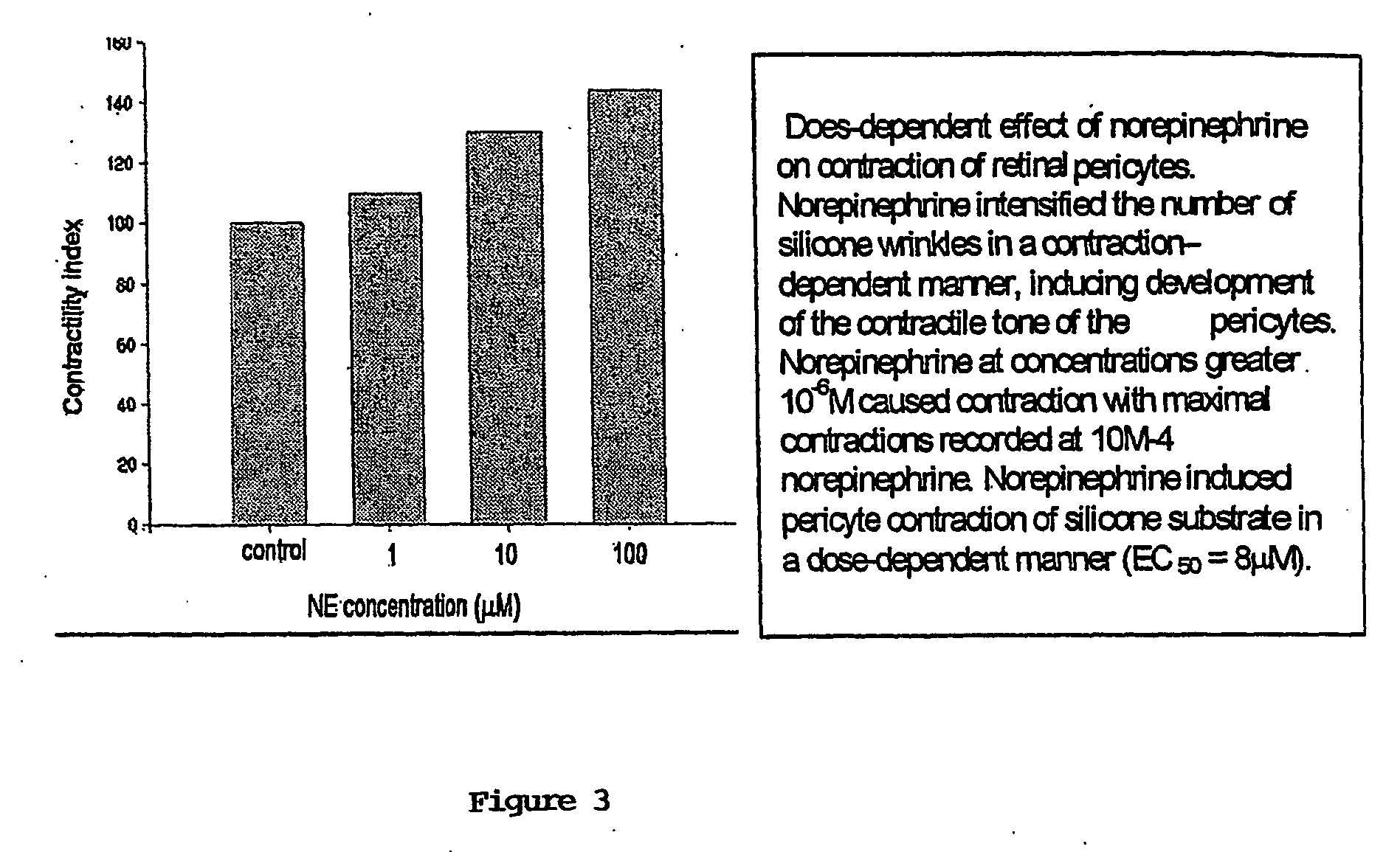
[0216] The response of the cells to the antagonists was evaluated using phase contrast microscopy at room temperature. The spontaneous contractile tone of pericytes induced tension wrinkles that could be observed after 24 h (FIG. 1). Wrinkles were only measured when associated with a pericyte, as shown in FIG. 1. Cells were identified as relaxed when the tension wrinkles associated with them diminished in size, and completely relaxed when the wrinkles disappeared. Conversely, a pericyte contracted when there was an increase in the number and length of the wrinkles associated with the pericyte. During each experiment, an image of the pericytes was captured (videocamera and framegrabber) by the computer every minute. The wrinkles were analysed after the experiment from these captured images.

[0217] In each of the experiments, the length of each clearly discerned wrinkle associated with the cell was measured 3 times, and the average of the leng...


PUM

PropertyMeasurementUnit
thickaaaaaaaaaa
diametersaaaaaaaaaa
diametersaaaaaaaaaa

Abstract

The present invention provides methods for determining or identifying compounds that modulate the function of an isolated retinal pericyte or blood vessel, wherein a change in the contractile state of said pericyte or blood vessel is determined in the presence of a test compound, said change indicating that the test compound modulates the function of pericytes and / or blood vessels.

Description

FIELD OF THE INVENTION [0001] The present invention is in the field of drug screening, such as, for example agents that modulate cellular contractility or blood flow. More particularly, the present invention relates to novel screens for agents that modulate retinal blood vessel function, such as, for example, permeability, integrity, and contractility. Further the present invention relates to novel screens for agents that modulate pericyte function, such as, for example, the contraction of pericytes, cell growth, differentiation, ion channel conductivity, neurotransmitter release, or gene transcription. The agents identified using such screens are useful for the diagnosis or therapeutic treatment of disorders involving impaired retinal blood vessel function or pericyte function, such as for example retinal oedema, glaucoma, retinopathy, or retinal neovascularisation in human or animal subjects. BACKGROUND OF THE INVENTION [0002] The retina of the eye is a seven-layered structure inv...

Claims


Application Information

Patent Timeline
Patent Type & Authority Applications(United States)
IPC IPC(8): A61K31/192A61K31/196A61K38/22A61K45/00A61K49/00A61P9/10A61P27/02A61P27/06A61P43/00C12Q1/02G01N33/15G01N33/50
CPCA61K31/192A61K31/196A61K38/2278G01N33/5088A61K49/0004A61P27/02A61P27/06A61P43/00A61P9/00A61P9/10
Inventor MARTIN, DONALD KEITHMARKHOTINA, NATALYA
Owner MARTIN DONALD KEITH
Learn More