Human cytoplasmic polyadenylation element binding protein and uses thereof

a technology of cytoplasmic polyadenylation and binding protein, which is applied in the field of molecular biology and gene cloning, can solve the problems of insufficient understanding of the mechanisms regulating human oocyte maturation, insufficient determination of the cytoplasmic polyadenylation of the maternal mos mrna during human oocyte maturation, and insufficient determination of the cytoplasmic polyadenylation of the maternal mos mrna

Inactive Publication Date: 2005-05-05
THE BOARD OF TRUSTEES OF THE UNIV OF ARKANSAS
View PDF0 Cites 3 Cited by
  • Summary
  • Abstract
  • Description
  • Claims
  • Application Information

AI Technical Summary

Benefits of technology

[0030]FIGS. 7A-7D shows the human cytoplasmic polyadenylation element binding protein (hCPEB1) specifically interacts with the CPE in the human Mos 3′ UTR. FIG. 7A shows GST western blot to demonstrate the relative expression levels of the GST moiety or GST-hCPEB1 fusion protein in programmed rabbit reticulocyte lysates. The GST moiety alone was expressed to slightly higher levels than the GST-hCPEB1 fusion protein. UP, unprogrammed lysate. Arrowheads indicate position of the expressed proteins. FIG. 7B is schematic representation of the mutant human Mos 3′ UTRs generated in this study. FIG. 7C shows RNA EMSA using a radiolabelled wild-type human Mos 3′ UTR and either unprogrammed reticulocyte lysate (UP), or reticulocyte lysate programmed with mRNA encoding the GST moiety alone (GST) or an mRNA encoding a GST-hCPEB1 fusion protein. Where indicated, a 50 fold excess of unlabelled wild-type human Mos 3′ UTR (Specific competitor) or a 50 fold excess of human Mos mutant 2 UTR (Non-specific competitor) RNA probe were also added to the binding reaction. The specific complex could be super shifted by addition of GST antiserum but not by addition of an irrelevant antiserum (B-Raf). FIG. 7D shows radiolabelled b-globin 3′ UTR, wild-type human Mos 3′ UTR, or various mutant human Mos 3′ UTR probes (see FIG. 7B) were analyzed for interaction with the GST-hCPEB1 fusion protein by RNA EMSA as described for FIG. 7C. Specific complex formation was only observed with the wild-type and mutant 1 human Mos 3′ UTR probes. For both FIGS. 7C and 7D, the position of the specific complex is indicated by an arrowhead, free probe by a filled circle and background non-specific complexes by an open circle. It should be noted that in the case of the mutant 1 UTR probe in (FIG. 7D, lane 5), an additional non-specific high molecular weight complex near the top of the gel was observed regardless of whether unprogrammed, GST moiety-alone or GST-hCPEB1 lysates were used. Representative results are shown.
[0031]FIGS. 8A-8B shows the human Mos 3′ UTR directs cytoplasmic polyadenylation in Xenopus oocytes. Xenopus immature oo...

Problems solved by technology

Thus, the prior art is deficient in identifying a human cytoplasmic polyadenylation element binding protein which is essential for the study of mRNA translation control in human.
Despite a critical role in the control of human fertility, the mechanisms regulating human oocyte mat...

Method used


Image

  • Human cytoplasmic polyadenylation element binding protein and uses thereof
  • Human cytoplasmic polyadenylation element binding protein and uses thereof
  • Human cytoplasmic polyadenylation element binding protein and uses thereof

Examples


example 1

Plasmids Constructs and RNA Synthesis

[0060] Plasmid pGEM XeMos wt UTR was constructed as follows. The terminal 321 nucleotides of the Mos 3′ UTR was cloned from immature oocytes by reverse transcription polymerase chain reaction (RT-PCR) to make pGEM Mos 321 UTR (Howard et al., 1999). A primer with a 5′ BamHI site (+) GCGGG ATCCA TTCCA TATGT GAATA TATAG (SEQ ID NO. 11) was designed to amplify the last 48 nucleotides of the Mos 3′ UTR from pGEM Mos 321 UTR. The reverse primer was the T7 promoter primer TAATA CGACT CACTA TAGGG (SEQ ID NO. 12). The PCR product was cut with BamHI / HindIII and inserted into BamHI / HindIII digested pGEM4Z. The size of the probe after in vitro transcription with SP6 RNA polymerase was 82 nt (48 nt that correspond to Mos UTR, and 34 nt that derive from the pGEM polylinker). The integrity of the probe was confirmed by DNA sequencing.

[0061] Plasmid pGEM XeMos mut UTR was constructed as follows. The terminal 321 nucleotides of the Mos 3′ UTR was cloned from i...

example 2

Northern and Southern Blot Analyses

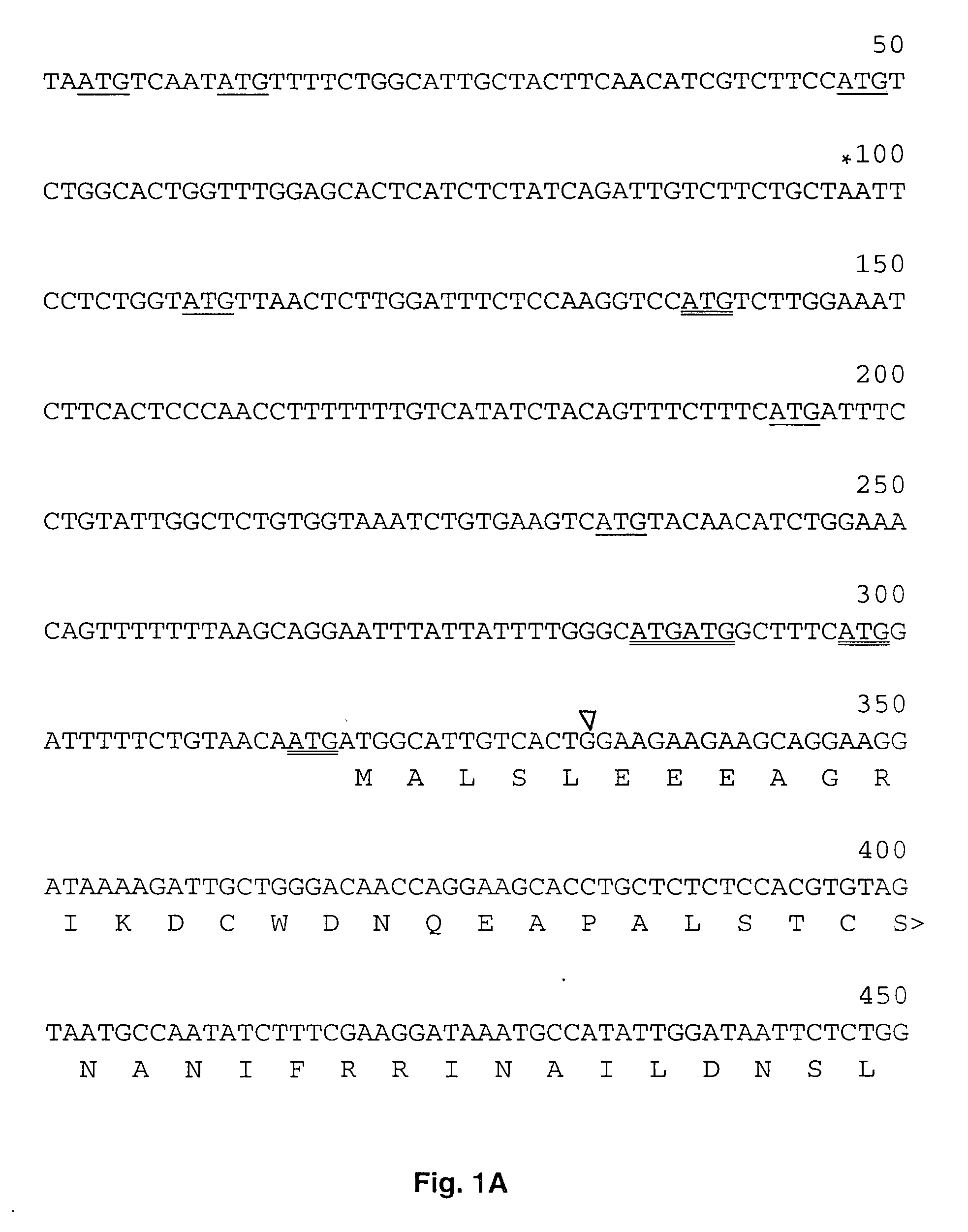
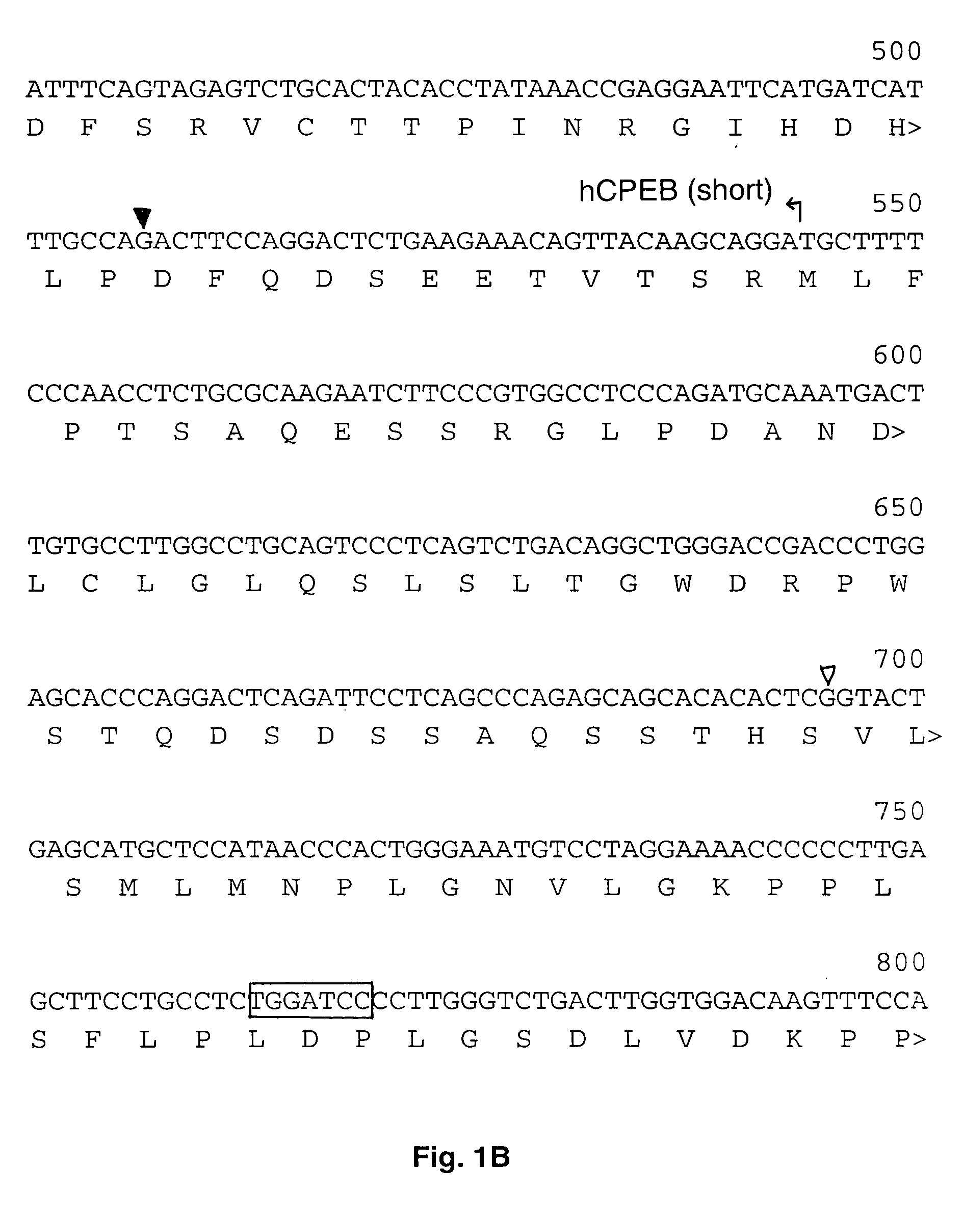
[0063] Human multiple tissue northern blots (2 mg polyA+RNA per lane) (Clontech) were probed with a 1326 bp BamHI / XcmI fragment of clone H12139 radioactively labeled using the random primer labeling kit (Pharmacia) and [a-32P]dCTP (Amersham). Following overnight hybridization at 55° C., the membranes were washed twice in 1×SSC (0.15 M NaCl, 0.015 M sodium citrate)-0.5% SDS for 10 min at 55° C. and twice in 0.1×SSC-0.1% SDS for 30 min at 55° C. and analyzed by autoradiography. For Southern analysis, 10 ug of human genomic DNA was digested with the indicated enzymes, transferred to nitrocellulose and probed with a multimerized probe corresponding to nucleotides 543-765 of hCPEBL. Filters were washed twice with 2×SSC, 0.1% SDS for 10 min at room temperature and twice 0.1× SSC, 0.1% SDS for 30 min at 65° C. and analyzed by autoradiography.

example 3

Human Oocyte Harvest and RT-PCR Analysis

[0064] Germinal vesicle-intact human oocytes were collected from consenting patients following a protocol approved by the Institutional Review Board at the University of Michigan. Oocyte-cumulus masses were collected into HEPES-buffered human tubule fluid media containing 3% (vol / vol) human serum albumin (HTF-H3; Irvine Scientific, Santa Ana, Calif.) by transvaginal follicular aspiration after controlled ovarian stimulation (Pool and Martin, 1994). Cumulus and corona radiata cells were removed by brief exposure to HTF-H3 containing 80 IU hyaluronidase (Sigma, St. Louis, Mo.) followed by mechanical pipetting through flame-pulled Pasteur pipettes. Complete absence of non-gamete cells and presence of germinal vesicle were confirmed using an inverted microscope with Hoffman optics at 400×. Germinal vesicle-intact oocytes were pooled, frozen in liquid nitrogen and stored at −80° C. until analyses were performed. RT-PCR was performed as described ...


PUM


Abstract

The present invention provides genomic and cDNA encoding human cytoplasmic polyadenylation element binding protein, expression vectors comprising human cytoplasmic polyadenylation element binding protein cDNA and host cells that contain the expression vectors. Also provided are recombinant human cytoplasmic polyadenylation element binding protein and polypeptides derived thereof. In addition, the present invention reports that the human Mos 3′ untranslated region (3′ UTR) contains a functional cytoplasmic polyadenylation element (CPE), interacts with the human CPEB1 protein and directs maturation-dependent cytoplasmic polyadenylation of the endogenous Mos mRNA.

Description

CROSS-REFERENCE TO RELATED APPLICATION [0001] This is a continue-in-part application of patent application U.S. Ser. No. 10 / 349,852, filed Jan. 23, 2003, which claims the benefit of priority of provisional patent application U.S. Ser. No. 60 / 351,121, filed Jan. 23, 2002, now abandoned.FEDERAL FUNDING LEGEND [0002] This invention was produced in part using funds obtained through a grant from the National Institutes of Health. Consequently, the federal government has certain rights in this invention.BACKGROUND OF THE INVENTION [0003] 1. Field of the Invention [0004] The present invention relates generally to the fields of molecular biology and gene cloning. More specifically, the present invention relates to the identification and characterization of the gene encoding human cytoplasmic polyadenylation element binding protein and uses thereof. [0005] 2. Description of the Related Art [0006] During meiotic maturation of human oocytes gene tran-scription is repressed (Braude et al., 1988...

Claims


Application Information

Patent Timeline
IPC IPC(8): C07H21/04C07K14/47
CPCC07H21/04G01N33/689C07K14/4702
Inventor MACNICOL, ANGUS
Owner THE BOARD OF TRUSTEES OF THE UNIV OF ARKANSAS
Why Eureka
Try Eureka