
Method for identification and enumeration of nucleic acid sequence, expression, copy, or DNA methylation changes, using combined nuclease, ligase, polymerase, and sequencing reactions

a nucleic acid sequence and methylation technology, applied in the field of nucleic acid sequence identification and methylation change identification and enumeration, can solve the problems of not being able to meet the best treatment, technical challenges, and beyond the reach of current sequencing technology, and achieve high-sensitivity detection, reduce false positives, and maximize the sensitivity of capture

Active Publication Date: 2019-09-10
CORNELL UNIVERSITY
View PDF22 Cites 3 Cited by
  • Summary
  • Abstract
  • Description
  • Claims
  • Application Information

AI Technical Summary

Benefits of technology

The present invention provides a collection of circular chimeric single-stranded nucleic acid constructs that can be used for rolling circle amplification and sequencing of nucleic acid molecules. These constructs have a unique identifier sequence that distinguishes them from each other. The invention also provides a system comprising these constructs and a method for identifying target ribonucleic acid molecules in a sample by hybridizing them with oligonucleotide probes and detecting the resulting circular ligated products. The technical effects of this invention include improved accuracy and sensitivity in identifying target nucleic acid molecules and the ability to distinguish them from other molecules in a sample.

Problems solved by technology

Currently, patients with predisposing or early disease are not identified, and those with disease are not given the best treatment—all because of failures at the diagnostic level.
There are a number of challenges for developing reliable diagnostic and screening tests.
The first challenge is to distinguish those markers emanating from the tumor or fetus that are indicative of disease (i.e. early cancer) vs. presence of the same markers emanating from normal tissue.
This is a challenge that needs to address the biological variation in diseases such as cancer.
Finally, there is the technical challenge to distinguish true signal resulting from detecting the desired disease-specific nucleic acid differences vs. false signal generated from normal nucleic acids present in the sample vs. false signal generated in the absence of the disease-specific nucleic acid differences.
This would require sequencing 300,000 GB of DNA, a task beyond the reach of current sequencing technology, not to mention the enormous data-management issues.
Sequence capture has suffered from dropout, such that maybe 90-95% of the desired sequences are captured, but desired fragments are missing.
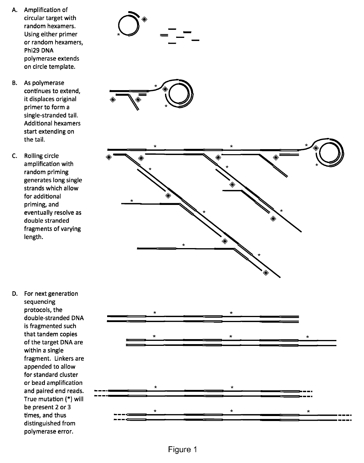
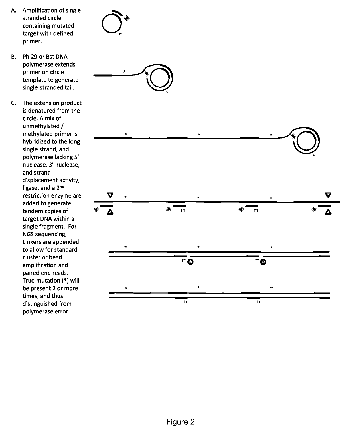
Alternatively, PCR amplification provides the risk of introducing a rare error that is indistinguishable from a true mutation.
Further, PCR loses methylation information.
While bisulfite treatment has been traditionally used to determine the presence of promoter methylation, it is also destructive of the DNA sample and lacks the ability to identify multiple methylation changes in cell-free DNA.
However, a high error rate makes it extremely difficult to identify a sequence variant in low abundance, for example when trying to identify a cancer mutation in the presence of normal DNA.
Therefore, a low error rate is required to detect a mutation in relatively low abundance.
The disadvantage is that this approach is unable to distinguish a true mutation from a PCR error in the early rounds of amplification.
Thus while the sensitivity of 2% (i.e. detecting one mutant allele in 50 wt alleles) is sufficient for evaluating late-stage cancers prior to making a treatment decision, it is not sensitive enough for early detection.
Both of these methods requires extending across the target DNA, and thus it would be impossible to distinguish true mutation, from polymerase generated error, especially when copying across a damaged base, such as deaminated cytosine.
Finally, these methods do not provide information on methylation status of CpG sites within the fragment.
Mutations present in only one or a few family members represent sequencing mistakes or PCR-introduced errors occurring late in amplification.
Mutations occurring in many or all members of one family in a pair arise from PCR errors during the first round of amplification such as might occur when copying across sites of mutagenic DNA damage.
The disadvantage of this approach is that many fragments need to be sequenced in order to get at least five members of each strand in a family pair (i.e. minimum of 10 sequence reads per original fragment, but often requiring far more due to fluctuations).
Further, the method has not been tested on cfDNA, which tend to be smaller then fragments generated from intact genomic DNA, and thus would require sequencing more fragments to cover all potential mutations.
Finally, the method does not provide information on methylation status of CpG sites within the fragment.
The disadvantage of this approach is that many fragments need to be sequenced in order to get at least five members of each strand in a family pair (i.e. minimum of 10 sequence reads per original fragment, but often requiring far more due to fluctuations).
Also, the method requires extending across the target DNA, and thus it would be impossible to distinguish true mutation, from polymerase-generated error, especially when copying across a damaged base, such as deaminated cytosine.
Further, the method has not been tested on cfDNA, which tend to be smaller then fragments generated from intact genomic DNA, and thus would require sequencing more fragments to cover all potential mutations.
Finally, the method does not provide information on methylation status of CpG sites within the fragment.
However, the method does not provide the ability to determine copy number changes, nor provide information on methylation status of CpG sites within the fragment.

Method used


Image

  • Method for identification and enumeration of nucleic acid sequence, expression, copy, or DNA methylation changes, using combined nuclease, ligase, polymerase, and sequencing reactions
  • Method for identification and enumeration of nucleic acid sequence, expression, copy, or DNA methylation changes, using combined nuclease, ligase, polymerase, and sequencing reactions
  • Method for identification and enumeration of nucleic acid sequence, expression, copy, or DNA methylation changes, using combined nuclease, ligase, polymerase, and sequencing reactions

Examples


case 10

[0751 shows inherited Duchenne's muscular dystrophy, where the mother is a carrier. Under these conditions, the amount of the total amount of two X-chromosome alleles appears half of other positions. In case 11, the fetus does not have the disease, and in case 12 the disease is a sporadic mutation that appears in the fetus. The Y chromosome marker confirms the fetus in male. The above shows how genotyping would be performed at the DMD locus. If there is prior knowledge that the mother is a carrier, than phasing of the deletion with neighboring polymorphisms can be determined (see below), and then these neighboring polymorphisms may also be used to verify if the fetus also carries the deletion, and if the fetus is male, and susceptible to the disease. This approach may be used to find both X-linked and autosomal dominant changes.

[0752]To determine if the fetus contains an inherited or sporadic mutation on one of the roughly 3,500 other disorders, including deletions, point mutations,...


PUM

PropertyMeasurementUnit
temperatureaaaaaaaaaa

Abstract

The present invention relates to a method for the highly specific, targeted capture of regions of human genomes and transcriptomes from the blood, i.e. from cell free circulating DNA, exosomes, microRNA, circulating tumor cells, or total blood cells, to allow for the highly sensitive detection of mutation, expression, copy number, translocation, alternative splicing, and methylation changes using combined nuclease, ligation, polymerase, and massively parallel sequencing reactions. The method generates a collection of different circular chimeric single-stranded nucleic acid constructs, suitable for sequencing on multiple platforms. In some embodiments, each construct of the collection comprised a first single stranded segment of original genomic DNA from a host organism and a second single stranded synthetic nucleic acid segment that is linked to the first single stranded segment and comprises a nucleotide sequence that is exogenous to the host organism. These chimeric constructs are suitable for identifying and enumerating mutations, copy changes, translocations, and methylation changes. In other embodiments, input mRNA, IncRNA, or miRNA is used to generate circular DNA products that reflect the presence and copy number of specific mRNA's, IncRNA's splice-site variants, translocations, and miRNA.

Description

[0001]This application is a national stage application under 35 U.S.C. § 371 of PCT Application No. PCT / US2015 / 034724, filed Jun. 8, 2015, which claims the benefit of U.S. Provisional Patent Application Ser. Nos. 62 / 009,047, filed Jun. 6, 2014, and 62 / 136,093, filed on Mar. 20, 2015, each of which is hereby incorporated by reference in its entirety.FIELD OF THE INVENTION[0002]The present invention relates to a method for the highly specific, targeted capture of regions of human genomes and transcriptomes from the blood, i.e. from cell free circulating DNA, exosomes, microRNA, circulating tumor cells, or total blood cells, to allow for the highly sensitive detection of mutation, expression, copy number, translocation, alternative splicing, and methylation changes using combined nuclease, ligation, polymerase, and sequencing reactions.BACKGROUND OF THE INVENTION[0003]Advances in DNA sequencing hold the promise to standardize and develop non-invasive molecular diagnosis to improve pren...

Claims


Application Information

Patent Timeline
Patent Type & Authority Patents(United States)
IPC IPC(8): C12Q1/68C12N15/10C12Q1/6874C12Q1/6816C12Q1/6869C12Q1/6844C12Q1/6848C12Q1/6855C12Q1/6806C12Q1/6809
CPCC12Q1/6874C12N15/1068C12Q1/6806C12Q1/6809C12Q1/6816C12Q1/6844C12Q1/6869C12Q1/6855C12Q1/6848C12Q2531/125C12Q2535/122C12Q2537/143C12Q2565/501C12Q2521/319C12Q2521/325C12Q2521/331C12Q2521/501C12Q2525/155C12Q2525/161C12Q2525/179C12Q2525/191C12Q2525/307C12Q2563/179
Inventor BARANY, FRANCISEFCAVITCH, JOHN WILLIAM
Owner CORNELL UNIVERSITY
Learn More