
Preparation method of decellularized amnion intrauterine scaffold carrying estradiol microspheres

An estradiol-loaded, decellularized technology, applied in the field of biomedical equipment preparation, can solve the problems of high recurrence rate and poor treatment effect of intrauterine adhesions

Active Publication Date: 2020-09-25
ATTACHED OBSTETRICS & GYNECOLOGY OSPITAL MEDICALCOLLEGE ZHEJIANG UNIV
View PDF5 Cites 1 Cited by
  • Summary
  • Abstract
  • Description
  • Claims
  • Application Information

AI Technical Summary

Problems solved by technology

[0004] The purpose of the present invention is to provide a preparation method of acellular amniotic uterine cavity stent loaded with estradiol microspheres, which is a kind of estradiol-loaded poly The preparation method of the acellular amniotic cavity stent made of lactic acid-glycolic acid copolymer (PLGA) microspheres, the stent can restore the shape of the uterine cavity, effectively repair the endometrium, reduce adverse drug reactions, and realize precise drug delivery in the uterine cavity

Method used


Image

  • Preparation method of decellularized amnion intrauterine scaffold carrying estradiol microspheres
  • Preparation method of decellularized amnion intrauterine scaffold carrying estradiol microspheres
  • Preparation method of decellularized amnion intrauterine scaffold carrying estradiol microspheres

Examples


Embodiment 1

[0028] A. Preparation of acellular amnion matrix:

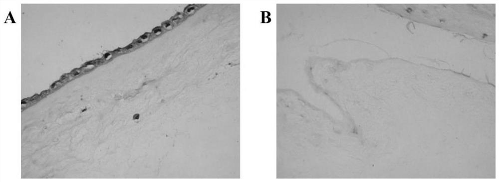
[0029] (1) Take amniotic membrane tissue: take the placenta negative for hepatitis B, hepatitis C, AIDS, and syphilis under sterile conditions, bluntly separate the amniotic membrane about 10cm×10cm, remove the chorionic membrane tissue, and use 50mg / ml penicillin, 50mg / ml streptomycin Stored after washing with phosphate buffered saline (pH 7.2-7.4).

[0030] (2) Preparation of acellular amniotic matrix: the amniotic membrane was washed repeatedly with normal saline, soaked in the volume fraction polyethylene glycol octylphenyl ether (TritonX-100) solution, placed in a constant temperature incubator at 37°C and shaken overnight, and taken out Then rinse with normal saline, add 25g / L trypsin and 0.2g / L ethylenediaminetetraacetic acid (EDTA), fully shake, and incubate at 37°C for 4h. After taking it out, rinse it repeatedly with normal saline, then place the amniotic membrane tissue in 8.8% sodium chloride solution, incubate a...

Embodiment 2

[0050] A. Preparation of acellular amnion matrix:

[0051] (1) Take amniotic membrane tissue: take the placenta negative for hepatitis B, hepatitis C, AIDS, and syphilis under sterile conditions, bluntly separate the amniotic membrane about 10cm×10cm, remove the chorionic membrane tissue, and use 50mg / ml penicillin, 50mg / ml streptomycin Stored after washing with phosphate buffered saline (pH 7.2-7.4).

[0052] (2) Preparation of acellular amnion matrix: the amnion was rinsed repeatedly with normal saline, soaked in 0.1% TritonX-100 solution by volume fraction, placed in a constant temperature incubator at 37°C and shaken overnight, rinsed with normal saline after taking it out, and added 25g / L trypsin and 0.2g / L EDTA, shake well, and incubate at 37°C for 4h. After taking it out, rinse it repeatedly with physiological saline, then place the amnion tissue in 8.8% sodium chloride solution, incubate at room temperature for 4 hours, take it out, wash it repeatedly with 0.01M phos...

Embodiment 3

[0075] A. Preparation of acellular amnion matrix:

[0076] (1) Take amniotic membrane tissue: take the placenta negative for hepatitis B, hepatitis C, AIDS, and syphilis under sterile conditions, bluntly separate the amniotic membrane about 10cm×10cm, remove the chorionic membrane tissue, and use 50mg / ml penicillin, 50mg / ml streptomycin Stored after washing with phosphate buffered saline (pH 7.2-7.4).

[0077] (2) Preparation of acellular amnion matrix: the amnion was rinsed repeatedly with normal saline, soaked in 0.1% TritonX-100 solution by volume fraction, placed in a constant temperature incubator at 37°C and shaken overnight, rinsed with normal saline after taking it out, and added 25g / L trypsin and 0.2g / L EDTA, shake well, and incubate at 37°C for 4h. After taking it out, rinse it repeatedly with physiological saline, then place the amnion tissue in 8.8% sodium chloride solution, incubate at room temperature for 4 hours, take it out, wash it repeatedly with 0.01M phos...


PUM


Abstract

The invention provides a preparation method of a decellularized amnion intrauterine scaffold carrying estradiol microspheres. A human amnion cell extracellular matrix is combined with PLGA estradiol microspheres to construct a biologically active intrauterine drug sustained-release scaffold. By regulating the molecular weight of a PLGA polymer and the matrix concentration of the amnion scaffold, estradiol can be completely released within 21 days and synchronizes with the fluctuation of the estrogen level in a female menstrual cycle. The decellularized amnion intrauterine scaffold carrying estradiol microsphere can be used to prevent adhesion recurrence after adhesion separation under a hysteroscope, and to prevent intrauterine adhesions and promote endometrial repair after intrauterine operation. The intrauterine scaffold provided by the invention can not only effectively provide an intrauterine barrier support function, but also increase the concentration of intrauterine estradiol and effectively promote the regeneration of endometria. The intrauterine scaffold has a simple preparation process and is prone to enlarged production, and is expected to become a novel pharmaceutical preparation for the prevention and treatment of intrauterine adhesions.

Description

technical field [0001] The invention belongs to the field of preparation of biomedical equipment, and relates to a method for preparing an acellular amniotic uterine cavity support loaded with estradiol microspheres, which can be used in repairing endometrium and preventing intrauterine adhesions. Background technique [0002] Intrauterine adhesions (IUA), also known as Asherman syndrome, refers to the injury of the basal layer of the endometrium due to trauma or infection, which causes all or part of the uterine cavity wall and (or) cervical canal to be atresiad, and the uterus loses its original structure. A disease with a physiological function. At present, the incidence of IUA in my country remains high, and with the increase of uterine cavity surgery, it shows a trend of increasing year by year. The risk factors for the incidence of IUA include: reproductive system infection, low estrogen environment in the body, history of uterine cavity operations such as abortion cu...

Claims


Application Information

Patent Timeline
IPC IPC(8): A61L31/00A61L31/14A61L31/16
CPCA61L31/005A61L31/16A61L31/146A61L31/148A61L31/14A61L2300/222A61L2300/43A61L2300/622A61L2430/40
Inventor 陈玥郑彩虹费伟东叶轶青赵梦丹赵云春张骁吴凡
Owner ATTACHED OBSTETRICS & GYNECOLOGY OSPITAL MEDICALCOLLEGE ZHEJIANG UNIV
Learn More