Chimeric antigen receptor mechanocyte as well as building method and application thereof
A technology of chimeric antigen receptors and fibroblasts, applied in the fields of medicine, biomedicine, and genetic engineering
- Summary
- Abstract
- Description
- Claims
- Application Information
AI Technical Summary
Problems solved by technology
Method used
Image
Examples
Embodiment 1
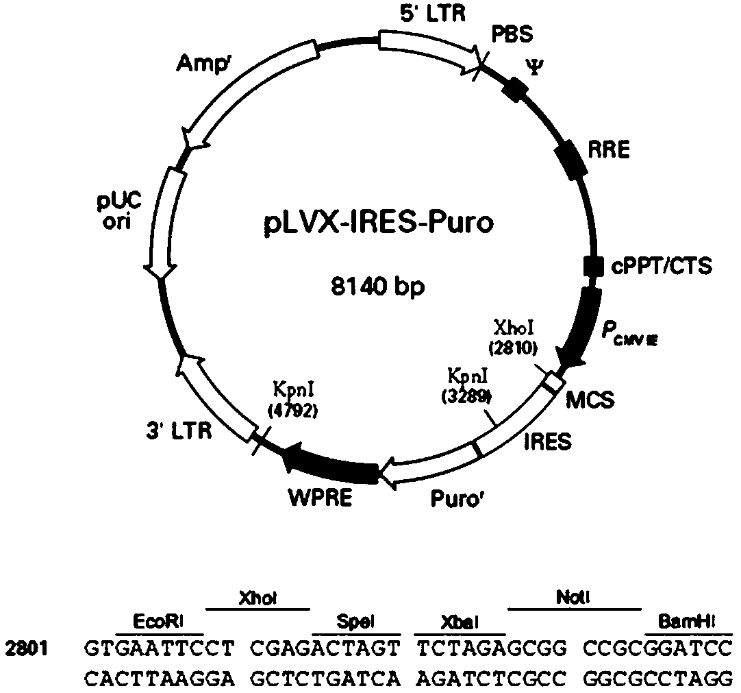
[0068] Example 1: Construction of a plasmid comprising a chimeric antigen receptor protein of an anti-human EpCAM single-chain antibody
[0069] The chimeric antigen receptor protein comprising the single-chain antibody of anti-human epithelial cell adhesion molecule (EpCAM) comprises from N-terminal to C-terminal sequence: 1) signal peptide sequence; 2) anti-human EpCAM single-chain antibody sequence; 3) including trans The junction sequence of membrane sequence; 4) mouse CD8 intracellular sequence or GFP sequence; 5) HAtag sequence ( figure 1 ). The sequence information involved is detailed in the table below:
[0070]
[0071]
[0072] The nucleotide and amino acid sequences of the signal peptide are shown in SEQ ID NO.1 and 2; the encoded nucleotide and amino acid sequences of the anti-human EpCAM single-chain antibody are shown in SEQ ID NO.11-22; including the transmembrane sequence The nucleotide and amino acid sequences of the connecting sequence are shown in ...
Embodiment 2
[0075] Example 2: Lentivirus packaging, concentration and titer determination
[0076] 1. Experimental materials:
[0077] 1) Virus packaging plasmids: pCMVΔR.89 plasmid (Pcg41), pVSV-Gplasmid plasmid (Pcg10).
[0078] 2) Packaging cells: Hek-293T cells (DuBridge, et. al. 1987, Pear, et al. 1993).
[0079] 3) Virus packaging medium: DMEM complete medium (Dulbecco's Modified Eagle Medium, Invitrogen, catalog #11995); fetal bovine serum (FBS, Hyclone, catalog #: SH30084.03); non-essential amino acids (Gibcocatalog #1140-050); Antibiotics (PenStrep, Gibic, Cat. No. 15070063).
[0080] 4) Cell transfection reagent: Lipofectamine2000 (Invitrogene, product number: 11668-019); serum-free medium Opti-MEM (Gibco, catalog #11058);
[0081] 5) Virus purification and infection: 0.45 μM filter (Corning, catalog #430314); sucrose (Sigma, catalog #: S7903); polybrene (Polybrene, Sigma, catalog #: H9268).
[0082] 6) Detection antibody: HAtag antibody (GenScript, catalog number: A01244). ...
Embodiment 3
[0094] Example 3: Infection of Target Cells and Screening of Chimeric Antigen Receptor Stable Expression Cell Lines
[0095] 1. Mouse embryonic fibroblast (MEF) preparation
[0096] 1) Sexually mature female mice were caged with male mice, and the vaginal opening of the mice was observed to have jelly-like substances. The morning was defined as 0.5 days of pregnancy.
[0097] 2) Execute the female mouse at 12.5-14.5 days pregnant, take out the embryos with fetal membranes, place them on a sterile plate, wash with PBS and discard the red blood cells. The fetal membranes were peeled off, the fetal mice were taken out, and washed three times with PBS. Remove the head, viscera and limbs of the embryo, place the trunk in a sterile plate, wash with PBS at least three times, and discard the red blood cells. Cut mouse embryo torso to 1mm 3 The following fragments are sucked into the centrifuge tube.
[0098] 3) Add an appropriate amount of trypsin, incubate at 37°C for 10-20 minut...
PUM
| Property | Measurement | Unit |
|---|---|---|
| molecular weight | aaaaa | aaaaa |
Abstract
Description
Claims
Application Information
 - R&D
- Intellectual Property
- Life Sciences
- Materials
- Tech Scout
- Unparalleled Data Quality
- Higher Quality Content
- 60% Fewer Hallucinations
Browse by: Latest US Patents, China's latest patents, Technical Efficacy Thesaurus, Application Domain, Technology Topic, Popular Technical Reports.
© 2025 PatSnap. All rights reserved.Legal|Privacy policy|Modern Slavery Act Transparency Statement|Sitemap|About US| Contact US: help@patsnap.com


