
A method for in vitro expansion of umbilical cord blood Treg cells

An in vitro expansion and cell technology, which is applied in the field of in vitro expansion of cord blood Treg cells, can solve the problems of unsatisfactory expansion multiples and expansion cycles, cumbersome operations, and reduced cell numbers, so as to achieve immunosuppressive function and simplify the Amplification method, the effect of increasing the amplification factor

Active Publication Date: 2020-03-27
深圳市沃英达生命科学有限公司
View PDF3 Cites 0 Cited by
  • Summary
  • Abstract
  • Description
  • Claims
  • Application Information

AI Technical Summary

Problems solved by technology

However, these methods reported so far require purification of cells with immunomagnetic beads before in vitro amplification, which is cumbersome to operate and increases the possibility of cell contamination, which is not conducive to large-scale clinical application.
Moreover, existing studies have shown that CD4+CD25-T cells can be transformed into CD4+CD25+Tregs under the induction of interleukin 7, interleukin 15 and estrogen, etc. Pre-purification, complicated operation
[0006] In addition, there are other limitations in the clinical application of adult peripheral blood Tregs. For example, in patients with autoimmune diseases, the number of cells may be significantly reduced or dysfunctional, so it is impossible to extract their own Tregs for treatment

Method used


Image

  • A method for in vitro expansion of umbilical cord blood Treg cells
  • A method for in vitro expansion of umbilical cord blood Treg cells
  • A method for in vitro expansion of umbilical cord blood Treg cells

Examples


Embodiment 1

[0040] The process of isolating umbilical cord blood mononuclear cells is as follows: newborn umbilical cord blood is collected from full-term healthy newborns in the Department of Obstetrics and Gynecology. After the fetus is delivered, 100ml of blood is collected from the umbilical vein of the placenta and put into a blood collection bag containing citrate anticoagulant. Blood was sent to the laboratory for processing within 4 hours after collection. Take 100ml of cord blood and centrifuge at 750g / min for 15min. Collect the upper layer of plasma, inactivate the plasma at 56°C for 30 min, and centrifuge at 2500 rpm / min for later use; add an equal volume (1:1) of PBS to dilute the remaining blood cells, and separate mononuclear cells by conventional density gradient centrifugation.

Embodiment 2

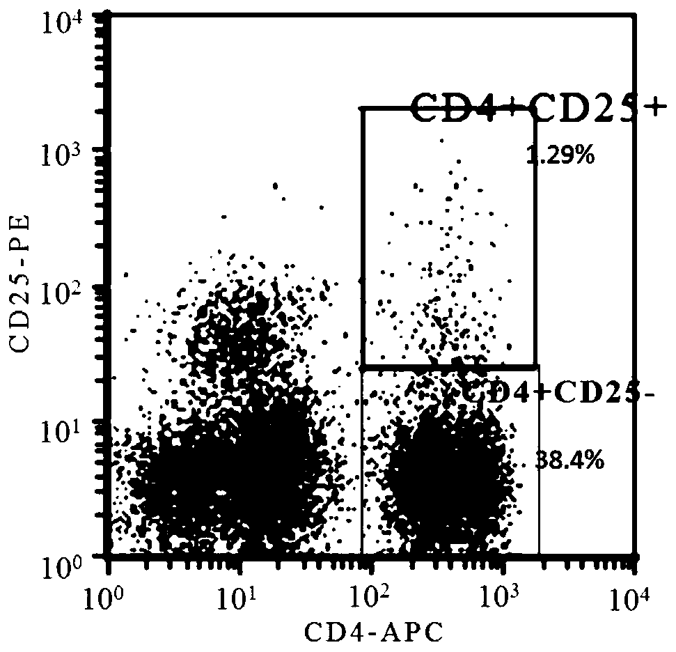
[0042] Flow cytometry detection of cell ratio in mononuclear cells: Flow cytometry detection of the ratio of CD4+CD25-T cells and CD4+CD25+Tregs in the mononuclear cells isolated in Example 3.

[0043] Collect the isolated mononuclear cells, 1x10^6 cells per group, centrifuge at 750g room temperature for 5min, discard the supernatant; wash the cells repeatedly with 2ml PBS for 3 times, resuspend the cells with 100ulPBS; add 10ul APC-CD4 and 10ul PE-CD25 antibody double-stained group was used as the test group, and 10ul APC-CD4 and 10ul PE-CD25 antibody single-stained groups were set as fluorescence compensation, and the unstained cell group was used as negative background. Incubate at 4°C for 30 min in the dark. After incubation, add 2ml of PBS to each tube, centrifuge at 750g for 5min at room temperature, and discard the supernatant. And continue to wash with 2mlPBS repeatedly for 2 times, add 200ulPBS to resuspend the cells, and test on the machine.

[0044] It was found t...

Embodiment 3

[0046] A method for in vitro expansion of umbilical cord blood Treg cells, the extraction method comprising the following steps;

[0047] S1: Separation of mononuclear cells: Take 100ml of cord blood and centrifuge at 800g / min for 10min to collect the upper plasma, inactivate the plasma at 54°C for 40min, and centrifuge at 2500rpm / min for later use; then add an equal volume of PBS to dilute the remaining blood cells , the umbilical cord blood was separated by conventional density gradient centrifugation;

[0048] S2: Adjust the cell density to 0.5x10 with RPMI1640 6 / ml, 5ml per bottle was inoculated into a T25 culture flask; add inactivated plasma to make the final plasma concentration 5.5%.

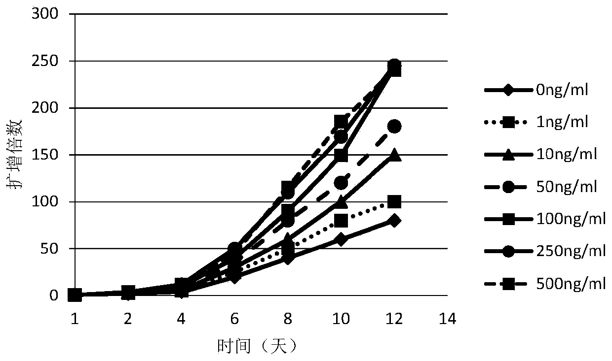
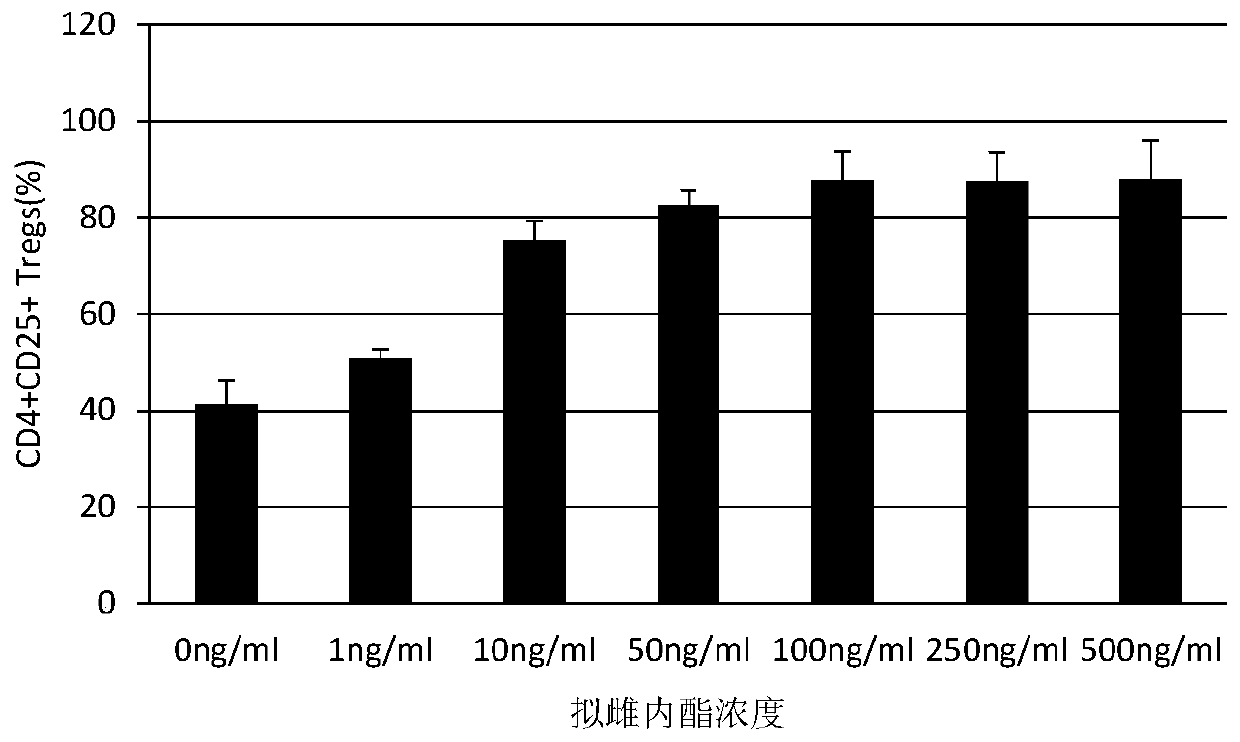
[0049] S3: The first day: Add pseudoestrolactone, TGF-β, CD3 / CD28 monoclonal antibody and IL-2, and make the final concentration of various components: pseudoestrolactone 100ng / ml, TGF-β5ng / ml , CD3 / CD28 monoclonal antibody 2μg / ml, IL-2 2000U / ml;

[0050] S4: The fourth day: add 3ml ...


PUM


Abstract

The invention relates to an in-vitro amplification method of cord blood Treg cells, comprising the steps of S1, isolating mononuclear cells; S2, adjusting cellular density, inoculating to a culture flask, and adding inactivated plasma; on the first day, adding coumestrol, TGF-beta, CD3 / CD28 monoclonal antibody and IL-2; S3, on the fourth day, supplementing a culture medium, adding inactivated plasma, coumestrol and TGF-beta, and keeping the final concentration constant; S4, on the sixth day, supplementing the culture medium, and keeping the final concentrations of other components unchanged; S5, on the ninth day, supplementing the culture medium, and keeping the final concentrations of other components unchanged; S6, on the twelfth day, collecting cells. The amplification of CD4, CD25 and Tregs is induced under the coaction of coumestrol and TGF-beta, the amplifying efficiency is high, and high-purity Treg cells can be acquired without pre-purification.

Description

technical field [0001] The invention relates to the field of biotechnology, in particular to an in vitro expansion method of umbilical cord blood Treg cells. Background technique [0002] Regulatory cells (Tregs for short) are a type of T cell subsets discovered in recent years that can control autoimmune reactivity in the body. Because of their immunosuppressive effect, they were also called suppressor Tcells in the early days. Tregs are a subset of CD4+ T cells that highly express CD25 and depend on IL-2 for survival. According to the source of Tregs cell development, it can be divided into two types of CD4+CD25+Tregs, natural Tregs (nTregs) and inducible Tregs (iTregs). nTregs develop in the thymus and iTregs develop in the periphery. What we usually refer to as regulatory T cells are nTregs that develop in the thymus. So far, people have studied Tregs for more than 20 years. Although the factors that affect the development of Tregs in vivo are still not very clear, th...

Claims


Application Information

Patent Timeline
Patent Type & Authority Patents(China)
IPC IPC(8): C12N5/0783
CPCC12N5/0637C12N2501/15C12N2501/2302C12N2501/392C12N2501/51C12N2501/515
Inventor 林词雄王旭李陶林洁璇朱刚
Owner 深圳市沃英达生命科学有限公司
Learn More