A kind of corneal stroma, preparation method and application
A technology of corneal stroma and cornea, applied in medical science, tissue regeneration, prostheses, etc., can solve problems such as failure to meet clinical requirements, inability to resist mechanical traction of intraocular pressure, poor collagen toughness, etc., and achieve dense matrix tissue , restore elasticity, and reduce corneal thickness
- Summary
- Abstract
- Description
- Claims
- Application Information
AI Technical Summary
Problems solved by technology
Method used
Image
Examples
Embodiment 1
[0047] The preparation method of a kind of corneal stroma of the present embodiment comprises the following steps:
[0048] (1) Raw material handling:
[0049] The eyeballs of the pigs within 4 hours of death were taken, the blood was rinsed with normal saline, the cornea was drilled with a trephine with a diameter of 8.5mm, rinsed with purified water, and then soaked in purified water.
[0050] (2) freeze-thaw swelling:
[0051] Soak the cornea with a small amount of water, freeze it in a -40°C freezer for 1 hour, take it out and rinse it with purified water for 15 minutes after it is completely thawed, then soak it in sufficient purified water for 30 minutes, repeat the freezing and thawing process 3 times, and obtain a cornea that absorbs water and swells.
[0052] (3) Virus inactivation:
[0053] Virus inactivation is performed by immersing the cornea in a peracetic acid-ethanol solution, which can be performed in a stainless steel barrel. The concentration (percentage ...
Embodiment 2
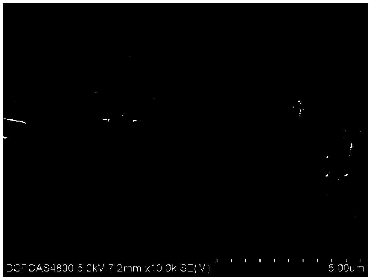
[0072] The sample in embodiment 1 is observed by a scanning electron microscope, as attached figure 1 and 4 As shown, it can be observed that after the immunogen removal treatment, the Bowman's membrane of the cornea after immunogen removal is intact, and is not affected or damaged by the enzyme solution, which is conducive to the migration, adhesion and proliferation of corneal epithelial cells, and is conducive to the rapid and complete cornea. Epithelialization to avoid infection. If the epithelium can grow normally and cover the surface of the corneal stroma, it can effectively isolate the degradation of its collagen components by various hydrolytic enzymes, prolong the degradation time, and facilitate the reconstruction of the corneal stroma.
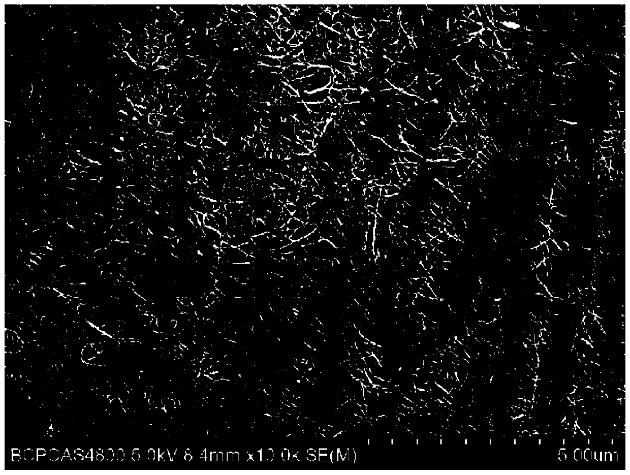
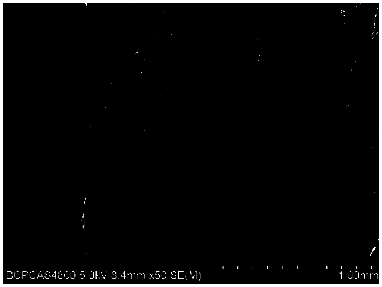
[0073] After removing the pre-elastic layer figure 2 and 3 As shown, it can be seen that the inside of the cornea presents a three-dimensional network structure, and the three-dimensional network structure forms a biological sc...
Embodiment 3
[0076] The sample obtained by the preparation method of the present invention is tested for physical properties, as follows:
[0077] a. Suture tensile strength: Prepare 5 samples according to Example 1, use a 3-0 non-absorbable suture at the edge of one end of the patch at 2 mm, fix the suture and the other end of the patch on the tension meter, and use 20mm Stretch at a speed of 1 / min until the suture point is torn, and record the maximum force value. The results show that the average value of the maximum value can reach 3.2N.
[0078] b. Light transmittance: 5 samples were prepared according to Example 1, and the light transmittance of air-dried samples, swollen samples, and glycerol dehydrated samples were measured respectively. Air-dried sample preparation: paste the swollen sample directly on the inner wall of the light-transmitting side of the cuvette, and send it to a blast oven to air-dry for 24 hours; preparation of the swollen sample: paste the swollen sample direct...
PUM
| Property | Measurement | Unit |
|---|---|---|
| diameter | aaaaa | aaaaa |
| thickness | aaaaa | aaaaa |
| clearance rate | aaaaa | aaaaa |
Abstract
Description
Claims
Application Information
 - R&D
- Intellectual Property
- Life Sciences
- Materials
- Tech Scout
- Unparalleled Data Quality
- Higher Quality Content
- 60% Fewer Hallucinations
Browse by: Latest US Patents, China's latest patents, Technical Efficacy Thesaurus, Application Domain, Technology Topic, Popular Technical Reports.
© 2025 PatSnap. All rights reserved.Legal|Privacy policy|Modern Slavery Act Transparency Statement|Sitemap|About US| Contact US: help@patsnap.com


