Preparation method of bionic porous tissue-engineered cartilage scaffold
A porous tissue and engineering technology, applied in the field of biomedicine, can solve problems such as compound ratio transformation, achieve the effect of improving cartilage quality and good application prospects
- Summary
- Abstract
- Description
- Claims
- Application Information
AI Technical Summary
Problems solved by technology
Method used
Image
Examples
preparation example Construction
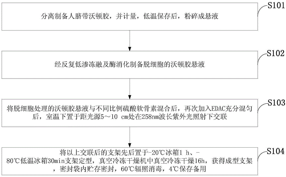
[0024] Such as figure 1 As shown, a kind of biomimetic porous tissue engineering cartilage scaffold preparation method of the embodiment of the present invention comprises the following steps:
[0025] S101: separate and prepare human umbilical cord Wharton's jelly, measure it, store it at low temperature, and crush it into a suspension;
[0026] S102: Prepare decellularized Wharton's gum suspension through repeated hypotonic freezing-thawing and enzymatic digestion;
[0027] S103: Mix the decellularized Wharton gum suspension with chondroitin sulfate in different proportions, add EDAC again and mix thoroughly, place at room temperature at a distance of 5-10 cm from the light source, and cross-link under the irradiation of 258nm wavelength ultraviolet light;
[0028] S104: Place the above cross-linked scaffolds in a -20°C refrigerator and a -80°C low-temperature refrigerator successively to shape the scaffolds, vacuum freeze-dry them in a vacuum freeze dryer to obtain shaped...
Embodiment 1
[0031] Step 1, separate and prepare human umbilical cord Wharton gum, measure it, store it at low temperature, and crush it into a suspension;
[0032] Step 2, preparing decellularized Wharton gum suspension through repeated hypotonic freeze-thaw and enzyme digestion;
[0033] Step 3: Mix the decellularized Wharton’s gum suspension with chondroitin sulfate, Wharton’s gum suspension: chondroitin sulfate = 2:0.25, add EDAC again and mix well, place at room temperature 5-10cm away from the light source Cross-linking under 258nm wavelength ultraviolet light irradiation;
[0034] Step 4: Place the above cross-linked scaffolds in a -20°C refrigerator and a -80°C low-temperature refrigerator successively to shape the scaffolds, vacuum freeze-dry them in a vacuum freeze dryer to obtain shaped scaffolds, store and seal them in sealed bags, and irradiate at 60°C Sterilize and store at 4°C for later use.
Embodiment 2
[0036] Step 1, separate and prepare human umbilical cord Wharton gum, measure it, store it at low temperature, and crush it into a suspension;
[0037]Step 2, preparing decellularized Wharton gum suspension through repeated hypotonic freeze-thaw and enzyme digestion;
[0038] Step 3, mix the decellularized Wharton’s gum suspension with different proportions of chondroitin sulfate, the mass ratio of Wharton’s gum suspension to chondroitin sulfate is 2.0:0.75; add EDAC again and mix well, place at room temperature Cross-linking under the irradiation of 258nm wavelength ultraviolet light at a distance of 5-10cm from the light source;
[0039] Step 4: Place the above cross-linked scaffolds in a -20°C refrigerator and a -80°C low-temperature refrigerator successively to shape the scaffolds, vacuum freeze-dry them in a vacuum freeze dryer to obtain shaped scaffolds, store and seal them in sealed bags, and irradiate at 60°C Sterilize and store at 4°C for later use.
PUM
| Property | Measurement | Unit |
|---|---|---|
| density | aaaaa | aaaaa |
| porosity | aaaaa | aaaaa |
| water absorption | aaaaa | aaaaa |
Abstract
Description
Claims
Application Information
 - R&D Engineer
- R&D Manager
- IP Professional
- Industry Leading Data Capabilities
- Powerful AI technology
- Patent DNA Extraction
Browse by: Latest US Patents, China's latest patents, Technical Efficacy Thesaurus, Application Domain, Technology Topic, Popular Technical Reports.
© 2024 PatSnap. All rights reserved.Legal|Privacy policy|Modern Slavery Act Transparency Statement|Sitemap|About US| Contact US: help@patsnap.com


