
Preparation of fluorescent microsphere immunochromatographic test strip and quantitative detection method

A technology of immunochromatography test paper and fluorescent microspheres, which is applied in the field of biomedical detection, can solve the problems of protein molecules falling off easily again, limited application range, poor ability to resist solution interference, etc., to increase fluorescence stability and fluorescence life, expand Scope and type, the effect of overcoming the easy leakage of dyes

Inactive Publication Date: 2015-10-14
刘宏飞
View PDF2 Cites 17 Cited by
  • Summary
  • Abstract
  • Description
  • Claims
  • Application Information

AI Technical Summary

Problems solved by technology

[0004] (1) The colloidal gold labeling process is an electrostatic adsorption process, which belongs to physical adsorption, and has poor stability in the liquid phase, and the protein molecules on the label are easy to fall off again
[0005] (2) Only when the gold particles reach a certain amount, the naked eye can distinguish the difference between the detection band and the background, so the detection sensitivity is not high
[0006] (3) The matrix effect of different materials is obvious, and the background interference is very large
The methods disclosed in these patents can improve the sensitivity of immunochromatographic detection, but the reported fluorescent nanoparticles have certain defects, such as dyes are easy to leak, and the ability to resist solution interference is poor, so the scope of application is largely limited.

Method used


Image

  • Preparation of fluorescent microsphere immunochromatographic test strip and quantitative detection method
  • Preparation of fluorescent microsphere immunochromatographic test strip and quantitative detection method
  • Preparation of fluorescent microsphere immunochromatographic test strip and quantitative detection method

Examples


specific Embodiment approach

[0052] 1. Preparation of rhodamine B n-octyl ester-polystyrene fluorescent microspheres

[0053] Weigh 0.009g potassium persulfate, 0.004gNaHCO 3 Dissolve in 7ml of deionized water, dissolve 1mg of rhodamine B n-octyl ester in 2ml of absolute ethanol and 1ml of styrene, add it to the aqueous solution, shake well, ventilate nitrogen for 5min, put it in a 70°C constant temperature water bath and shake for 24h , taken out, centrifuged (10000rpm × 10min) separated polymer was washed 3 times with ethanol and deionized water, respectively, to obtain rhodamine B n-octyl ester-polystyrene fluorescent microspheres.

[0054] 2. Preparation of fluorescent microspheres labeled with CRP monoclonal antibody S1 (EDC method)

[0055] Take 1mg of fluorescent microspheres coated with rhodamine B n-octyl fluorescein and centrifuge at 1000×g for 10-15min, collect the precipitate, and adjust the concentration of the microspheres to OD with 0.01M borate buffer solution of pH 4.8 450 =0.2. Then a...

Embodiment 2

[0075] 1. Preparation of rhodamine B methyl ester-polystyrene fluorescent microspheres

[0076] Weigh 0.009g potassium persulfate, 0.004g NaHCO 3 Dissolve in 7ml of deionized water, dissolve 1mg of rhodamine B methyl ester in 2ml of absolute ethanol and 1ml of styrene, add to the aqueous solution, shake well, ventilate nitrogen for 5min, put in a constant temperature water bath at 70°C and shake for 24h, Take it out, centrifuge (10000rpm×10min) and separate the polymer with ethanol and deionized water and centrifuge wash three times respectively to obtain rhodamine B methyl ester-polystyrene fluorescent microspheres.

[0077] 2. Preparation of fluorescent microspheres labeled with S100 monoclonal antibody:

[0078] Take 1mg rhodamine B methyl ester-polystyrene fluorescent microspheres and centrifuge at 1000×g for 10-15min, collect the precipitate, and adjust the concentration of the microspheres to OD with 0.01M pH4.8 borate buffer 450 =0.2. Then add 90 μL of 50 mg / mL p-eth...


PUM

PropertyMeasurementUnit
diameteraaaaaaaaaa

Abstract

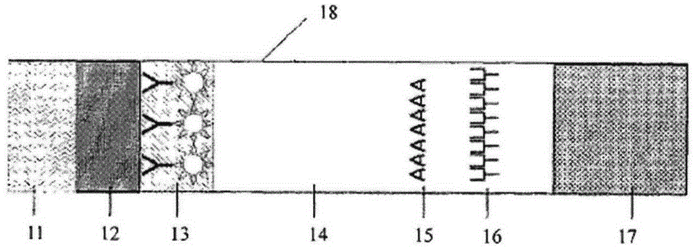
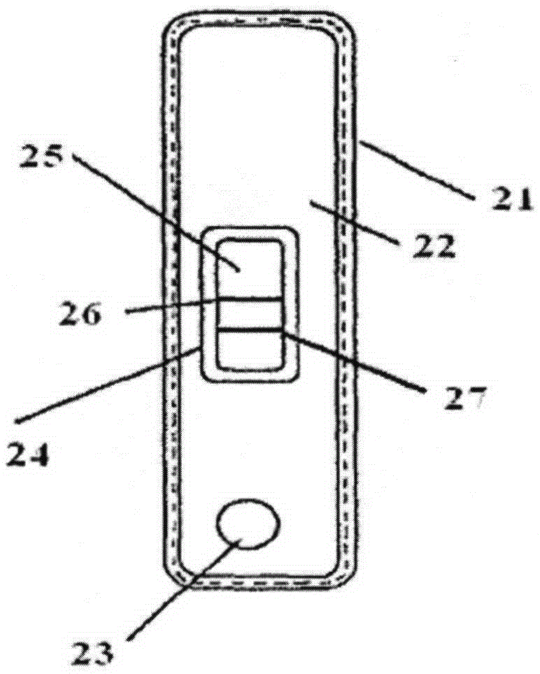
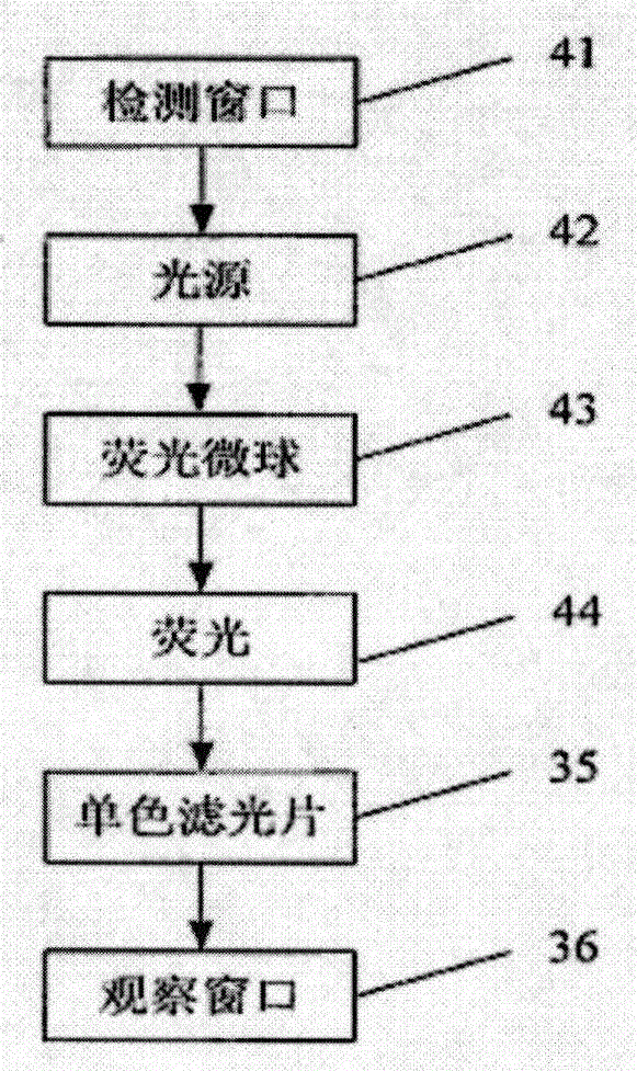
The invention discloses a preparation method of a fluorescent microsphere immunochromatographic test strip and a quantitative detection method. The fluorescent microsphere immunochromatographic test strip is prepared by taking light emitting nanoparticles prepared by a soap-free emulsion polymerization method as a label and is prepared through an immunochromatographic technology; and then, the fluorescent microsphere immunochromatographic test strip is prepared into a detection card comprising a sample cushion, a glass fibre film, a nitrocellulose film and a piece of absorbent paper, wherein a detection line and a quality control line are fixed on the nitrocellulose film. The quantitative detection method disclosed by the invention comprises the following steps: stimulating by adopting an optimal excitation light source of a fluorescent microsphere, enabling emitted fluorescence to pass through a light filter, collecting, gathering and multiplying an emission spectrum by using a CCD scanning technology or an optical fibre technology, converting into a numerical signal, multiplying the tested fluorescence intensity of a detection line by a correction coefficient, substituting the corrected fluorescence intensity in a pre-set standard curve in a fluoroanalyzer, and then, automatically calculating through the fluoroanalyzer so as to obtain the concentration of an object to be detected in a sample. The detection method disclosed by the invention is high in sensitivity, accurate in quantification and convenient to operate.

Description

technical field [0001] The invention relates to the field of biomedical detection, in particular to a preparation method and a quantitative detection method of a fluorescent microsphere immunochromatography test strip. technical background [0002] In biological and medical testing, immunoassays are often used, including radioimmunoassays, enzyme-linked immunoassays, and immunochromatography. Radioimmunoassay and enzyme-linked immunoassay require expensive equipment, professional operators and complicated operation steps, and it is difficult to obtain test results quickly. Immunochromatography is often used for rapid qualitative or semi-quantitative detection due to its simple operation, rapidity and low cost, such as colloidal gold test strips such as clenbuterol hydrochloride, hepatitis B surface antigen, aflatoxin and luteinizing hormone. At present, the incidence of common infectious diseases such as AIDS, hepatitis B, hepatitis C, and syphilis in my country continues t...

Claims


Application Information

Patent Timeline
Patent Type & Authority Applications(China)
IPC IPC(8): G01N33/577G01N33/533
CPCG01N33/577G01N33/533
Inventor 刘宏飞
Owner 刘宏飞
Learn More