Preparation method of functionalized retinal pigment epithelial cell graft
A technology of retinal pigment and epithelial cells, applied in the field of preparation of functionalized retinal pigment epithelial cell transplants, can solve the problems of poor mechanical properties, difficult to meet clinical application, long time, etc., to promote the formation of microstructure and morphology , avoid RPE cell death, good cell affinity effect
- Summary
- Abstract
- Description
- Claims
- Application Information
AI Technical Summary
Problems solved by technology
Method used
Image
Examples
Embodiment 1
[0037] Embodiment 1 prepares ultra-thin porous tussah silk fibroin / polycaprolactone / gelatin composite nanofiber bionic Bruch's membrane preparation method comprises the following steps:
[0038] 1. Tussah silk treatment: degumming tussah silk, then dissolving, dialysis, purification and freeze-drying to obtain pure tussah silk fibroin;
[0039] 2. Preparation of electrospinning solution: Weigh 0.125g of tussah silk fibroin, 2.125g of polycaprolactone, and 0.25g of gelatin into a 50mL conical flask, add 10mL of 98% formic acid solution, and stir overnight at room temperature to obtain static electricity. spinning solution;
[0040] 3. Electrospinning process: put the electrospinning solution into the syringe, and the syringe is equipped with a blunt needle with a diameter of 0.6mm. Electrospinning is performed under the conditions of voltage 12-20kv, collection distance 10-20cm, and extrusion speed 1-5mL / h ;The preferred voltage is 18kv, the collection distance is 15cm, and th...
Embodiment 2
[0045] Example 2 Nano-bionic Bruch's membrane pretreatment of the present invention; cell planting and cultivation; and preparation of functionalized retinal pigment epithelial cell transplant sheet of the present invention
[0046] The nano-bionic Bruch's membrane prepared in Example 1 was further studied as a carrier for RPE cell transplantation. Pretreatment such as disinfection must be carried out before co-culture with cells. In addition, factors such as cell planting density, method, and cell culture conditions were also optimized.
[0047] 1. Pretreatment before use such as disinfection of the ultra-thin porous nano-bionic Bruch’s membrane prepared in the above-mentioned embodiment 1: cutting the ultra-thin porous nano-bionic Bruch’s membrane into 2×2cm 2 sized diaphragm and place it in a Petri dish. After soaking in 75% ethanol for 30-120min, suck out the ethanol, wait for the ethanol to completely volatilize on the ultra-clean bench, add PBS or HBSS buffer and soak ...
Embodiment 3
[0051] Adhesion evaluation of RPE cells on each nano-bionic Bruch's membrane in embodiment 3 of the present invention
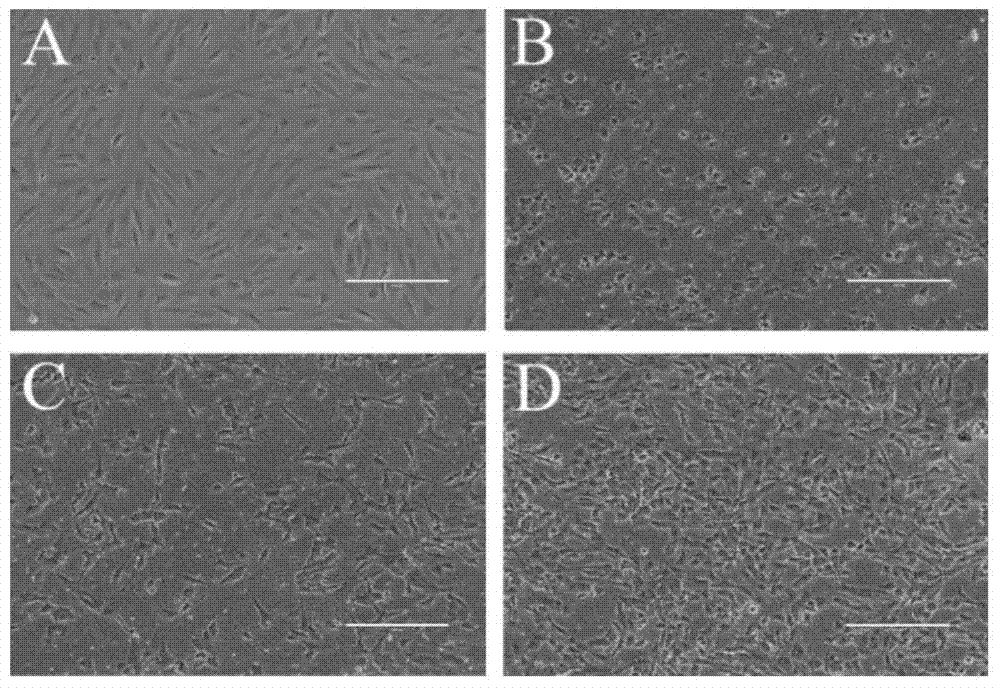
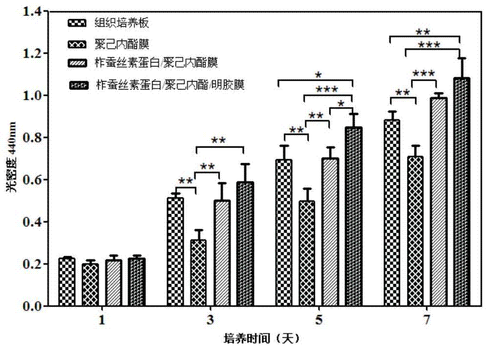
[0052] According to the following steps to study the influence of each nano-bionic Bruch's membrane on the cell adhesion ability, such as figure 1 shown.
[0053] 1. according to the step 2 in embodiment 2,3,4 operation, RPE cell is inoculated to each nano-bionic Bruch's film (respectively polycaprolactone nano-bionic Bruch's film, tussah silk fibroin / polycaprolactone nano-bionic Bruch's film) membrane, tussah silk fibroin / polycaprolactone / gelatin nano-bionic Bruch's membrane) and control TCP, remove the culture medium after 24 hours, and wash 4 times with PBS or HBSS to remove non-adherent cells;
[0054] 2. Observe cell adhesion under an inverted microscope, such as figure 1 As shown, among them, A indicates that RPE cells are planted on TCP, B indicates that RPE cells are planted on polycaprolactone nano-bionic Bruch's membrane, and C indicates that RPE ...
PUM
 Abstract
Description
Claims
Application Information
 - R&D
- Intellectual Property
- Life Sciences
- Materials
- Tech Scout
- Unparalleled Data Quality
- Higher Quality Content
- 60% Fewer Hallucinations
Browse by: Latest US Patents, China's latest patents, Technical Efficacy Thesaurus, Application Domain, Technology Topic, Popular Technical Reports.
© 2025 PatSnap. All rights reserved.Legal|Privacy policy|Modern Slavery Act Transparency Statement|Sitemap|About US| Contact US: help@patsnap.com


