Simple separation and identification method of human placenta myofibroblasts

A fibroblast and separation method technology, which is applied in the field of simple separation and identification of human placental myofibroblastic cells, can solve the problems of complicated experimental steps, waste of resources and the like, and achieves the advantages of convenient cell purification, short time-consuming and pollution reduction. effect of risk

Active Publication Date: 2013-06-26
BEIJING JING MENG STEM CELL TECH
View PDF5 Cites 0 Cited by
  • Summary
  • Abstract
  • Description
  • Claims
  • Application Information

AI Technical Summary

Problems solved by technology

[0005] However, there are few reports on the separation of placental fibroblasts in China at present. Even if there are reports, they mainly focus on the separation of MSCs or stem cells, such as CN 102586184 A (Chinese Patent Application No. 201210044638.6, published on July 18, 2012 ) published an invention entitled "Method for Establishing Placental Mesenchymal Stem Cell Bank"; CN 101395266 A (Chinese Patent Application No. 200680053575.3, date of publication March 25, 2009)
[0006] In the international reports on placental fibroblasts, the isolation methods are all focused on enzyme digestion, but there are many enzymes involved in the digestion process: for example, when Zuzana Strakova of the University of Illinois in the United States isolated placental fibroblasts, collagenase, Deoxyribonuclease, hyaluronidase, and pronase are mixed with four enzymes to digest and obtain fibroblasts. The participation of multiple enzymes will inevitably lead to complex experimental procedures and unnecessary waste of resources.

Method used


Image

  • Simple separation and identification method of human placenta myofibroblasts
  • Simple separation and identification method of human placenta myofibroblasts
  • Simple separation and identification method of human placenta myofibroblasts

Examples


Embodiment 1

[0044] Example 1 Isolation and culture of placental myofibroblasts

[0045] Take fresh placental tissue (within 12 hours) from a normal delivery baby in a sterile biological safety cabinet, use a scalpel to take a tissue about 5 cm long and wide from the center of the placenta, wash it twice with 250 mL of normal saline, and squeeze the placental tissue vigorously. The blood in the tissue was fully washed out, and the blood transfusion tissue on the amniotic membrane and decidua was peeled off with tweezers and a scalpel; the fetal decidua tissue was collected, washed once with 250 mL of normal saline, and the fetal decidua tissue was removed with a scalpel. Cut into 1.5 cm in length and 0.5 cm in width; 75% alcohol was quickly cleaned and disinfected for 10 seconds, and then washed twice with 250 mL of normal saline; DMEM digestion solution containing 1% collagenase I equal to the volume of the tissue fragments was used to incubate at 37 °C Digest for 16-18 h, stop digestion ...

Embodiment 2

[0047] Example 2 Morphological identification of placental myofibroblasts

[0048] Cultivate the myofibroblasts isolated according to the method of Example 1. After 2 days of induction of adherence, scattered spindle-shaped adherent cells can be seen under the microscope. The culture supernatant is completely poured out, and the impurity cells that are not adhered to the wall are removed at the same time, and cultured for 5- After 7 days, radial monoclonal cells can be seen to form, and the monoclonal cells are scraped with a cell scraper and cultured in a 12-well plate until the number of cells reaches 5×10 6 When left or right, it is used for other follow-up identifications.

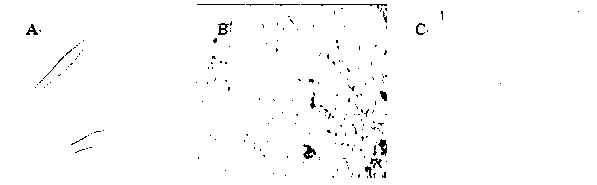
[0049] It can be seen under the microscope ( figure 1 ) The cells are spindle-shaped or spindle-shaped, have two poles, and are small in size. With the increase of culture time, the cells become larger and grow in fusion. These characteristics are in line with the growth characteristics and morphologica...

Embodiment 3

[0050] Example 3 Identification of surface markers of placental myofibroblasts

[0051] The 3rd passage cells cultured according to Example 1 from different placenta sources were collected respectively, and the cell surface markers were detected by flow cytometry, and the changes of the cell surface markers from different placenta sources were observed. Digest and collect cells, take 1×10 after counting 6 For each cell, wash once with PBS, centrifuge at 1500 rpm for 10 min; discard the supernatant, leave 200 uL, mix the cells by pipetting, add FITC-labeled CD90, CD45, APC-labeled CD29, HLA-DR and PE-labeled CD44 antibodies 10 each μL, and set 1 tube as a blank control, react at 4 °C in the dark for 30 min, wash once with PBS, centrifuge at 1500 rpm for 10 min, discard the supernatant, leave 200 uL, and test on the machine.

[0052] Flow test results figure 2 It shows that the expressions of CD90 (about 99%), CD44 (about 98%), and CD29 (about 97%) in different placental-deri...


PUM


Abstract

The invention relates to a separation and identification method of human placenta myofibroblasts. The separation and identification method comprises the following steps of: mechanically crushing the decidual tissue of the placenta fetal surface, digesting the decidual tissue by DMEM digestive juice containing collagenase I and thereby separating the decidual tissue into individual karyocytes; cultivating the karyocytes by using a DMEM / F12 culture medium containing 10% of fetal calf serum under the conditions of 37 DEG C, 5% of CO2 and saturated humidity; inducing adherence to wall and completely replacing the liquid in 48 hours, removing cells not adhering to the wall, adding fresh culture medium, and after the cells adhering to the wall is mono-cloned, cultivating each monoclonal cell separately; and when the cells are fused by about 90%, performing pancreatin passage, thereby obtaining the myofibroblasts. The method further identifies the obtained myofibroblasts by the combined method of cell morphology, cell cycle, cell surface marker detection, cell adipogenesis and osteogenesis differentiative capacity detection, cell multipotential stem cell factor detection and cell desmin expression detection.

Description

field of invention [0001] The invention relates to a method for isolating myofibroblasts from the decidua surface of human placenta and identifying the myofibroblasts. Background technique [0002] The placenta is considered to be the most attractive "adult stem cell", which can differentiate into muscle cells, liver cells, osteoblasts, chondrocytes, adipocytes and other cells in a suitable in vivo or in vitro environment. Because the source of placental stem cells is relatively convenient, easy to separate and purify, it still has the characteristics of stem cells after multiple passages and expansion, and does not have the characteristics of immune rejection; therefore, it has become a hot spot in stem cell research in recent years. Transplantation, tissue engineering, genetic engineering and other fields have good application prospects. [0003] Myofibroblasts refer to muscle tissue precursor cells containing myofilaments in the cytoplasm, derived from mesoderm stem cell...

Claims


Application Information

Patent Timeline
Patent Type & Authority Applications(China)
IPC IPC(8): C12N5/074C12Q1/68G01N15/14
Inventor 王云虹李志刚丁炜王颖颖高长青杨海莲武晓云黄芳蕾聂晨飞刘洁李大为安志达吴兰兰马瑞吕岩康会彦刘学敏柏小丽
Owner BEIJING JING MENG STEM CELL TECH
Why Eureka
Try Eureka