A fluorescent quantitative PCR detection method for detecting Ebola virus and its primers and kit
A technology of Ebola virus and kit, which is applied in the field of Ebola virus SYBRGreenI fluorescence quantitative PCR detection, can solve the problems of no EBOV detection technology patent announcement, and achieve the effects of stable source, time saving and high sensitivity
- Summary
- Abstract
- Description
- Claims
- Application Information
AI Technical Summary
Problems solved by technology
Method used
Image
Examples
Embodiment 1
[0058] Example 1 Primer Design
[0059] 1. Experimental steps
[0060] EBOVSYBRGreenI fluorescent quantitative PCR primers refer to: oligonucleotide chains with a length of 25±5nt, which are completely identical or complementary to the NP gene sequences of the five subtypes of EBOV, Z, S, B, C, and R.
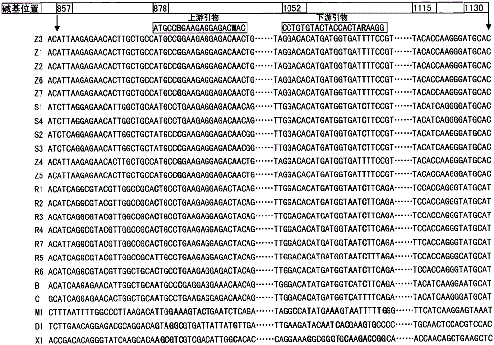
[0061] According to the NP gene sequences of EBOV, MARV, XFHV, and DHFV published in the NCBI gene database (see Table 1 for reference strain information), the primer and probe design software PrimerExpress2. S, B, C, R 5 subtype specific primers of EBOV. The primer sequence is shown in Table 2, and the primer gene position in Table 2 refers to the position on the gene of ZaireEbolavirusstrainMayinga (NCBI accession number AF499101.1). The results of comparing the designed primers with the corresponding gene sequences of EBOV, MARV, XFHV, and DHFV are shown in figure 1 .
[0062] Table 1. Strain information for designing primers and probe references
[0063]
[0064] ...
Embodiment 2
[0069] The preparation of embodiment 2RNA positive standard molecule and negative standard molecule
[0070] 1. Experimental steps
[0071] 1. Five subtypes of EBOV, as well as Marburg virus (MARV), dengue virus (DHFV), Xinjiang hemorrhagic fever virus (XHFV) (see Table 3 for reference strains), according to the accession numbers The sequence of their full-length NP gene cDNA was completely artificially synthesized by Nanjing Jinsite Technology Co., Ltd. MARV, XHFV, and DHFV were used as negative standard molecules.
[0072] Table 3. The strain information of artificially synthesized full-length NP gene cDNA reference
[0073]
[0074] 2. According to embodiment 1, design the synthetic primers (see Table 1), extend outwards to 273nt respectively along the EBOVNP gene sequence primer amplification region, design upstream primers and downstream primers connected to the T7 promoter sequence, and the primer sequences are shown in Table 4. The primer sequence is located on th...
Embodiment 3
[0084] The extraction of embodiment 3 sample RNA
[0085] 1. Experimental steps
[0086] Tissue sample processing: Take about 100 mg of the tissue sample to be tested (such as liver, kidney) and put it into a grinder and add 1000 μL DEPC water for grinding. Take 100 μL of the supernatant of the tissue to be tested that has been ground, put it in a 1.5 mL sterilized centrifuge tube, add 1000 μL Trizol, mix well, and let it stand for 10 min.
[0087] Liquid sample processing: Take 100 μL of the liquid sample to be tested (such as blood, physiological saline dilution of nasal swab, and physiological saline dilution of respiratory secretions), put it into a 1.5mL sterilized centrifuge tube, add 1000 μL Trizol, mix well, and statically Set for 10min. Add 200μL of chloroform, shake vigorously for 15s, let stand at room temperature for 2-3min, and centrifuge at 12000g for 15min at 4°C. Carefully pipette 450 μL of the supernatant into another clean 1.5 mL centrifuge tube free of DN...
PUM
 Abstract
Description
Claims
Application Information
 - R&D
- Intellectual Property
- Life Sciences
- Materials
- Tech Scout
- Unparalleled Data Quality
- Higher Quality Content
- 60% Fewer Hallucinations
Browse by: Latest US Patents, China's latest patents, Technical Efficacy Thesaurus, Application Domain, Technology Topic, Popular Technical Reports.
© 2025 PatSnap. All rights reserved.Legal|Privacy policy|Modern Slavery Act Transparency Statement|Sitemap|About US| Contact US: help@patsnap.com


