Porotic bone scaffold and preparation method thereof
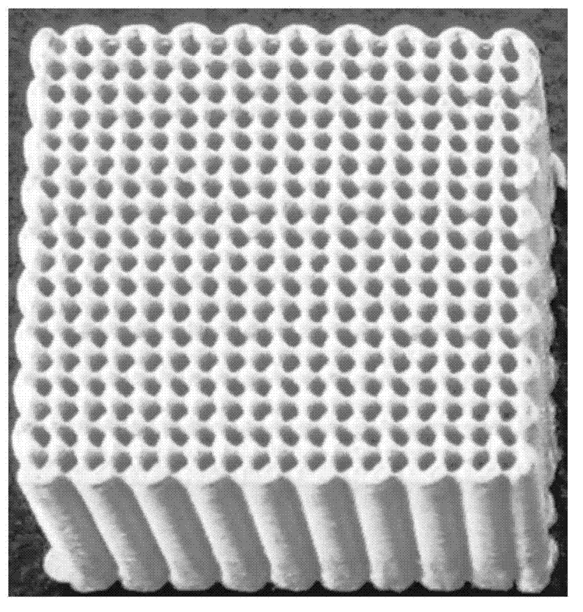
A bone scaffold and porosity technology, applied in the field of biomedical tissue engineering, can solve the problems of unfavorable cell migration, difficulty in meeting the requirements of pore size, porosity, pore wall specific surface area, and low controllability of porous bone scaffolds
- Summary
- Abstract
- Description
- Claims
- Application Information
AI Technical Summary
Problems solved by technology
Method used
Image
Examples
preparation example Construction
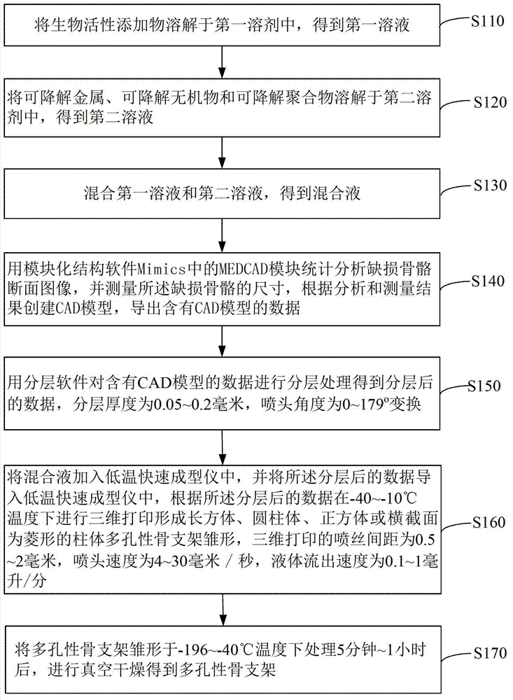
[0046] see figure 2 , the preparation method of the porous bone scaffold of an embodiment, comprises the steps:
[0047] Step S110: dissolving the bioactive additive in the first solvent to obtain a first solution.
[0048] The bioactive additive is at least one selected from chitosan, collagen, bone morphogenic protein, icariin and epimedium.
[0049] The first solvent is selected from one of 1,4-dioxane, acetonitrile, cyclohexane, acetone, ethylene glycol, cyclohexanone and dichloromethane, preferably 1,4-dioxane. The amount of the first solvent is such that the biologically active additive is sufficiently dissolved.
[0050] Step S120: dissolving the degradable metal, the degradable inorganic substance and the degradable polymer in a second solvent to obtain a second solution.
[0051] The degradable metal is selected from one of magnesium (Mg), iron (Fe), aluminum (Al), zinc (Zn), strontium (Sr) and manganese (Mn) or magnesium (Mg), iron (Fe), aluminum An alloy formed...
Embodiment 1
[0088] 1. At room temperature, dissolve 1 part of collagen in 1,4-dioxane to obtain the first solution; weigh 25 parts of metal magnesium powder with a particle size of 300 mesh, 25 parts TCP with a particle size of 300 mesh and 100 parts of PLGA were dissolved in 1,4-dioxane to obtain a second solution. The first solution and the second solution were mixed and stirred overnight to obtain a homogeneous mixed solution.
[0089] 2. Use the MEDCAD module in the modular structure software Mimics to statistically analyze the cross-sectional image of the leg tibial defect bone, measure the size of the defect bone, and create a CAD model based on the analysis and measurement results. The CAD model is length×width×height It is a cuboid porous bone scaffold of 3×3×4cm. Export the data containing the CAD model as an STL format file, and then use the layering software that comes with the low-temperature rapid prototyping instrument to perform layering processing to determine the thickness...
Embodiment 2
[0096] 1. At room temperature, dissolve 1 part of bone morphogenic protein in acetonitrile to obtain the first solution; weigh 100 parts of metal iron powder with a particle size of 500 mesh and 100 parts of metal iron powder with a particle size of 500 mesh at a mass ratio of 1:1:5. The objective TCP and 500 parts of PCL were dissolved in acetonitrile to obtain a second solution, the first solution and the second solution were mixed, and stirred overnight to obtain a homogeneous mixed solution.
[0097] 2. Use the MEDCAD module in the modular structure software Mimics to statistically analyze the sectional image of the defective hand ulnar bone, measure the size of the defective bone, and create a CAD model based on the analysis and measurement results. The CAD model is 2 cm in diameter and 4 cm in height Export the data containing the CAD model as an STL format file, and then use the layering software that comes with low-temperature rapid prototyping for layering processing. ...
PUM
| Property | Measurement | Unit |
|---|---|---|
| Aperture | aaaaa | aaaaa |
| Aperture | aaaaa | aaaaa |
| Aperture | aaaaa | aaaaa |
Abstract
Description
Claims
Application Information
 - R&D
- Intellectual Property
- Life Sciences
- Materials
- Tech Scout
- Unparalleled Data Quality
- Higher Quality Content
- 60% Fewer Hallucinations
Browse by: Latest US Patents, China's latest patents, Technical Efficacy Thesaurus, Application Domain, Technology Topic, Popular Technical Reports.
© 2025 PatSnap. All rights reserved.Legal|Privacy policy|Modern Slavery Act Transparency Statement|Sitemap|About US| Contact US: help@patsnap.com


