A multi-modal imaging microbubble structure, preparation method and application
A multi-modal imaging and micro-bubble technology, applied in the field of materials and nano-medicine, biomedical, can solve the problem that the resolution and accuracy of ultrasonic imaging cannot meet the requirements of advanced clinical diagnosis, so as to facilitate repeated inspections and enhance ultrasonic imaging. Simple effect of image and synthesis process
- Summary
- Abstract
- Description
- Claims
- Application Information
AI Technical Summary
Problems solved by technology
Method used
Image
Examples
Embodiment 1
[0035] Example 1 Preparation of multimodal microbubbles of the present invention
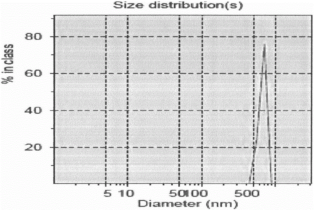
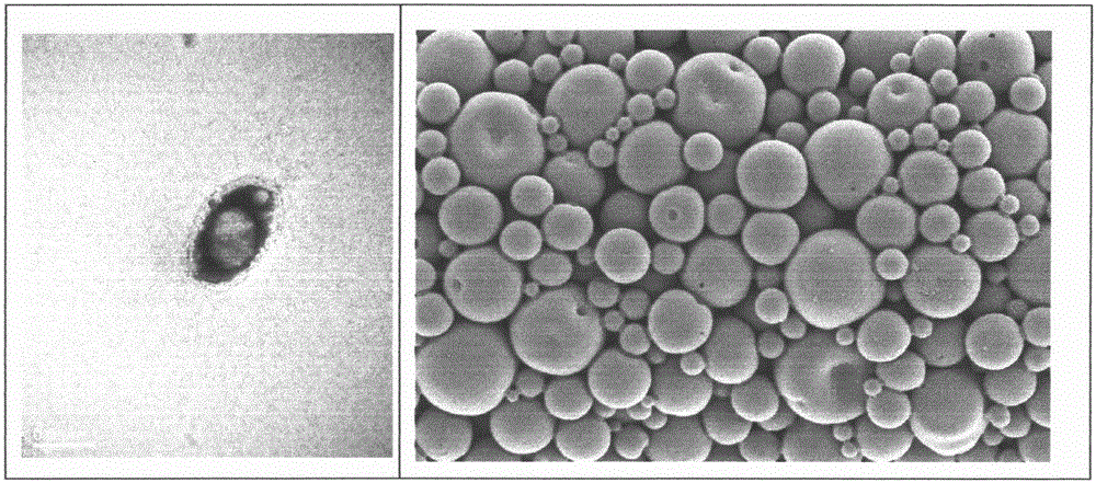
[0036]Dissolve 50mg PLGA (50:50, MW 12,000) and 0.15mg PE in 2ml dichloromethane solvent with an electrolytic balance, add 100μl water-soluble quantum dots after complete dissolution, and vibrate for 35-45s to obtain the primary emulsion (W / O ); the primary emulsion is mixed with 6ml 4% polyvinyl alcohol solution, and acts on 9600r / min dispersion homogeneous equipment 5min, obtains microsphere (W / O / W), adds 5ml2% isopropanol solution, room temperature magnetic force The mixer stirred at a constant speed for 2-5 hours to solidify the surface of the microspheres, and the dichloromethane volatilized naturally as much as possible. The above liquid was divided into 5ml centrifuge tubes, and the supernatant was discarded by high-speed centrifugation (3500rpm, 5min). Add an appropriate amount of double distilled water again, mix thoroughly with a vortex mixer, wash, centrifuge, and discard the superna...
Embodiment 2A10
[0038] Example 2A10-PLGA QDs Coupling experiments targeting microbubbles
[0039] Get the white powdery PLGA that makes in embodiment 1 QDs Dissolve the microbubbles in 10 μg / μL RNase-free aqueous solution (DNase RNase-free water), add 400 μL EDC / NHS (4:1 molar ratio), and incubate for 45 minutes on a constant temperature incubation shaker . The microbubble suspension activated by NHS was washed three times by centrifugation with buffer (centrifugal speed: 3000r / min), then dissolved in DNase RNase-free water with a concentration of 1 μg / μL, and 50 μL 3’-NH2 modified A10PSMA was added aptamer, incubated at low temperature for 2-4 hours, and centrifuged and washed three times with ribonuclease-free deoxyribonuclease aqueous solution to obtain the covalent coupling product of A10 aptamer and microbubbles, which was stored in the form of suspension. Detect 0.5mg / mL 3'-NH2 modified A10PSMA and PLGA by flow cytometry and fluorescence microscope QDs -COOH microbubble coupling pro...
Embodiment 3
[0041] Example 3 In vivo MRI imaging experiment of multimodal microbubbles according to the present invention
[0042] After the SD rats were anesthetized by intraperitoneal injection of 3% pentobarbital sodium (1ml / kg), they were scanned with a clinical GE 3.0T superconducting magnetic resonance instrument, using a head orthogonal circle with a relatively uniform internal magnetic field, SE sequence, T1W1, parameters: TR / TE=824ms / 10ms, field of view FOV=80mm*80mm. Using the control method of two rats with a body weight of 220g, inject the multimodal microbubble PLGA prepared in Example 1 through the tail vein of the rats at a dose of 5ml / kg during imaging QDs and blank microbubble PLGA, observe the imaging time and imaging effect after liver and kidney contrast (such as image 3 As shown), it can be clearly seen that the multimodal non-targeting microbubbles protected by the present invention have the purposes of preparing MRI contrast agents, and predictably the multimodal ...
PUM
 Abstract
Description
Claims
Application Information
 - R&D Engineer
- R&D Manager
- IP Professional
- Industry Leading Data Capabilities
- Powerful AI technology
- Patent DNA Extraction
Browse by: Latest US Patents, China's latest patents, Technical Efficacy Thesaurus, Application Domain, Technology Topic, Popular Technical Reports.
© 2024 PatSnap. All rights reserved.Legal|Privacy policy|Modern Slavery Act Transparency Statement|Sitemap|About US| Contact US: help@patsnap.com


