GDNF-carrying microbubble preparation and method for making the same
A technology of microbubbles and lipid bilayers, applied in pharmaceutical formulations, medical preparations containing active ingredients, nuclear magnetic resonance/magnetic resonance imaging contrast agents, etc., to increase effective drug concentration and promote blood-brain barrier penetration Effect
- Summary
- Abstract
- Description
- Claims
- Application Information
AI Technical Summary
Problems solved by technology
Method used
Image
Examples
Embodiment 1
[0040] Example 1. Preparation of GDNF-loaded microbubble contrast agent and its research on opening the blood-brain barrier
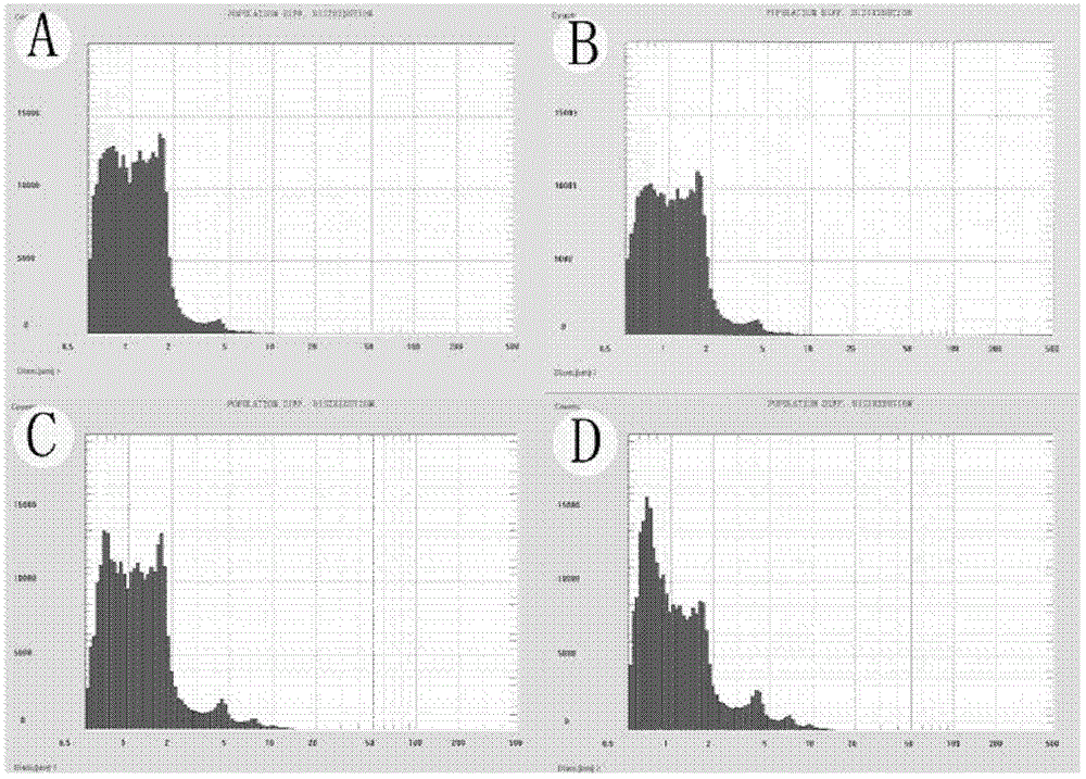
[0041] 1) Preparation of GDNF-loaded microbubbles
[0042] A certain amount of DSPC (distearoylphosphatidylethanolamine, 10.64mg), DSPE-PEG2k (polyethylene glycol-distearoylphosphatidylethanolamine, 2.10mg), DSPE-PEG2k-Biotin (polyethylene glycol) was added in a certain proportion. Ethylene glycol-distearoylphosphatidylethanolamine-biotin, 2.26 mg) was dissolved in 0.5 ml of chloroform and mixed on a vortex mixer. in dry N 2 Remove chloroform under the action of flow to form a uniform film of phospholipid on the test tube wall, and dry it in a vacuum oven for more than 2 hours. Then add the Tris buffer solution of 5ml degassed pH7.4 in the test tube that contains dry phospholipid film: containing glycerol (10% volume fraction) and propylene glycol (10% volume fraction) obtain the phospholipid solution of certain concentration. Heat the phospholipid s...
Embodiment 2
[0053] Example 2. Determining the optimal parameters of MRI-guided low-frequency focused ultrasound combined with targeted microbubbles for local opening of the BBB
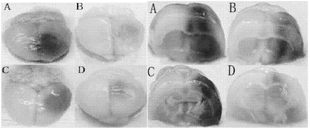
[0054] Taking ultrasonic irradiation time, microbubble dose, delay time (interval between microbubble injection time and acoustic shock time), ultrasonic frequency, and sound pressure as influencing factors, three levels were selected for each factor, and an orthogonal experimental design was adopted (see Table 1). , taking Evans blue (EB, Evens blue, Sigma company, E2129) exudation as an indicator of the blood-brain barrier passage rate (EB exudation), to determine the local opening of the BBB by MRI-guided low-frequency focused ultrasound combined with targeted microbubbles, Optimal parameters for increasing central GDNF levels.
[0055] Test method: The permeability is measured by the quantitative estimation of the exudation of EB. Take the rat brain tissue perfused with PBS, immerse the partially blue-staine...
Embodiment 3
[0068] Embodiment 3, the mensuration of protein content in brain
[0069] The ultrasonic conditions of this embodiment are the optimal parameter conditions obtained in Example 2, that is, the probe frequency is 1 MHz, the amount of microbubbles is 0.5 ml, the irradiation time is 60 s, the sound pressure is 0.8 MPa, and the time delay is 60 s.
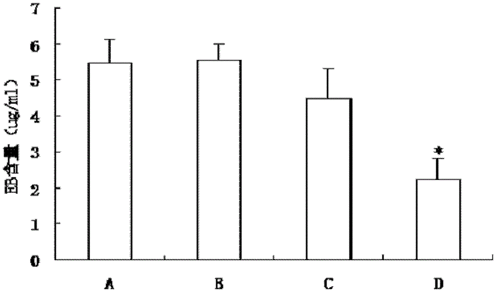
[0070] Ultrasound combined with microbubbles to open the blood-brain barrier, after intravenous injection of protein, the determination of protein content in the brain and the comparison of the two protein transport methods. Detect the amount of protein and the amount of protein entering the brain tissue after ultrasound combined with microbubbles to open the BBB.
[0071] (1) Due to the high price of GDNF, another low-priced alternative protein BSA was used to simulate the experimental conditions to determine the amount entering the brain tissue.
[0072] Divided into two groups: ultrasound + microbubbles + protein (group A); ultrasou...
PUM
| Property | Measurement | Unit |
|---|---|---|
| particle size | aaaaa | aaaaa |
| molecular weight | aaaaa | aaaaa |
Abstract
Description
Claims
Application Information
 - R&D
- Intellectual Property
- Life Sciences
- Materials
- Tech Scout
- Unparalleled Data Quality
- Higher Quality Content
- 60% Fewer Hallucinations
Browse by: Latest US Patents, China's latest patents, Technical Efficacy Thesaurus, Application Domain, Technology Topic, Popular Technical Reports.
© 2025 PatSnap. All rights reserved.Legal|Privacy policy|Modern Slavery Act Transparency Statement|Sitemap|About US| Contact US: help@patsnap.com


