
Antisense compounds targeted to connexins and methods of use thereof

a technology of anti-convulsive compounds and anti-inflammatory agents, which is applied in the direction of anti-inflammatory agents, drug compositions, applications, etc., can solve the problems of corneal tissue reconstruction, tissue or organ failure, and inability to meet the needs of patients, so as to prevent tissue damage in cells, improve corneal thickness, and promote healing

Active Publication Date: 2011-10-11
CODATHERAPEUTICS NZ LTD
View PDF8 Cites 13 Cited by
  • Summary
  • Abstract
  • Description
  • Claims
  • Application Information

AI Technical Summary

Benefits of technology

"The patent describes compounds and methods for using them to treat tissue damage caused by ophthalmic procedures. The compounds, called antisense compounds, can inhibit the expression of certain proteins that are involved in the healing process. By targeting these proteins, the antisense compounds can reduce tissue damage and promote healing. The methods for using the antisense compounds involve administering them to the eye or other tissues in various ways, such as through local or topical application or systemic or intraocular injection. The antisense compounds can be administered before, during, or after the ophthalmic procedure. Overall, the invention provides a way to protect and improve the health of the eye during ophthalmic procedures."

Problems solved by technology

Tissue or organ failure due to illness or injury is a major health problem worldwide with little option for full recovery other than organ or tissue transplantation.
However, problems finding a suitable donor mean that this option is not available to the majority of patients, tissue engineering or remodeling whereby synthetic or semi synthetic tissue or organ mimics that are either fully functional or which are grown in a desired functionality is currently being investigated as replacements.
The prevalence of refractive surgery for correction of myopia such as photorefractive keratectomy (PRK) and laser in situ keratomileusis (LASIK) has led to shortage of suitable cornea for transplant for tissue reconstruction after surgery or disease processes and for tissue manipulation in vivo to engineer changes.
In addition, approximately 5% of patients undergoing laser surgery experience unexpected outcomes.
Trauma to the surface of the cornea is highly prevalent; for example, minor scrapes, eye infections and diseases, chemical or mechanical accidents and surgical practice can all damage the cornea.
One major complication in post corneal-trauma wound healing is the loss of visual acuity due to tissue reorganization.
Surgery disrupts the forces which anchor the cornea in its normal configuration.
The cornea contracts when it heals, causing a local distortion of the tissue and a concomitant distortion in the visual field in the affected region (astigmatism).
This technique, however, is limited due to a lack of predictable results and significant fluctuations in vision, both of which are related to the nature and extent of wound healing (Jester et al., Cornea (1992) 11: 191).
Wounds in the cornea also heal slowly, and incomplete healing tends to be associated with instability of visual acuity (with fluctuations in vision from morning to evening, as well as drifting visual acuity occurring over a period of weeks to months).
Also, if a corneal wound fails to heal completely, a wound “gape” can occur leading to a progressive hyperopic effect.
Corneal regeneration after trauma is complex and not well understood.
The corneal regeneration process post-trauma can result in the loss of corneal clarity and therefore influence the outcome of refractive surgery.
However, post-operative trauma to the corneal and the surrounding soft tissue following surgical procedures such as, for example, excimer laser photorefractive keratectomy, often results in scarring due to hypercellularity associated with modification of the extracellular matrix; including changes in epithelial cell patterning, myofibroblast differentiation, stromal remodeling, and epithelial hyperplasia at the site of a laser induced lesion.
Within 24-48 hours after injury, the damage spreads and significantly increases the size of the affected area.
Although progress has been made with some current therapeutic modalities, major constraints to spinal cord repair still remains, including the invasive intervention itself can further lesion spread and glial scar formation, impeding the repair process and risk further loss of neural function (Raisman, G J.
However, many difficulties remain that need to be overcome.
It is often the case, for example, that the down regulation of a particular gene product in a non-target cell type can be deleterious.
Additional problems that need to be overcome include the short half life of such ODN's (unmodified phosphodiester oligomers) typically have an intracellular half life of only 20 minutes owing to intracellular nuclease degradation (Wagner 1994, supra) and their delivery consistently and reliably to target tissues.

Method used


Image

  • Antisense compounds targeted to connexins and methods of use thereof
  • Antisense compounds targeted to connexins and methods of use thereof
  • Antisense compounds targeted to connexins and methods of use thereof

Examples


example 1

In Vivo Analysis

Materials and Methods

Laser Treatment

[0194]Female Wistar rats (d32-34) were raised under conditions consistent with the ARVO Resolution on the Use of Animals in Research. Animals were anaesthetized by administrating a 1:1 mixture of Hypnorm™ (10 mg / ml, Jansen Pharmaceutica, Belgium) and Hypnovel® (5 mg / ml, Roche products Ltd, New Zealand) at a dose of 0.083 ml / 100 g body weight in the peritoneum of the animal.

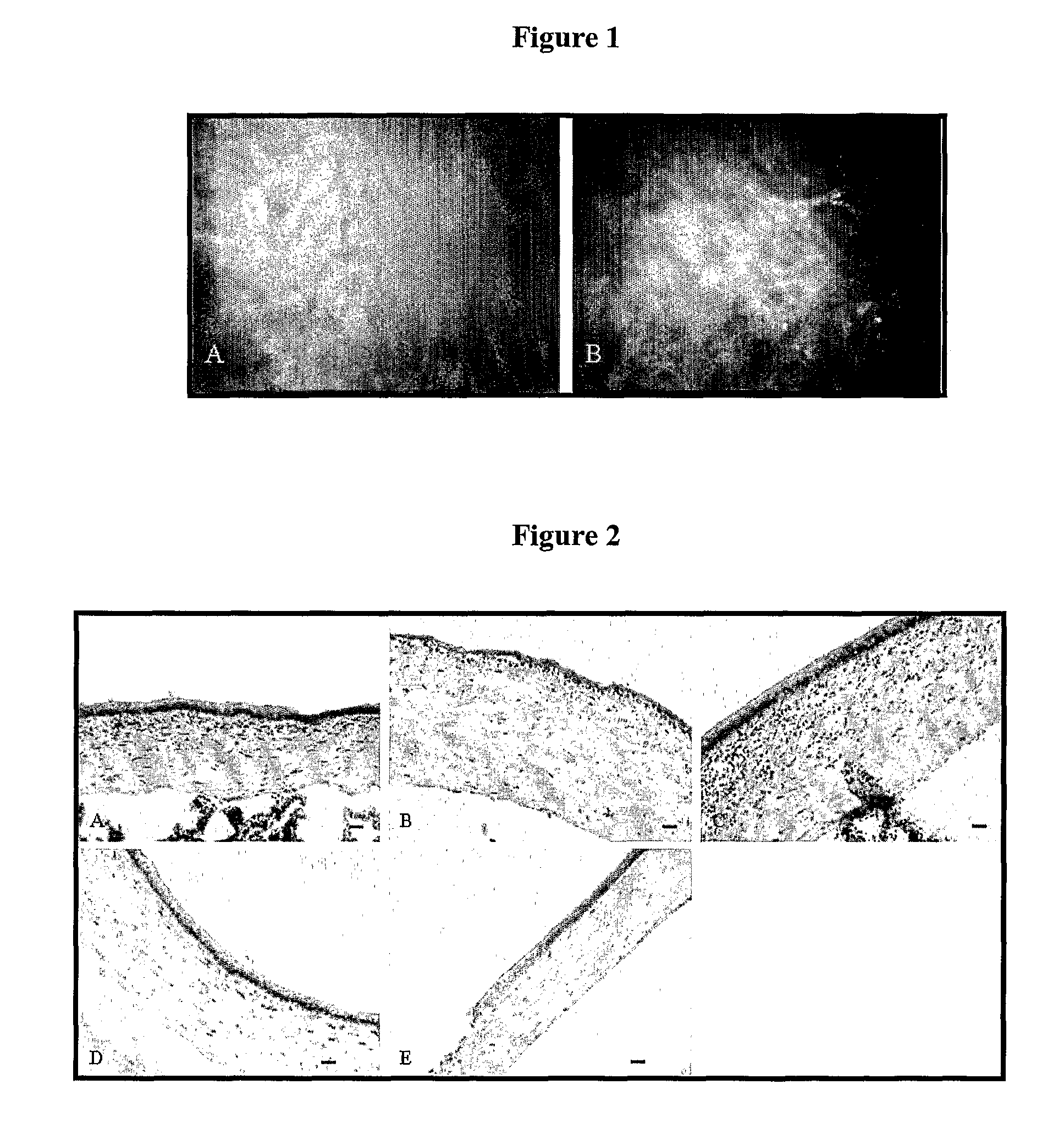
[0195]Excimer laser treatment was performed through the intact epithelium using a Technolas 217 Z excimer laser (Bausch & Lomb Surgical, USA). The eye was centered at the middle of the pupil and ablation was performed with the following parameters: treatment area was of 2.5 mm diameter and of 70 μm depth. This resulted in the removal of a small thickness of the anterior stroma and of the whole epithelium. Excimer laser treatment was preferentially used to produce reproducible lesions and investigate the effects of connexin43 AS ODNs on corneal remodeling and engi...

example ii

Ex Vivo Analysis

Materials and Methods

Histology: Tissue Collection and Fixation

[0212]Appropriate numbers of animals (Wistar rats) were terminated at selected time points following photorefractive keratectomy and DB1 anti-connexin43 ODNs were administered to anaesthetized rats as described in experiment 1 above and corneal sections were prepared for histological analysis. Control rats had received laser surgery only. Whole eyes and control tissues were rinsed in Oxoid PBS prior to embedding in Tissue-Tek® OCT (Sakura Finetek, USA) and freezing in liquid nitrogen. When necessary (for the use of some antibodies), frozen tissues were later fixed in cold (−20° C.) acetone for 5 min after being cryocut.

Tissue Cutting

[0213]The procedure for cryosectionning was as follows: frozen blocks of unfixed tissue were removed from −80° C. storage and placed in the Leica CM 3050S cryostat for about 20 min to equilibrate to the same temperature as the cryostat (i.e. −20° C.). When equilibration of the ...

example iii

Ex Vivo Tissue Engineering

[0225]Corneas were placed into an ex vivo organ culture model and specific connexin modulated using antisense ODNs. Two connexins were targeted in these experiments, connexin43 and connexin31.1. Connexin43 downregulation is used to demonstrate that connexins can be regulated in vitro, and connexin31.1 was targeted because this connexin is expressed in the outer epithelial layers of the cornea in cells about to be shed from the cornea. The aim was to engineer a thickening of epithelial tissue by reducing connexin31.1 expression.

Materials and Methods

[0226]30-34 day old Wistar rats were euthanized with Nembutal or carbon dioxide and whole rat eyes dissected. The ocular surface was dissected, disinfected with 0.1 mg / ml penicillin-streptomycin for 5 minutes and rinsed in sterile PBS. The whole eye was then transferred onto a sterile holder in a 60 mm culture dish with the cornea facing up. The eyes were mounted with the corneal epithelium exposed at the air-medi...


PUM

PropertyMeasurementUnit
thicknessaaaaaaaaaa
melting temperatureaaaaaaaaaa

Abstract

Methods and compositions for modulating the activities of connexins are provided, including, for example, for use in post-surgical, trauma, or tissue engineering applications. These compounds and methods can be used therapeutically, for example, to reduce the severity of adverse effects associated diseases and disorders where localized disruption in direct cell-cell communication is desirable.

Description

[0001]This application is a National Phase Application under 35 U.S.C. §371 of PCT / IB2004 / 004431 filed Dec. 3, 2004 which claims priority to New Zealand Patent Application No. 529936, filed Dec. 3, 2003. The disclosures of both are incorporated herein by reference.FIELD[0002]The present disclosure relates to and describes agents, compositions and methods of using compounds for modulation of gap-junction-associated protein expression. These agents, compositions, and methods are useful, for example, for tissue engineering in vivo and in vitro, including for example in the skin, in corneal tissue, and conjunction with surgical procedures of the eye.BACKGROUND[0003]Tissue or organ failure due to illness or injury is a major health problem worldwide with little option for full recovery other than organ or tissue transplantation. However, problems finding a suitable donor mean that this option is not available to the majority of patients, tissue engineering or remodeling whereby synthetic...

Claims


Application Information

Patent Timeline
Patent Type & Authority Patents(United States)
IPC IPC(8): A61K31/70C07H21/04C07H21/02C12Q1/68C12N15/113
CPCC12N15/111C12N15/1138C12N15/113A61K31/7105A61K31/711C12N2310/11C12N2310/12C12N2320/31C12N2310/315Y02P20/582A61P17/00A61P17/02A61P17/12A61P25/00A61P27/02A61P27/06A61P27/10A61P27/12A61P29/00A61P41/00A61P43/00A61K48/00C12N15/11Y10S977/907
Inventor LAUX, WILDAGREEN, COLIN
Owner CODATHERAPEUTICS NZ LTD
Learn More