Assay method for group transfer reactions

a group transfer and reaction technology, applied in the field of group transfer reaction methodologies, can solve the problems of cumbersome post-reaction separation steps, serious limits on the utility of the assay, and slowed or prevented incorporation of group transfer enzymes into pharmaceutical hts programs

Active Publication Date: 2008-05-27
BELLBROOK LABS
View PDF3 Cites 4 Cited by
  • Summary
  • Abstract
  • Description
  • Claims
  • Application Information

AI Technical Summary

Problems solved by technology

Despite their importance from a drug discovery perspective, the incorporation of group transfer enzymes into pharmaceutical HTS programs is being slowed or prevented for a number of reasons: a) homogenous assay methods are not currently available, b) the detection methods used place serious limits on the utility of the assay, and c) the non-generic nature of the assay requires the development of many specific detection reagents to test diverse acceptor substrates.
The approach currently used to identify sulfotransferase substrates or inhibitors requires the use of radioactivity and involves cumbersome post-reaction separation steps, such as precipitation or chromatography.
This approach is not desirable in a high throughput screening (HTS) format because of the high radiation disposal costs and because the incorporation of separation steps complicates the automation process.
Other SULT assays have been developed using colorimetric and fluorescent means, but they are dependent on the use of a specific acceptor substrate for detection, so their use is limited to a single SULT isoform, and they cannot be used to screen for diverse substrates (Burkart, M. D. and Wong, C. H., Anal Biochem, 1999, 274:131-7; Frame, L. T., Ozawa, S. et al., Drug Metab Dispos, 2000, 28:1063-8).
As a result, SULT interaction studies are currently not included during the preclinical development of drugs.
However, since this method requires a separation step such as filtering or centrifugation, it cannot be easily adapted to an automated HTS format.
Surface proximity assays (SPA) allow radioassays in a multiwell format with no separation (Mallari, R., Swearingen, E. et al., J Biomol Screen, 2003, 8:198-204) but their use by pharmaceutical companies is declining because of the disposal and regulatory costs of handling radioisotopes.
Though generic phosphotyrosine antibodies are common, phosphoserine and phosphothreonine antibodies are notoriously difficult to produce and only recognize phospho-serine or -threonine in the context of specific flanking amino acids (Eglen and Singh, 2003).
This greatly complicates the incorporation of new kinases into HTS, especially if their substrate specificity is not well defined.
It also creates analysis problems in comparing data among kinases with different substrate selectivities, because the output of the assay depends on the particular antibody(Ab)-phosphopeptide pair used.
Although efforts to develop generic phospho-serine antibodies and identify more generic kinase substrates continue (Sills, M. A., Weiss, D. et al., J Biomol Screen, 2002, 7:191-214), research in this direction has not been very successful to date.
Chemical phosphate binding reagents suffer from background binding to nucleotide phosphates, requiring the use of very low, non-physiological levels of ATP and limiting assay flexibility.
Modified nucleotides do not provide a generic format because the ability to use the ATP analogs as donors varies among kinases, requiring the development of a number of different analogs.
Differences in protease sensitivity caused by peptide phosphorylation have also been exploited in developing fluorescence based kinase assays (Kupcho, K., Somberg, R. et al., Anal Biochem, 2003, 317:210-7), but these assays are not truly homogenous; i.e. they require the post-reaction addition of developing protease reagents.
In addition, the applicability of this method is limited to peptides where kinase and protease specificity overlap.
There are also a few methods that are dependent on interaction of reaction products with specific multiwell plate chemistries, but these are not truly homogenous in that they require post reaction reagent additions and / or processing.
Also the requirement for specialized instrumentation for processing and / or detection does not fit with the open architecture desired by most pharmaceutical HTS platforms.
However, in practice, the kinase assays are often run in multiwell plates and then the products are transferred to microfluidic devices for separation—a cumbersome process for an HTS format.
In summary, the non-generic nature of the current group transfer assays is resulting in significant expense and delays for drug discovery because of the need to develop assays for individual enzymes or small subgroups within a family.
Also, because many of the current assays are based on modification and detection of specific tagged acceptors, there is limited ability for testing different acceptor substrates.
Often the tagged acceptor substrates used are different from the substrates that are phosphorylated in vivo, thus the physiological relevance of the assay is questionable.
However, studies have shown that there are significant differences in the pharmaceutical targets identified using different assays methods (Sills, M. A. et al., J Biomol Screen, 2002, 7:191-214), which is a significant problem for profiling inhibitor selectivity across several kinases.
These shortcomings of the existing HTS assay methods for group transfer reactions are hampering the rapid analysis of important enzyme families in pharmaceutical drug discovery programs.
Detection of these products, however, is complicated because the cleaved mono- and di-nucleotides cannot be differentiated from the activated donor molecules based on absorbance or fluorescence properties because for example ADP has the same fluorescence and absorbance properties as ATP.
Separation of the donor product from the donor can be effected using chromatographic methods, such as thin layer chromatography or high pressure liquid chromatography, but incorporation of these methods into an HTS format is cumbersome.
However, the optical interference of drug compounds with absorbance assays, especially in the ultra violet, is a widely recognized problem with this approach.
However this assay is not optimal because it relies on small decreases in a high initial signal.
The need to keep ATP concentrations low to minimize background results in nonlinear reaction kinetics if assay conditions are not carefully controlled.
In a related method, competition binding assays using fluorescent ATP analogs have also been developed, but these do not give a measure of enzyme catalytic activity, thus are of limited utility.
Enzymes that catalyze group transfer reactions where water is the acceptor, hydrolases, present an even more difficult assay challenge because generally the products are all small molecules, and thus more difficult to detect than a covalently adducted peptide or nucleic acid, which can be detected easily with an antibody.
This method is problematic for HTS because the colorimetric readout is not very sensitive and is also subject to interference from colored test compounds.
Though fluorescence is preferable to absorbance as a detection mode for HTS, any method that relies upon phosphate detection is going to suffer from background signal because of the high phosphate concentrations found in biological samples and its use as a common biological buffer reagent.
Detection of ADP using a coupled enzyme assay linked to NADPH formation is also used as an HTS assay approach for ATPases (DeBonis, S., Skoufias, D. A. et al., Mol Cancer Ther, 2004, 3:1079-90), but this method again relies on absorbance detection and so suffers from low sensitivity and interference from colored compounds.
The coupled enzyme approach has also been adapted for fluorescence detection (Zhang, B., Senator, D. et al., Anal Biochem, 2005, 345:326-35), but the requirement for several enzymes for signal generation greatly increases the potential for assay interference from test compounds.
In general, the use of enzymes that catalyze group transfer reactions into HTS assays has been hampered by the lack of universal, homogenous assay methods that are not subject to interference from molecules in pharmaceutical drug libraries.

Method used


Image

  • Assay method for group transfer reactions
  • Assay method for group transfer reactions
  • Assay method for group transfer reactions

Examples


example 1

Uridine Glucuronide Transferase Assay

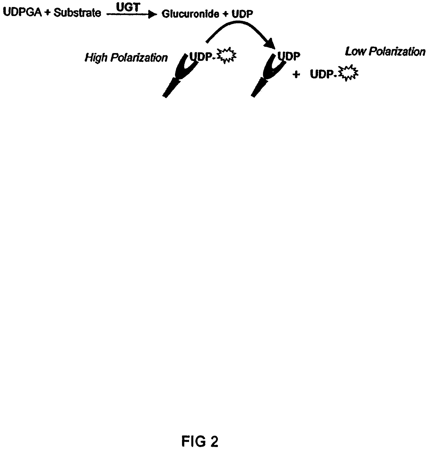
[0145]One embodiment of this application involves detecting the “donor product” of the UGT reaction using a competitive fluorescence polarization immunoassay where the antibody-bound tracer has a high polarization value which decreases when it is displaced by an analyte, such as UDP (as shown in FIG. 2). The main reagent required for this assay is the production of an antibody that binds UDP with high selectivity and has negligible binding to the donor, UDP-glucuronic acid (UDPGA). This highly selective antibody is used in combination with fluorescent UTP compounds (as substitutes for UDP tracers) to establish the assay. The polyclonal antibody produced against UDP required covalent binding of the nucleotide hapten to a carrier protein. UTP was used as the hapten only because reactive derivatives of the triphosphate, but not the diphosphate, were readily available that could be used for conjugation. It was reasoned that the majority of the tripho...

example 2

Assays for Other Types of Glycosyltransferases

[0156]Glucuronic acid is one type of carbohydrate molecule that is activated by UDP for enzymatic transfer to other biomolecules; there are many other types of group transfer reactions that use UDP-sugar as an activated donor. These include protein glycosyltransferases that modulate the cellular localization and / or secretion of proteins via their carbohydrate modifications and biosynthetic enzymes that incorporate sugar monomers into polymers such as glycogen and bacterial peptidoglycan. Many of these enzymes are potential targets for therapeutic intervention, both in humans or for antimicrobials.

[0157]One class of microbial glycosyltransferases that are of interest from a drug discovery perspective include transferases involved in bacterial cell wall synthesis such as the UDP-galactofurnosyltransferase of Mycobacterium tuberculosis (Cren, S., Gurcha, S. S., Blake, A. J., Besra, G. S., Thomas, N. R. Org. Biomol. Chem. 2; 2418-2420. A mam...

example 3

Kinase Assays

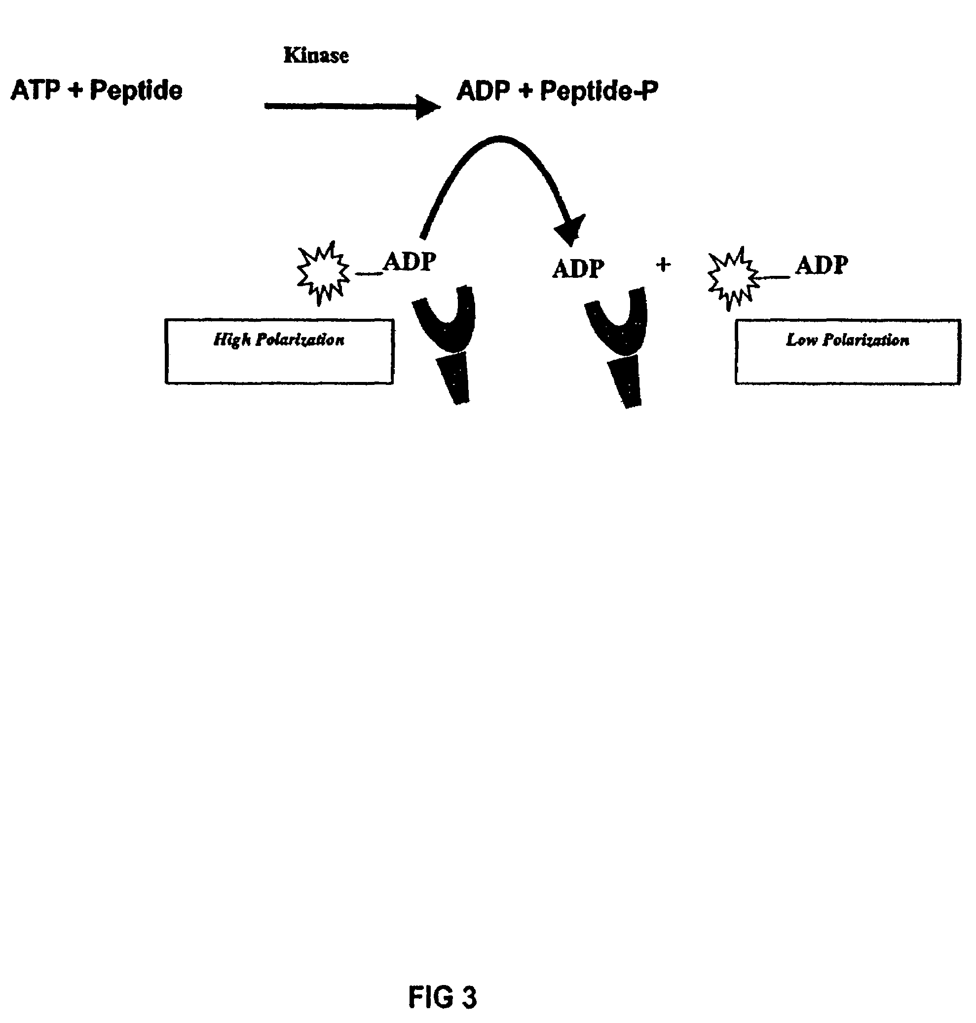
[0158]To develop an anti-ADP antibody, 6-aminobutyl-ADP (Biolog Life Science Inst., Bremen, Germany) was conjugated to keyhole limpet hemocyanin with a suitable reagent such as glutaraldehyde and used to immunize rabbits. Other carrier proteins such as BSA or ovalbumin and other conjugation chemistries may be used. Attachment to carrier protein via the adenine ring exposes the ribosyl-phosphate moiety, where selectivity is required, and minimizes exposure of the adenine ring. The resulting antisera was tested for ADP binding affinity and selectivity in competitive fluorescence polarization binding assays. Synthesis of the fluor-ADP conjugates used as tracers in these binding assays is described below. Crude serum from naïve rabbits can exhibit high background binding to ADP tracers, so the antiserum was purified using immobilized Protein G (Pierce, Rockford, Illinos) to isolate the IgG fraction. To show that ADP displaced tracer from the purified ADP polyclonal antibody...


PUM

PropertyMeasurementUnit
time periodaaaaaaaaaa
volumesaaaaaaaaaa
pHaaaaaaaaaa

Abstract

The present invention relates to methods for detecting, quantifying and high throughput screening of donor-products and the catalytic activities generating the donor-products in group-transfer reactions catalyzed by adenosine triphosphatase (ATPase) or guanine triphosphatase (GTPase). The invention further provides immunoassays, antibodies and kits that may be used to practice the methods of the invention.

Description

CROSS-REFERENCE TO RELATED APPLICATIONS[0001]This application is a continuation-in-part of U.S. application Ser. No. 10 / 769,578 filed Jan. 30, 2004 now U.S. Pat. No. 7,332,278 which claims the benefit of U.S. Provisional Application No. 60 / 443,746 filed Jan. 30, 2003. This application also claims priority to co-pending U.S. application Ser. No. 11 / 137,947 filed May 26, 2005. The contents of all of these applications is incorporated by reference here in their entirety.STATEMENT REGARDING FEDERALLY SPONSORED RESEARCH OR DEVELOPMENT[0002]This invention was made with United States government support awarded by the National Institutes of Health under grant numbers: GM59542. (5M69258 and CA10535. The United States government has certain rights in this invention.TECHNICAL FIELD[0003]The present invention relates to group transfer reaction methodologies. The invention provides methods for the detection and quantification of donor-products and the catalytic activities generating the donor-pr...

Claims


Application Information

Patent Timeline
Patent Type & Authority Patents(United States)
IPC IPC(8): C07K16/44
CPCC07K16/44C12Q1/34C12Q1/48G01N33/5308G01N33/542G01N33/5735G01N2800/52
Inventor LOWERY, ROBERTKLEMAN-LEYER, KARENSTAEBEN, MATTWESTERMEYER, THANE
Owner BELLBROOK LABS
Why Eureka
Try Eureka