Eureka AIR delivers breakthrough ideas for toughest innovation challenges, trusted by R&D personnel around the world.

Device for and method of rapid noninvasive measurement of parameters of diastolic function of left ventricle and automated evaluation of the measured profile of left ventricular function at rest and with exercise

a technology of diastolic function and measurement method, which is applied in the field of cardiac surgery, can solve the problems of inability to obtain high-fidelity tracings from the point of maximal pulse of the heart beat, current methods of measuring diastolic lv function are either extremely invasive or/and costly or/and noninvasive imaging approaches that require expertise and time-consuming measurements by highly skilled operators, and doppler techniques require highly and expensively developed laboratories as well as skilled interpretation. ,

Active Publication Date: 2006-09-12
MANOLAS JAN
View PDF1 Cites 49 Cited by
  • Summary
  • Abstract
  • Description
  • Claims
  • Application Information

AI Technical Summary

Benefits of technology

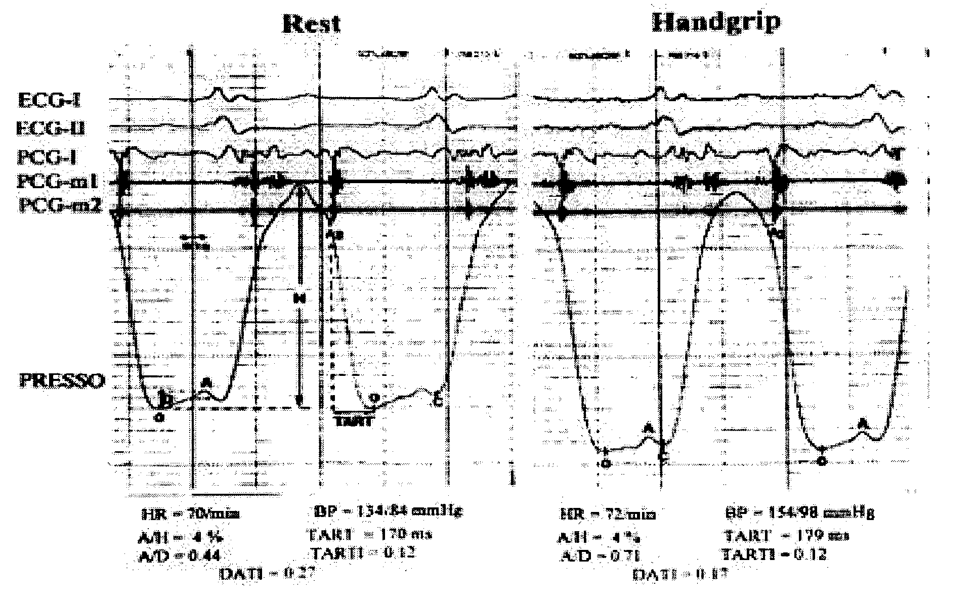
[0067]The introduction of new definitions and partly new indices as well as this new computer-based automated categorization of HAT result, represents a significant and clinically important improvement of pressocardiography, since the simplicity of the new system is providing a more practical and quicker readout without requirement for skilled interpretation. The invention provides, thus, both a new classification and instrumentation of HAT technique which is more suitable for a wider and more effective initial screening in the primary care. Additionally, this invention could also become a very useful novel diagnostic modality in sports centers for screening athletes for the presence of subclinic or atypical cardiomyopathies as well as in outpatient clinic—including Chest Pain Units (CPUs) for detecting or ruling out the presence of acute ischemia in patients with suspected unstable angina.
[0081]More particularly, the present invention is disclosed for discriminating myocardial disease patients with exercise-inducible ischemia from those without inducible ischemia; this separation helping potentially greatly in decision making about admission in the hospital and / or indication for performing a coronary angiography:
[0100]A further advantage of development of the present invention consists in a portable and handheld instrumentation—at best in pocket size—which includes the appropriate computer-based automation of HAT criteria and categories enabling us to evaluate a patient not only in a hospital, but also at the office or in sports centers and even at his home.
[0102]Thus, the result of the present invention is an user friendly small (even pocket sized) system which does not require expertise physicians and can be executed even by personnel which are not highly skilled and can be applied in almost every patient (including disabled and elderly) and everywhere (even at patient's home) providing useful information about diastolic left ventricular function changes at rest and during isometric exercise.

Problems solved by technology

Previous attempts to obtain high-fidelity tracings from the point of maximal pulse of the heart beat—referred as apexcardiography—have been handicapped mainly because of lack of an advanced instrumentation and automated evaluation of exactly defined diastolic indices as well as of exactly defined patterns, types and forms of isometric exercise-induced changes in diastolic profile.
On the other hand, current methods of measuring diastolic LV function are either extremely invasive or / and costly or / and noninvasively imaging approaches that require expertise and time-consuming measurements by highly skilled operators.
An example of the latter are all Doppler echocardiographic techniques providing ultrasonic dynamic images of the heart like transmitral flow velocities, pulmonary venous flow assessment, acoustic quantification, flow propagation assessment and the most recently introduced Tissue Doppler Imaging (TDI) and Strain-Rate Imaging (SRI); however, all these Doppler techniques require highly and expensively developed laboratories as well as skilled interpretation, whereas in most cases there is a lack of exact definition of normal limits and / or of standardization.
Consequently, there was hitherto a lack to assess LV diastolic function alterations, accurately as well as noninvasively, rapidly and safely; and above no simple and at best portable instrumentation—at best in pocket size—using an external pressure transducer with infinite time constant.
The diagnosis of coronary artery disease or other myocardial diseases based on symptoms is not accurate enough, since these symptoms occur rather late in the course of most myocardial diseases and atypical and above not infrequently entirely absent.
The probably greatest problem in cardiology but also in health care is still the early identification of subclinic myocardial diseases and especially of asymptomatic patients with coronary artery disease.
Alternatively, more sensitive and specific methods such as nuclear (Thallium) imaging or stress echocardiography (including transeosophageal echo) are time-consuming, not completely non-invasive, very costly and require expert interpretation.
Further, dynamic exercise or pharmacological stress tests are not convenient and not completely safe for the patients; resulting either to a decreased desire of the patients or even contraindication for performing them.
Therefore, these techniques can not be applied very widely and above they are rather excluded for use by primary care physicians.
This lack of appropriate, accurate and easy applicable techniques for screening healthy looking and feeling patients for “silent myocardial disease states, results in a great still unsolved problem in health care; namely, there are still today yearly >220.000 patients in US dying suddenly due to silent myocardial disease remaining mostly unidentified prior to their sudden death.
Thus, these asymptomatic patients suffering on subclinic myocardial disease could only being diagnosed by primary care physicians who, however, do not have enough diagnostic tools for such a screening.
All of them have one or more of the following limitations: they are time-consuming, costly, not standardized and require expert interpretation.
Thus, they can not be applied by primary care physicians.
Because of all these limitations and disadvantages, one should conclude that although there is no doubt about the existing and significantly increasing interest on and the clinical importance of diastolic function assessment, the clinical application of diastolic techniques in the daily practice remains rather limited.
Previous attempts at obtaining and evaluating (apex)pressocardiographic waveforms have been handicapped by the lack of advanced instrumentation, signal processing and pattern recognition techniques.
Moreover, they were obtained only at rest and / or after dynamic exercise, whereas the diastolic waves could not be exactly evaluated due to too high heart rate.
In past years, air-filled tubes were used for transmission of the pulse signal and transducers with too short time constant resulting in distorted tracings.
However, the received information needs a skilled further interpretation by a cardiologist and does not allow a rapid automated cardiac performance and diastolic behavior estimation by every non-expert physician or lab assistant, since an information about differentiation of underlying disease (ischemia vs non-ischemic or hypertensive myocardial disease vs Cardiomyopathy etc.) could not be derived from this previous technology.
There was a limited automated evaluation of the result and above a non-existent separation of patients with various myocardial diseases based on exactly defined patterns and forms of LV diastolic behavior.

Method used


Image

  • Device for and method of rapid noninvasive measurement of parameters of diastolic function of left ventricle and automated evaluation of the measured profile of left ventricular function at rest and with exercise
  • Device for and method of rapid noninvasive measurement of parameters of diastolic function of left ventricle and automated evaluation of the measured profile of left ventricular function at rest and with exercise
  • Device for and method of rapid noninvasive measurement of parameters of diastolic function of left ventricle and automated evaluation of the measured profile of left ventricular function at rest and with exercise

Examples


Embodiment Construction

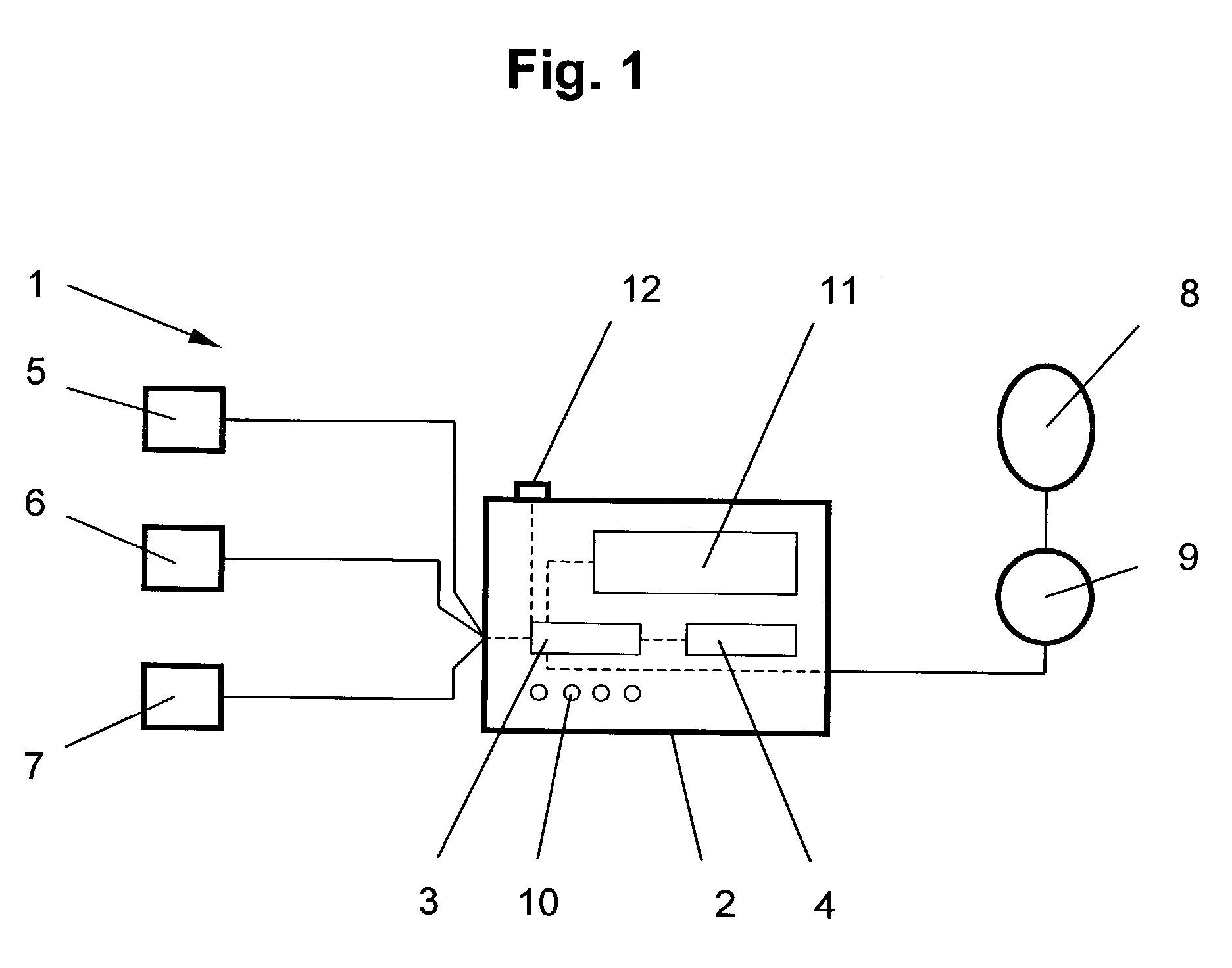
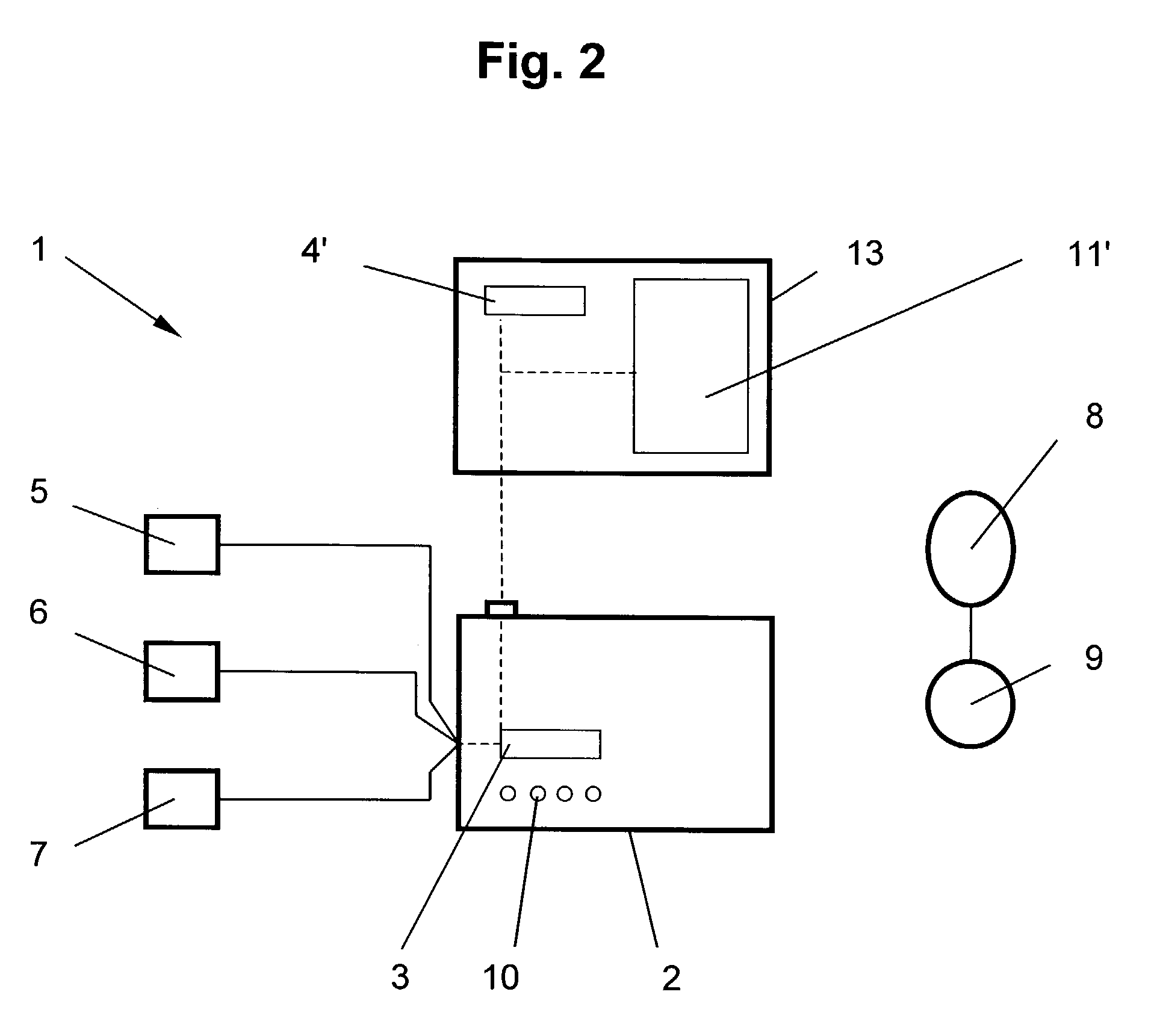
[0117]FIG. 1 shows a graphic illustration of an apparatus 1 for Handgrip-pressocardiographic test analysis and particularly for determining and calculating parameters of diastolic function of the left ventricle comprising a casing 2 which includes a control device 3 and interpretation unit 4. Further, casing 2 is connected to the following pick up units: the pressure transducer 5 for obtaining a tracing which mirrors left ventricular pressure, the heart sounds (phono) microphone 6 for recording the second heart sound and an electrical recorder 7 for recording ECG. Apparatus 1 includes also a device 8 for allowing the patient to perform an isometric exercise.

[0118]Control device 3 controls detection and processing of the parameters measured and data output; it also includes a programm for guiding a user. The interpretation unit 4 is connected with or implemented in control device 3. Both form an external utilization unit which determines and calculates characteristic diastolic parame...


PUM


Abstract

In a method of noninvasive measurement of parameters of diastolic function of left ventricle and automated evaluation of the measured profile at rest and with exercise, a patient performs an isometric exercise. An external pressure sensor and heart sounds microphone are applied in a non-invasive manner on the thoracic wall to obtain a left ventricular pressure mirroring curve (pressocardiogram) and simultaneously the heart sounds (phonocardiogram). An external unit determines and calculates characteristic diastolic parameters derived from the pressocardiographic curve and phonocardiogram at rest, during and after exercise, converts each said pressocardiogram into a digital waveform in the time domain, and automatically categorizes the mentioned characteristic parameters based on exact categorization criteria for defining several differentialforms of diastolic dysfunction of left ventricle in human beings.

Description

FIELD OF THE INVENTION[0001]This invention is in the cardiology and relates to the detection of heart dysfunction by use of pressocardiographic test in human beings. The so-called pressocardiograms are obtained from the chest wall by a pressure transducer in order to detect left ventricular (LV) diastolic dysfunction at rest and during short exercise. Specifically, this invention consists in a device—ideally portable and in pocket form—for assessing and calculating diastolic LV function indices as well as for evaluating the test results and above in differentiating characteristic patterns and differentialforms of diastolic behavior with exercise.BACKGROUND OF THE INVENTION[0002]Previous attempts to obtain high-fidelity tracings from the point of maximal pulse of the heart beat—referred as apexcardiography—have been handicapped mainly because of lack of an advanced instrumentation and automated evaluation of exactly defined diastolic indices as well as of exactly defined patterns, ty...

Claims


Application Information

Patent Timeline
IPC IPC(8): A61B5/02A61B5/04A61B5/021A61B7/00
CPCA61B5/021A61B7/00A61B5/4884A61B5/7239A61B5/742A61B5/339
Inventor MANOLAS, JAN
Owner MANOLAS JAN
Why Eureka
Eureka Blog
Learn More