
Selective labeling and isolation of phosphopeptides and applications to proteome analysis

a proteome analysis and labeling technology, applied in the field of selective labeling and isolation of phosphopeptides, can solve the problems of limiting the dynamic range of the technique, limiting the sample throughput, and no protein analytical technology approaching the throughput and level of automation of genomic technology, so as to facilitate separation, isolation and detection of phosphoproteins

Inactive Publication Date: 2006-05-30
UNIV OF WASHINGTON
View PDF30 Cites 32 Cited by
  • Summary
  • Abstract
  • Description
  • Claims
  • Application Information

AI Technical Summary

Benefits of technology

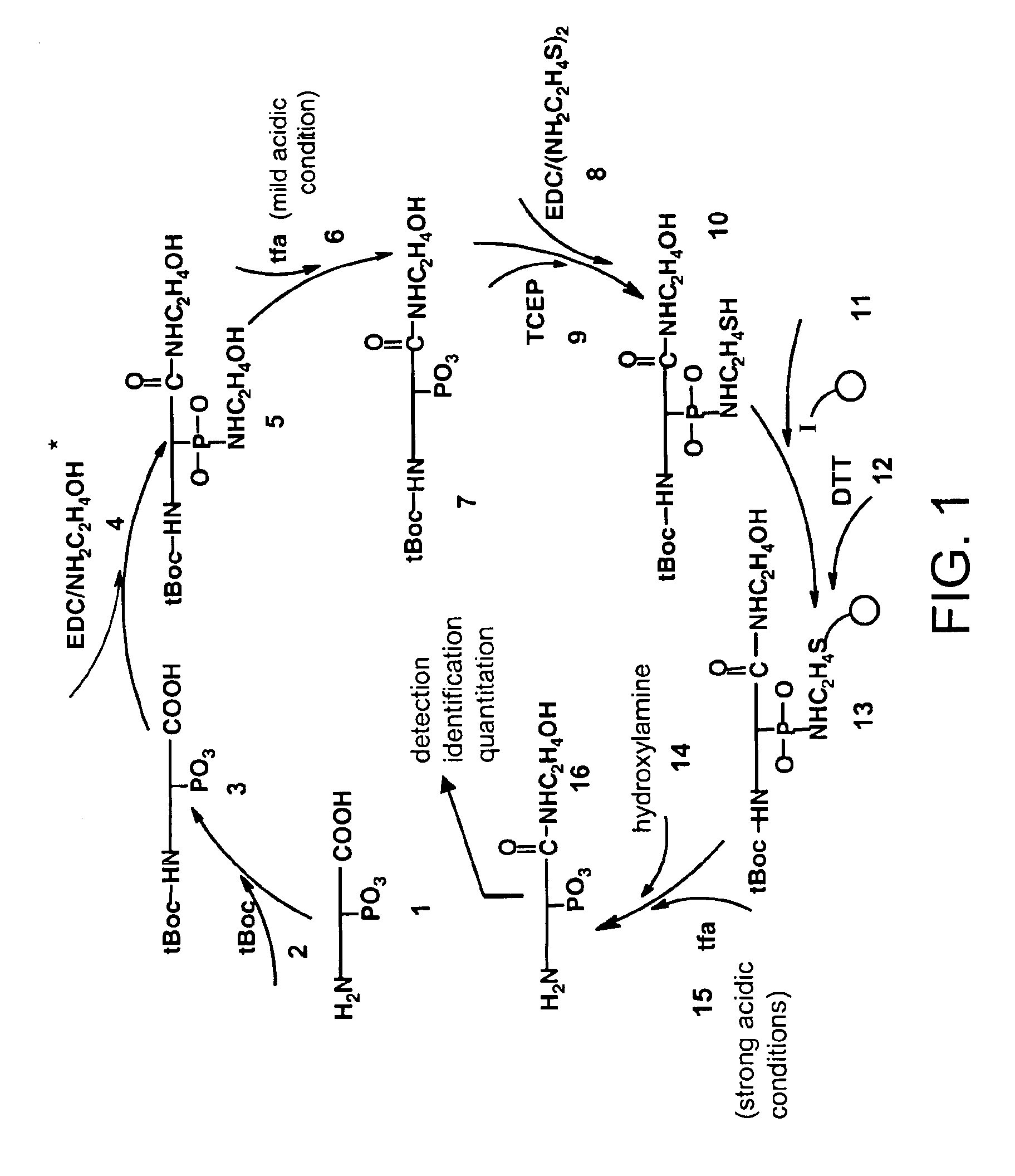
[0013]This invention provides a method for selective labeling of phosphate groups in natural and synthetic oligomers and polymers in the presence of chemically related groups such as carboxylic acid groups. The method is specifically applicable to biological oligomers and polymers, including phosphopeptides, phosphoproteins and phospholipids. Selective labeling of phosphate groups in proteins and peptides, for example, facilitates separation, isolation and detection of phosphoproteins and phosphopeptides in complex mixtures of proteins. Selective labeling can be employed to selectively introduce phosphate labels at phosphate groups in an oligomer or polymer, e.g., in a peptide or protein. Detection of the presence of the label is used to detect the presence of the phosphate group in the oligomer or polymer. The method is of particular use for the detection of phosphoproteins or phosphopeptides. The phosphate label can be a colorimetric label, a radiolabel, an isotope label, a fluorescent or phosphorescent label, an affinity label or a linker group carrying a reactive group (or latent reactive group) that allows selective attachment of the oligomer or polymer (protein or peptide) to a phosphate label, to an affinity label or to a solid phase material.
[0014]Selective attachment of a phosphate group to its affinity label or selective attachment to a solid support allows selective isolation of an oligomer or polymer (e.g., protein or peptide) that carries at least one phosphate group. The presence of an affinity label allows capture of the selectively labeled oligomer or polymer using a capture reagent that specifically bonds to the affinity label. The presence of a linker that allows selective covalent attachment of the oligomer or polymer to a solid surface allows physical separation of the selectively labeled oligomer or polymer from non-selectively labeled (non-phosphorylated) species in a sample. The method is particularly useful for the selectively labeling of proteins and peptides having a phosphate group (phosphoproteins and phosphoptides) in the presence of carboxylic acid groups. The method of this invention can be used specifically to separate phosphoproteins and phosphopeptides from mixtures of non-phosphorylated proteins mixtures of non-phosphorylated proteins and / or nonphosphorylated peptides and thus to overcome detection problems due to low levels of these species in samples.
[0016]When combined with well-known methods of mass spectrometry, the selective labeling method of this invention facilitates separation of phosphopeptides from mixtures and facilitates their detection by mass spectrometry and facilitating sequencing of the peptides by tandem mass spectrometry. Art-known methods can be applied to identify phosphoproteins in a sample from the sequences of phosphopeptides detected in a sample. When combined with methods for differential isotopic labeling, the method of this invention can be employed to quantify relative amounts of phosphopeptides and phosphoproteins in different samples. These quantitative methods allow a comparison of phosphorylation state in samples derived from different sources (e.g., from different cell-types or different organisms), samples that are differentially affected by a stimulus (e.g., administration of a drug or contact with a potentially toxic material), by a change in environment (e.g., nutrient level, temperature, passage of time) or by a change in condition or cell state (e.g., disease state, malignancy, site-directed mutation, gene knockouts) of a cell, tissue or organism from which the sample originated. The phosphoproteins identified in such a screen can function as markers for the changed state. Phosphopeptides and phosphoproteins from any naturally-occurring environment or artificially-controlled environment can be assessed by the methods herein. The method can be applied to mixtures of naturally-occurring proteins or peptides, as well as to mixtures of proteins or peptides derived from recombinant or synthetic methods.

Problems solved by technology

At present no protein analytical technology approaches the throughput and level of automation of genomic technology.
However, the sequential manner in which samples are processed limits the sample throughput.
The most sensitive methods have been difficult to automate and low abundance proteins, such as regulatory proteins, escape detection without prior enrichment, thus effectively limiting the dynamic range of the technique.
While these approaches dramatically accelerate protein identification, the quantities of the analyzed proteins cannot be easily determined due to the observation that mass spectrometers are inherently not quantitative devices.
Much of the difficulty in such studies lies in the fact that many phosphoproteins exist only at very low abundance.
Therefore, it is usually difficult to obtain sufficient amounts of pure phosphoprotein for such analyses.
Because cellular proteins are coordinately phosphorylated to control specific biological processes, the complex mechanisms that control biological systems by protein phosphorylation are difficult to investigate using current technology.
In addition, no MS-based method to quantify protein phosphorylation directly is currently available.

Method used


Image

  • Selective labeling and isolation of phosphopeptides and applications to proteome analysis
  • Selective labeling and isolation of phosphopeptides and applications to proteome analysis
  • Selective labeling and isolation of phosphopeptides and applications to proteome analysis

Examples


example 1

ptide Isolation Procedure

[0084]Peptide samples were dried, and then subjected to the method shown in FIG. 1A according to the following steps. 1) Peptide mixture was resuspended in 50% (v / v) of 0.1 M phosphate buffer (pH 11) / acetonitrile. 0.1 M of t-Butyl-dicarbonate (tBoc) was added for 4 hours at room temperature. 2) Acetonitrile was removed under reduced pressure. Samples were made to 1 M ethanolamine, 25 mM N-hydroxysuccinimide (NHS) and 0.5 M of N,N′-dimethylaminopropyl ethyl carbodiimide HCl (EDC) and incubated 2 hours at room temperature. 3) 10% trifluoroacetic acid (TFA) was added for 30 minutes at room temperature. Longer treatment under these conditions did not detrimentally affect the results. Samples can be neutralized at this point, but neutralization as found to have no significant effect on results. Samples were then desalted on and recovered from a C18 column (Waters Associates, Milford, Mass. WAT 023590) using elution with 80% acetonitrile, 0.1% TFA. 4) Peptides wer...

example 2

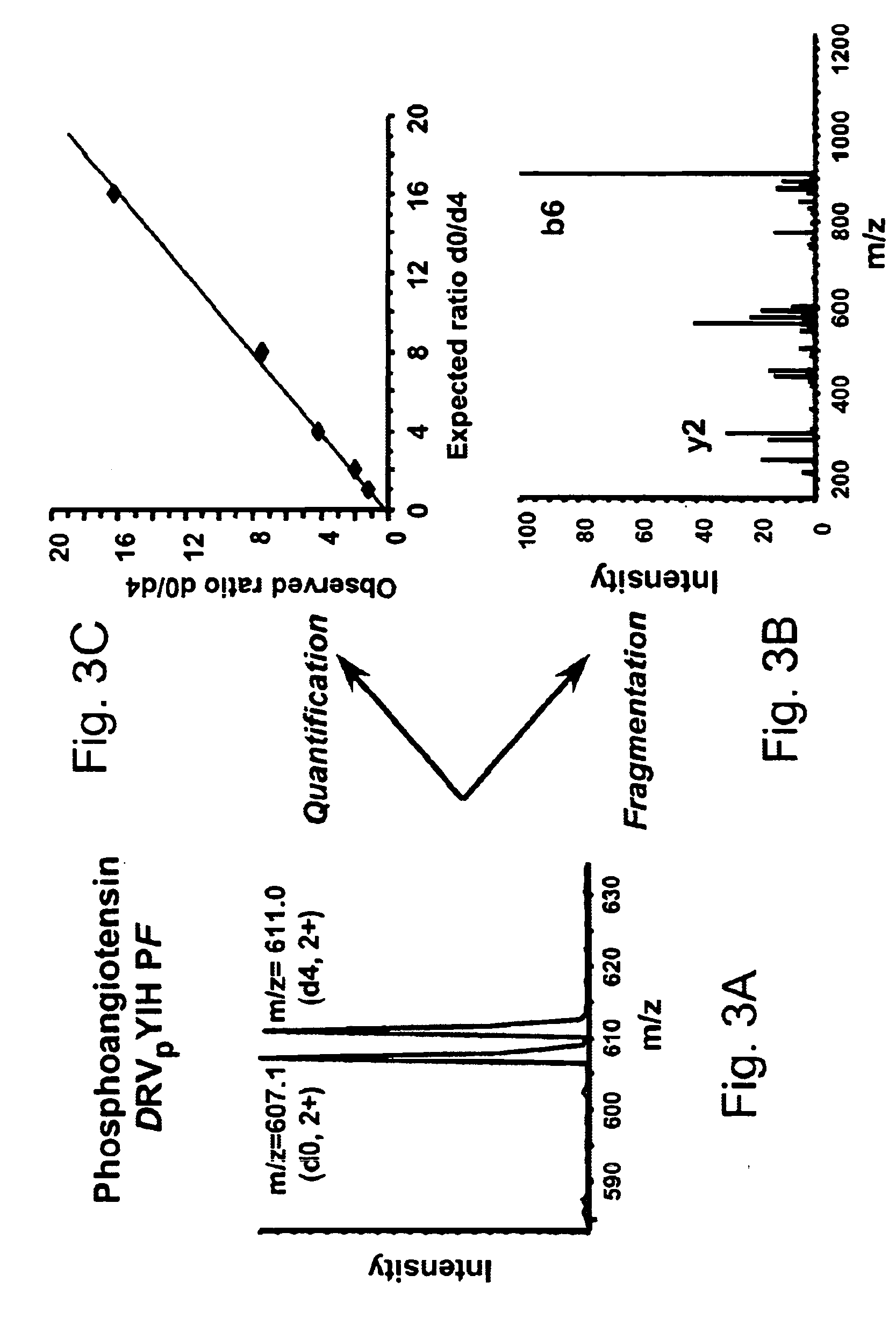
[0085]Two separate samples of equal amounts of phosphoangiotensin peptide were analyzed by the method of this invention. The carboxylic acid groups in the two different samples were blocked (leaving phosphate groups free as described above) by either light ethanolamine (d0-ethanolamine) or heavy ethanolamine (d4-ethanolamine, HOCD2CD2NH2). Phosphoangiotensin contains two carboxylic acid groups, so that the mass difference for the [M+2H]2+ ion is 4 for the differentially labeled peptides. The results of mass spectrometric analysis of the differentially labeled samples that were subjected to selective labeling and separation of phosphopeptides of this invention is illustrated in FIGS. 3A-C. A doublet of peaks [M+2H]2+ at m / z=607 and 611, due to light and heavy labeled samples, respectively, is observed as expected. Further the relative ratios of the two peaks is about 1:1 as expected. The CID spectrum of these peaks is similar to that of the unprotected peptide, except for the fragmen...

example 3

of Phosphopeptides from β-casein

[0089]The methods of this invention were also used to purify and detect phosphopeptides from bovine β-casein, a well-characterized phosphoprotein. The peptide was labeled as described in Example 2. A tryptic digest of the phosphoprotein was analyzed by microcapillary LC-MS / MS. As shown in FIG. 4A, numerous peptides were observed for the untreated β-casein digest. The peptide indicated in FIG. 4A was a doubly charged ion at m / z=1031.6. When selected for fragmentation via collision induced dissociation (CID) (FIG. 4C) (Papayannopoulos, I. A. (1995), “The interpretation of collision-induced dissociation tandem mass spectra of peptides,”Mass Spectrometry Rev. 14,49-73), its fragment ion spectrum exhibited mostly the y-ion series typical for low energy peptide fragmentation and an additional major signal at m / z=983.0 corresponding to a loss of 98 Da due to the loss of the H3PO4 group from the parent ion(Jonscher, K. R. and Yates, J. R. III (1997), “Matrix-...


PUM

PropertyMeasurementUnit
Acidityaaaaaaaaaa
Fluorescenceaaaaaaaaaa
Affinityaaaaaaaaaa

Abstract

A method for selective labeling of phosphate groups in natural and synthetic oligomers and polymers in the presence of chemically related groups such as carboxylic acid groups. The method is specifically applicable to biological oligomers and polymers, including phosphopeptides, phosphoproteins and phospholipids. In a specific embodiment, selective labeling of phosphate groups in proteins and peptides, for example, facilitates separation, isolation and detection of phosphoproteins and phosphopeptides in complex mixtures of proteins. Selective labeling can be employed to selectively introduce phosphate labels at phosphate groups in an oligomer or polymer, e.g., in a peptide or protein. Detection of the presence of the label, is used to detect the presence of the phosphate group in the oligomer or polymer. The method is useful for the detection of phosphoproteins or phosphopeptides. The phosphate label can be a colorimetric label, a radiolabel, a fluorescent or phosphorescent label, an affinity label or a linker group carrying a reactive group (or latent reactive group) that allows selective attachment of the oligomer or polymer (protein or peptide) to a phosphate label, to an affinity label or to a solid support. The method can be combined with well-known methods of mass spectrometry to detect and identify phosphopeptides and phosphoproteins.

Description

CROSS-REFERENCE TO RELATED APPLICATIONS[0001]This application takes priority from U.S. provisional application Ser. No. 60 / 210,972, filed Jun. 12, 2000, which is incorporated by reference herein in its entirety.STATEMENT REGARDING U.S. GOVERNMENT RIGHTS[0002]This invention was made with U.S. Government support under National Science Foundation grant No. BIR9214821 AM04 and under National Institutes of Health grant no. IR33CA84698. The United States government has certain rights in this invention.FIELD OF THE INVENTION[0003]A general method for selective chemical labeling of phosphate groups is provided which facilitates highly specific purification of molecules containing one or several phosphate group(s). The method is applicable to the selective labeling of phosphates in phosphoproteins and phosphopeptides. When combined with mass spectrometric techniques, the method can be employed to detect and identify phosphorylated proteins in complex mixtures and to precisely identify the ph...

Claims


Application Information

Patent Timeline
IPC IPC(8): G01N33/50C07K17/02C07K17/14G01N24/00G01N33/533G01N33/534G01N27/62C07K1/107C07K1/13C07K14/47G01N30/72G01N30/88G01N33/58G01N33/68
CPCC07K1/107C07K1/1075C07K1/13C07K14/4713G01N33/6803G01N33/6842C07K14/4732Y10T436/24Y10S530/811Y10S530/814
Inventor AEBERSOLD, RUEDIZHOU, HUILIN
Owner UNIV OF WASHINGTON
Learn More