Materials and methods for effective in vivo delivery of DNA nanostructures to atherosclerotic plaques

a technology of dna nanostructures and nanostructures, which is applied in the direction of capsule delivery, drug compositions, cardiovascular disorders, etc., can solve the problems of increasing cancer risk, difficult to accurately identify at-risk patients, and substantial residual risk still remains

Active Publication Date: 2019-02-28
THE CHINESE UNIVERSITY OF HONG KONG
View PDF2 Cites 4 Cited by
  • Summary
  • Abstract
  • Description
  • Claims
  • Application Information

AI Technical Summary

Benefits of technology

The nanoparticle-cored spherical nucleic acids (DNA-NPs) of this patent contain at least one therapeutic agent and can be used as in vivo imaging agents to selectively target and treat atherosclerotic lesions in patients with atherosclerotic disease. The DNA-SNPs can be superparamagnetic iron oxide nanoparticle-cored spherical nucleic acids (DNA-SPIONs) or superparamagnetic iron oxide nanoparticle-cored DNA-dendritic cells (DNA-SPION-DCs). The DNA-SPIONs contain a magnetic core, a polymeric spacer layer, and an outer DNA oligonucleotide shell. When systemically injected, the nanoparticles accumulate rapidly inside the atherosclerotic plaques and can be used as carriers of therapeutic and contrast agents to the plaque. The DNA-SPIONs can remain inside the plaque for up to 24 hours post-injection and can be useful in identifying and targeting plaque-related cells.

Problems solved by technology

Due to the slow and asymptomatic progression of atherosclerosis, an ongoing clinical challenge is to image and treat atherosclerotic plaques at the earliest possible stage.
Despite advances in drug development and stent design in angioplasty procedure, substantial residual risk still remains.
Currently it is challenging to accurately identify at-risk patients, as conventional imaging tools can only determine the degree of artery stenosis but not to detect early lesions or analyze plaque size, composition, activity or overall disease burden.
Most current noninvasive cardiovascular imaging methods, such as positron emission tomography (PET), single-photo emission computed tomography (SPECT) and X-ray angiography rely on radiation exposure to generate photos and is associated with an increased risk of cancer (8).
No clinically approved nanoparticle-based therapies are currently available for treating atherosclerosis.
One of the critical problems that remain in the clinical application is a lack of high accuracy, noninvasive methods for diagnosis and prognosis of atherosclerosis.
However, few of the nanomaterials have been approved in clinical trials for imaging atherosclerosis due to the lack of specificity, long waiting time and slow clearance rate.

Method used


Image

  • Materials and methods for effective in vivo delivery of DNA nanostructures to atherosclerotic plaques
  • Materials and methods for effective in vivo delivery of DNA nanostructures to atherosclerotic plaques
  • Materials and methods for effective in vivo delivery of DNA nanostructures to atherosclerotic plaques

Examples


example 1

Characterizations of PEG-NPs, PEG-SPNs and / or PEG-SPIONs and DNA-NPs, DNA-SPNs and / or DNA-SPIONs

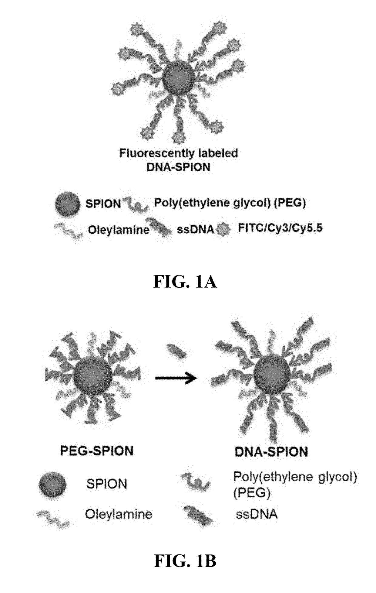
[0163]Although the core material of NP, SPN, and / or SPION-cored spherical nucleic acids does not affect their biological properties, their biocompatibility is still a great concern when NPs, SPNs, and / or SPIONs are used as in vivo delivery vehicles or imaging agents. Iron oxide nanoparticles were chosen as the model material due to their well-known biocompatibility and superior performances as MRI contrast agents. FIG. 1A shows the composition of fluorescently-labeled DNA-coated SPIONs (DNA-SPIONs), consisting of a SPION core, a dense poly(ethylene glycol) (PEG) layer and DNA conjugation in the outmost layer, which DNA is conjugated to a fluorescent molecule. Before DNA conjugation, the PEG-coated SPIONs (PEG-SPIONs) were synthesized from the high temperature decomposition of iron (III) acetylacetonate with PEG diacid and oleylamine as chelating agents (FIG. 1B, left). To the surface of a...

example 2

SR-A Expression on Different Cell Types and Cellular Uptake of DNA-SPIONs.

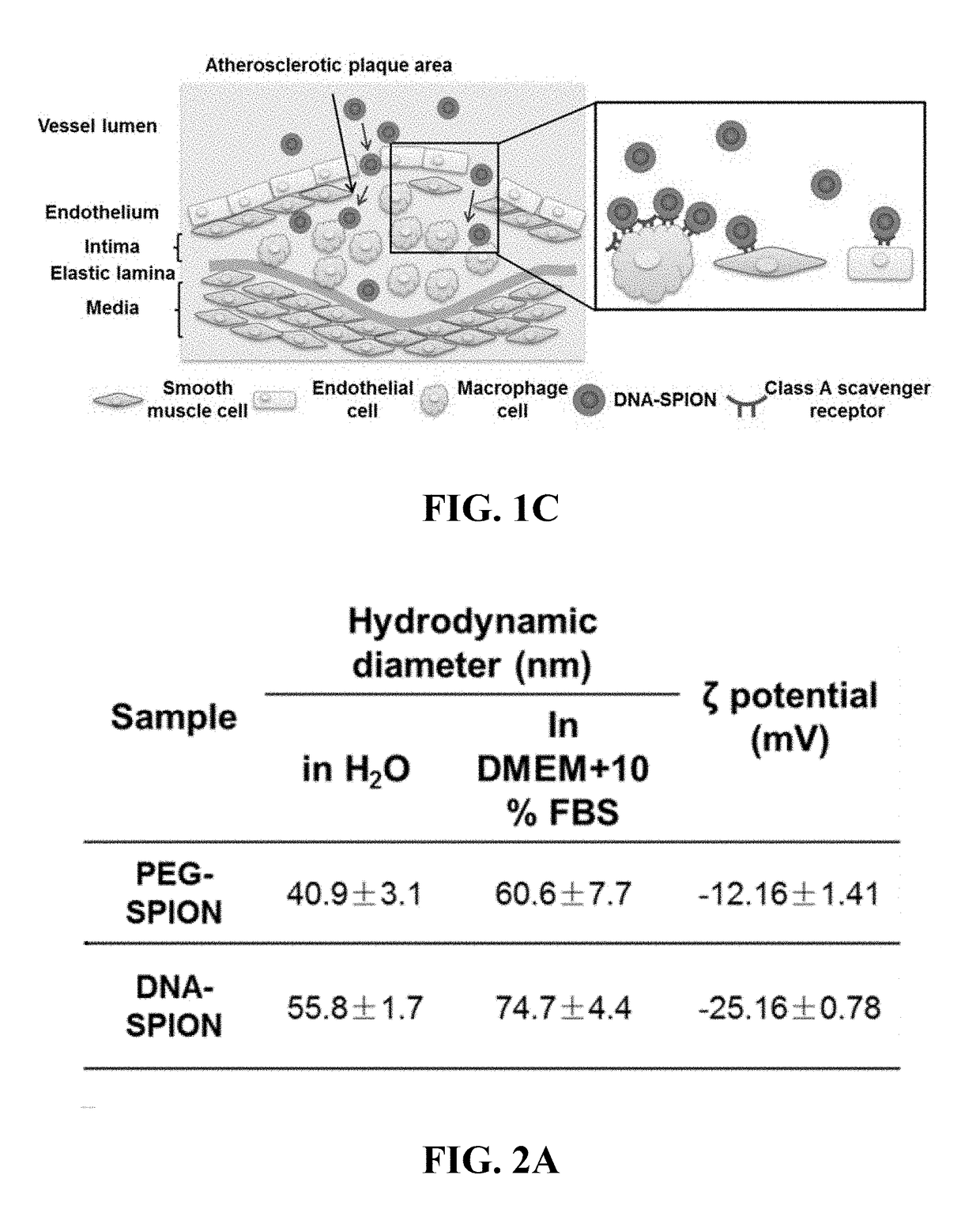
[0168]As shown in FIG. 1B, the major cell types in atherosclerotic plaques are monocyte / macrophage, smooth muscle cells and endothelial cells (40, 52). To probe the interaction of DNA-SPIONs with different types of cells representative of cells present inatherosclerotic plaques, the uptake of nanoparticles in four model cell types was studied in vitro, including mouse endothelial (C166), mouse smooth muscle (MOVAS) and mouse macrophage (RAW 264.7) cells. Since nanoparticles are known to be able to accumulate in organs of the mononuclear phagocyte system (MPS), including liver and spleen (55), mouse hepatocytes (AML-12) were also included for comparison. Before conducting cellular uptake studies, the cytotoxicity of PEG-SPIONs and DNA-SPIONs to the four types of cells was tested by MTT assay. In this MTT assay, PEG-SPIONs and DNA-SPIONs were incubated with cells for 24 hours (FIG. 4). No obvious cytotoxicity of...

example 3

Cellular Uptake Kinetics of PEG-SPIONs and DNA-SPIONs in RAW 264.7 Cells

[0173]The time-course uptake of both types of nanoparticles was studied by ICP-MS for measurement of iron content after incubation for different durations of time. During the first 8 hours of uptake, DNA-SPIONs associated with RAW 264.7 cells much faster than PEG-SPIONs (FIG. 6A). For example, 4 hours post-incubation, the iron content of cells treated with DNA-SPIONs was more than 5-fold higher than that of cells treated with PEG-SPIONs (0.89 pg / cell versus 0.17 pg / cell), validating the selective uptake of DNA-SPIONs over PEG-SPIONs by RAW 264.7 macrophages as shown in FIGS. 5C and 5D. The cellular association of DNA-SPIONs showed little increment in iron content between 8 hours and 16 hours, an observation potentially related to the doubling time of RAW 264.7 cells that is ˜12 hours for a standard plate set-up (56, 57).

[0174]Prussian blue staining, which turns content iron into blue signals and, thus, detects t...


PUM

PropertyMeasurementUnit
Fractionaaaaaaaaaa
Timeaaaaaaaaaa
Fractionaaaaaaaaaa

Abstract

Provided are DNA-coated nanoparticles (DNA-NPS), superparamagnetic nanoparticles (DNA-SPNs), and superparamagnetic iron oxide nanoparticles (DNA-SPIONs) as efficient imaging agents for targeting and imaging atherosclerotic lesions and treating atherosclerotic disease. The DNA-NS, DNA-SPNs, and DNA-SPIONs can enter macrophage cells via the Class A scavenger receptor (SR-A)-mediated pathways and can be used to specifically target atheroscleortic plaques.

Description

CROSS-REFERENCE TO RELATED APPLICATION[0001]This application claims the benefit of U.S. Provisional Application Ser. No. 62 / 550,995, filed Aug. 28, 2017, the disclosure of which is hereby incorporated by reference in its entirety, including all figures, tables and drawings.BACKGROUND OF THE INVENTION[0002]Cardiovascular diseases (CVD) are the leading cause of death globally, causing 17.3 million deaths per year (1). Initially triggered by chronic inflammation of the endothelium, atherosclerosis is characterized by persistent inflammation and accumulation of lipid-rich plaques or lesions in arterial walls and is the major cause of CVD (2). The hallmark feature of atherosclerosis is the development of atheromatous plaques or lesions characterized by the retention of lipids in the artery wall and infiltration of leukocytes (3). As the most populous cell type in the plaque, monocyte-derived macrophages readily internalize lipids and become foam cells (4). Macrophages also contriubute to...

Claims


Application Information

Patent Timeline
IPC IPC(8): A61K47/69A61K9/00A61K49/18A61P9/10A61K47/54A61K47/60A61K49/00
CPCA61K47/6935A61K9/0019A61K49/186A61K49/1845A61P9/10A61K47/549A61K47/60A61K49/0093A61K45/00A61K47/26A61K49/126A61K49/1854A61K9/513A61K47/6923
Inventor CHOI, CHUNG HANG JONATHANZHANG, LEI
Owner THE CHINESE UNIVERSITY OF HONG KONG
Why Eureka
Try Eureka