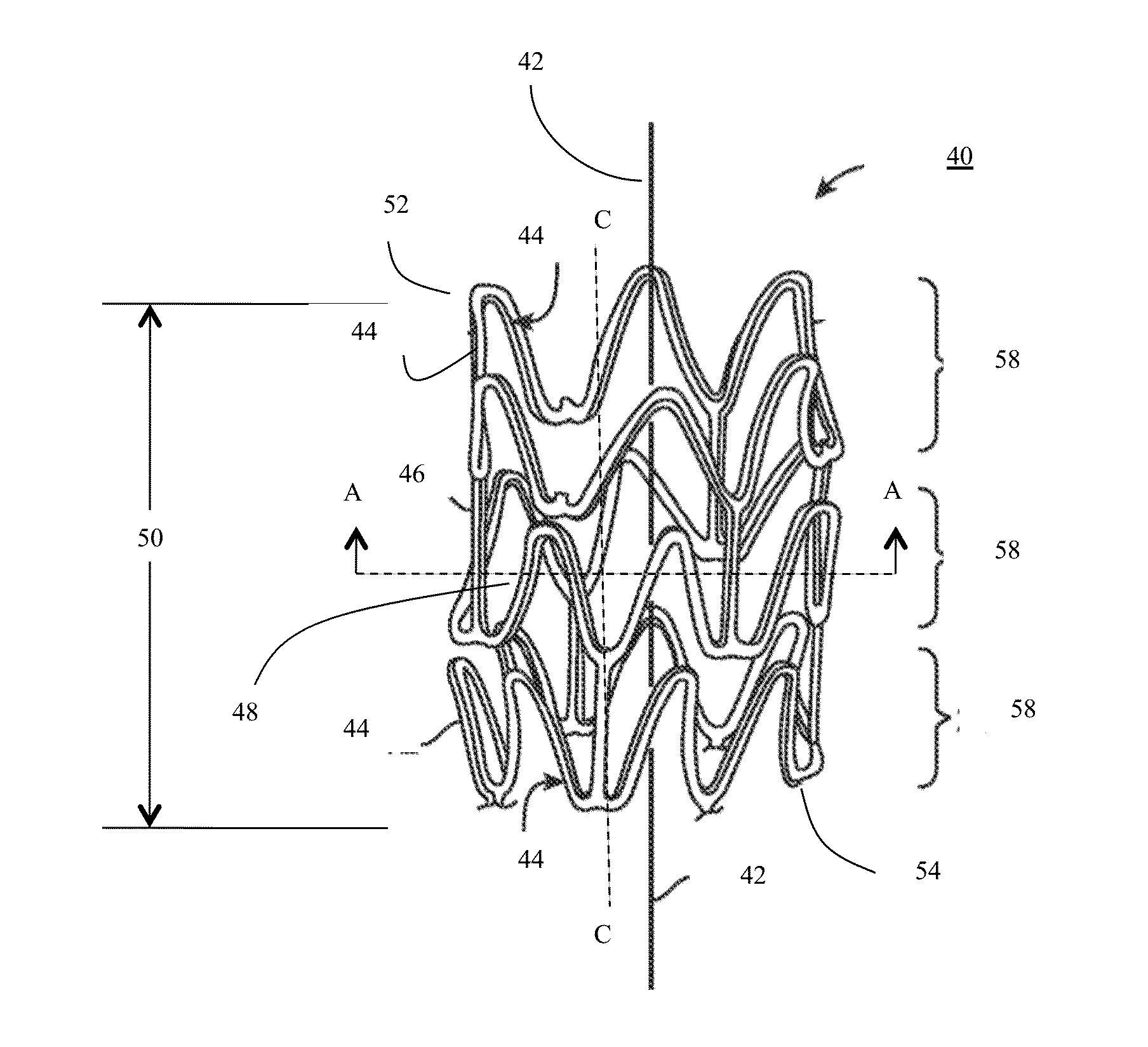
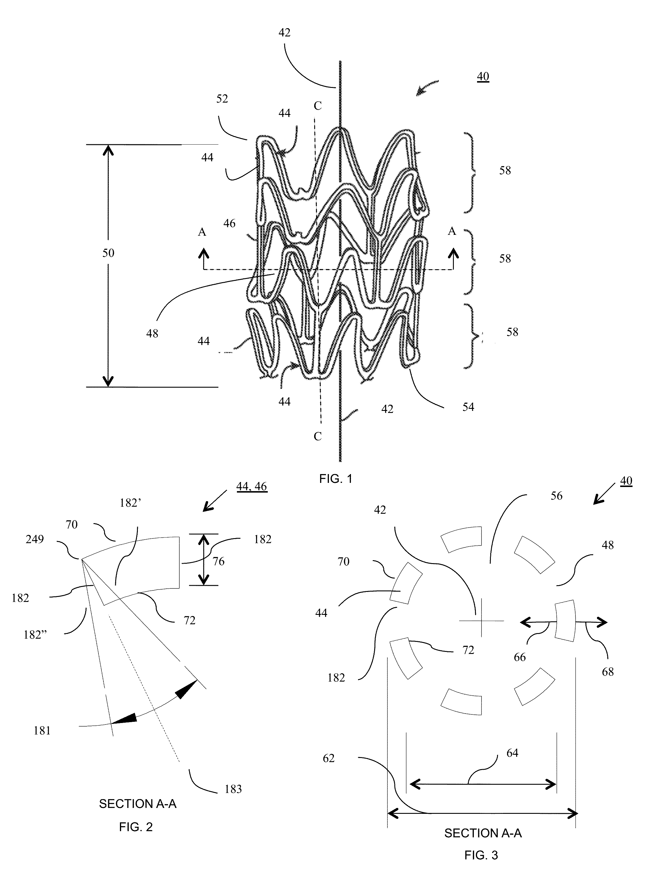
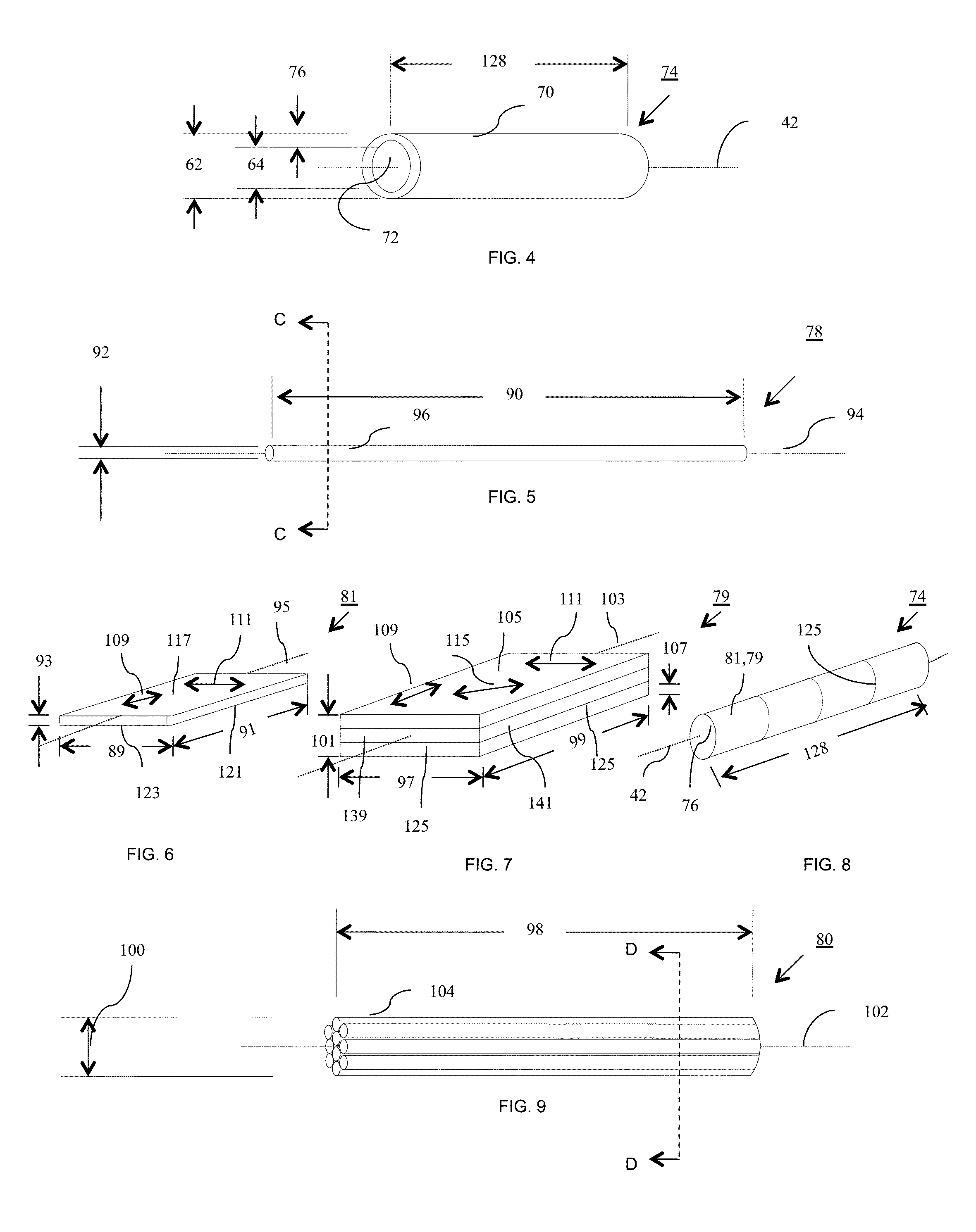

Bioresorbable Stent

Active Publication Date: 2015-10-29
SCANLON JOHN JAMES +1
View PDF4 Cites 43 Cited by
  • Summary
  • Abstract
  • Description
  • Claims
  • Application Information

AI Technical Summary

Benefits of technology

The invention provides an endoprosthesis that can be used as a bioresorbable stent, which has increased strength, thinner struts, and a more controlled degradation rate. The stent is made from a tubular precursor construct made of bioresorbable material, which is stronger and tougher than traditional bioresorbable materials due to the orientation of molecular chains during processing. These improvements lead to a reduction of strut fracturing, improved storage stability, and better radiopacity. The invention also blocks side artery branches and minimizes luminal wall contact surface area to prevent blockages. Overall, the invention provides a safer and more effective stent for the treatment of blood vessel disorders.

Problems solved by technology

Problems with PTCA include formation of intimal flaps or torn artery linings, both of which can create another occlusion in the lumen of the coronary artery.
Moreover, thrombosis and restenosis may occur several months after the procedure and create a need for additional angioplasty or a surgical bypass operation.
Thus, stents are often fabricated from biodegradable, bioabsorbable, and / or bioresorbable materials such that they completely erode only after the clinical need for them has ended.
These stent types are implanted in vessels that are closer to the surface of the body and are particularly vulnerable to crushing or pinching loads, which can partially or completely collapse the stent and thereby block fluid flow in the vessel.
A stent having suitable radial stiffness may not have sufficient crush resistance.
However, this variety of self-expanding stent has the problem of exerting a “chronic outward force (COF)” on the blood vessel supported by the stent.
However, plastically deformed stents have the drawback of higher potential for collapsing or becoming kinked in peripheral blood vessels and are generally not used for this reason.
A durable metallic stent has the drawbacks that it constrains the blood vessel after remodeling has occurred, is subject to frequent strut fractures, requires dual antiplatelet therapy (DAPT) to prevent subsequent thrombosis, sometimes requires revascularization, and sometimes results in the unsatisfactory outcome of late stent thrombosis which often results in patient death.
Relatively low radial strength potentially contributes to relatively high recoil of polymer stents after implantation into an anatomical lumen.
The higher stent-to-artery coverage may increase the probability of blocking anatomical lumen side branches or invoking an unfavorable response resulting in restenosis or thrombosis.
Another potential problem with polymer is that struts can crack or fracture during crimping, delivery, and deployment, especially with brittle polymers.
Some polymers like poly (L-lactide) (“PLLA”) are stiff and strong but can exhibit a brittle fracture mechanism at physiological conditions in which there is little or no plastic deformation prior to failure.
A stent fabricated from such polymers can have insufficient toughness for the range of use of a stent.
In particular, during deployment of a stent comprised of a material having insufficient ductility, strut fracture can occur when the stent is expanded during deployment with a balloon catheter beyond recommended expansion limits.
As a result, cracks can occur particularly in high strain regions, which can result in mechanical failure of the stent.
These prior art stents employ polymers with relatively low molecular orientation because the molecular orientation is limited to the random molecular orientation that is imparted on the polymer when it flows through the screw inside the barrel that conveys the material to the mold or die.
Instead of building strength in the polymer, these prior art process of molding or extruding can actually reduce the strength of the polymer because the polymer is subjected to relatively high shear stresses at temperatures above the melting point of the polymer which substantially reduces the molecular weight of the polymer.
Solid wall thickness polymeric stents are sometimes strengthened by radial or longitudinal plastic deformation of the precursor tubes used in manufacturing struts as described in U.S. Pat. No. 8,012,402 B2 filed 15 Apr. 2009, “Tube expansion process for semi-crystalline polymers to maximize fracture toughness.” The larger wall thickness is problematic in small diameter stents because the polymeric stents of the prior art reduce luminal capacity because the stent struts consume a significant portion of the orifice in which fluid flows.
Since the struts are typically 75-150 microns in width, it is necessary to use tubes having very little dimensional variation or it is impossible to accurately cut a strut pattern in a tube achieving these narrow strut widths.
The prior art methods of have difficulty producing tubes meeting these stringent tolerances, especially when manufacturing a tubular precursor construct having a length greater than 15 cm.
Stents produced from polymer tubes having a wall thickness comprised of filaments, fibers, fibers, and the like can suffer from insufficient stiffness.
The polymeric stents are also more difficult to plastically deform and to properly size in lumen after deployment.
Many polymers used for stents are relatively brittle under physiological conditions.
Polymers are susceptible to mechanical instability such as fracturing while in the body.
Bioresorbable stents are difficult to see during delivery and deployment because the polymers are not radiopaque, which leads to malposition of the stent.
Bioresorbable materials have considerably lower strength than prior art metallic materials used in the construction of stents.

Method used


Image

  • Bioresorbable Stent
  • Bioresorbable Stent
  • Bioresorbable Stent

Examples


example 1

Bioresorbable (PLLA) Stent Made by Solution Electrospinning Process

[0430]Particles of PURASORB PL-38 having a somewhat translucent to opaque appearance, which is a homopolymer of L-lactide (PLLA) having molecular weights in the range of 121-298 kg / mol and 298-727 kg / mol, were dissolved in hexafluoroisopropanol (HFIP) at 2.5, 5, 11, and 15 weight percent. The solutions were clear in appearance. The PLLA solutions were electrospun from a syringe equipped with a 22 gauge needle using 15 kV potential, a throw distance of 12-16 cm, and a syringe flow rate of 5 mL / h. To form a mesh wall thickness for use in a precursor tubular construct, having a wall thickness of different diameter fibers, the 2.7 mm diameter drum or mandrel having a length of twelve inches was rotated at 600, 1000, and 1500 rpm to achieve the different degrees of fiber orientation and a wall thickness of 150 microns. The precursor tubular construct tubes were dried under vacuum for two days to remove the residual HFIP a...

example 2

Wall Thickness Suitable for Production of Prosthesis—Flat Sheet

[0435]A blend of poly L-Lactide (Trade name PURASORB PL-38 blended with PURASORB PL-32 available from Purac, Gorinchem, The Netherlands) was dissolved in HPLC-grade dichloromethane (CAS 75-09-2) (available from Fischer Scientific, Boston, Mass., USA) at a 5% weight percent in a 20 ml scintillation vial (available from Fischer Scientific, Boston, Mass., USA) and electrospun. The polymer solution pumped through a stainless 21 gauge by ½ inch steel blunt needle (Part NE-211 PL-25, Available from Amazon.com) at a rate of 1.5 ml / hour to 0.2 ml / minute. A high voltage power supply (available from Gamma High Voltage, Ormond Beach, Fla., USA) was connected to the syringe needle and a three inch diameter by eight inch long rotating cylindrical-shaped mandrel or collector plate wherein the power supply was set at 90 milliamps (mA) and 10-12 kilovolts (KV). The throw distance, which is the distance between the end of the syringe nee...

example 3

Wall Thickness Suitable for Production of Prosthesis—Tube

[0438]The flat sheets approximately 8 inches by 9.43 inches prepared as described in EXAMPLE 2, were cut into longer strips 1 inch wide by 8 inches long and 1 inch wide by 9.43 inches long to obtain strips of fibrous material having fibers aligned in different directions. These strips were wound around a steel 5 mm diameter by 200 mm long steel shaft having dimensional variation of less than 0.008 mm (model 6112K37 available from McMaster Carr, Robbinsville, N.J., USA). The assembly was placed in an oven at 135-425 degrees Fahrenheit and baked until the fibrous material wrapped around the mandrel turned from an opaque white appearance to a translucent appearance. In some cases the assembly was removed from the heat prior to becoming fully translucent. In other cases the assembly was removed from the heat prior to losing it opaque white appearance. The heated fibrous material and shaft were then rolled over a flat plate having ...


PUM

PropertyMeasurementUnit
Temperatureaaaaaaaaaa
Fractionaaaaaaaaaa
Thicknessaaaaaaaaaa

Abstract

Disclosed herein is a high strength, bioresorbable wall thickness suitable for use in an endoprosthesis such as a stent that is produced by first forming a wall thickness by melt processing or solution processing one or more bioresorbable materials into a tubular shape; drawing the shape from shorter length to an optimum longer length and reducing the diameter from a larger diameter to a smaller diameter to orient the molecular chains of the material; fabricating a stent from the tube formed of the oriented material by cutting a strut pattern in its wall thickness; covering the stent's struts with at least one coating to delay degradation of the bioresorbable material; covering the stent's struts with one or more controlled release active ingredients to minimize the risk of restenosis or other side effects; crimping the stent onto a balloon catheter assembly; delivering the stent into an anatomical lumen via percutaneous methods to a treatment location; radially expanding the stent from a smaller size to a larger size at the treatment location wherein the stent temporarily supports the anatomical lumen; and removing the catheter from the lumen.

Description

PRIORITY CLAIMS[0001]This application claims benefits of Provisional Patent Application Ser. No. 61 / 816,800 filed Apr. 28, 2013 entitled “BIORESORBABLE STENT WITH STRUTS FORMED OF NANOFIBERS” and Provisional Patent Application Ser. No. 61 / 975,782 filed Apr. 5, 2014 entitled “BIORESORBABLE STENT.”FIELD OF INVENTION[0002]The present invention relates to drug-eluting medical devices. This invention especially relates to polymeric scaffolds that are expanded by a delivery balloon.BACKGROUND OF INVENTION[0003]In percutaneous transluminal coronary angioplasty (PTCA), a balloon catheter is inserted through a brachial or femoral artery, positioned across a coronary artery occlusion, and inflated to compress against atherosclerotic plaque to open, by remodeling, the lumen of the coronary artery. Problems with PTCA include formation of intimal flaps or torn artery linings, both of which can create another occlusion in the lumen of the coronary artery. Moreover, thrombosis and restenosis may o...

Claims


Application Information

Patent Timeline
IPC IPC(8): A61L27/58A61F2/95A61F2/82
CPCA61L27/58A61F2/95A61F2/82A61F2/91A61F2210/0004A61F2240/001A61L31/14A61L31/148A61L31/16A61L31/18
Inventor SCANLON, JOHN JAMESSCANLON, CATHERINE ANN
Owner SCANLON JOHN JAMES
Learn More