Same-day blood culture with digital microscopy
a technology of digital microscopy and blood culture, applied in the field of same-day blood culture with digital microscopy, can solve the problems of increasing frequency and complexity of bacteremia due to multiple drug resistant organisms (mdro), ineffective initial therapy, and inability to tolerate the delay of the initial treatment, so as to reduce the toxic condition, and inhibit the growth of microorganisms.
- Summary
- Abstract
- Description
- Claims
- Application Information
AI Technical Summary
Benefits of technology
Problems solved by technology
Method used
Image


Examples
example 1
Methods
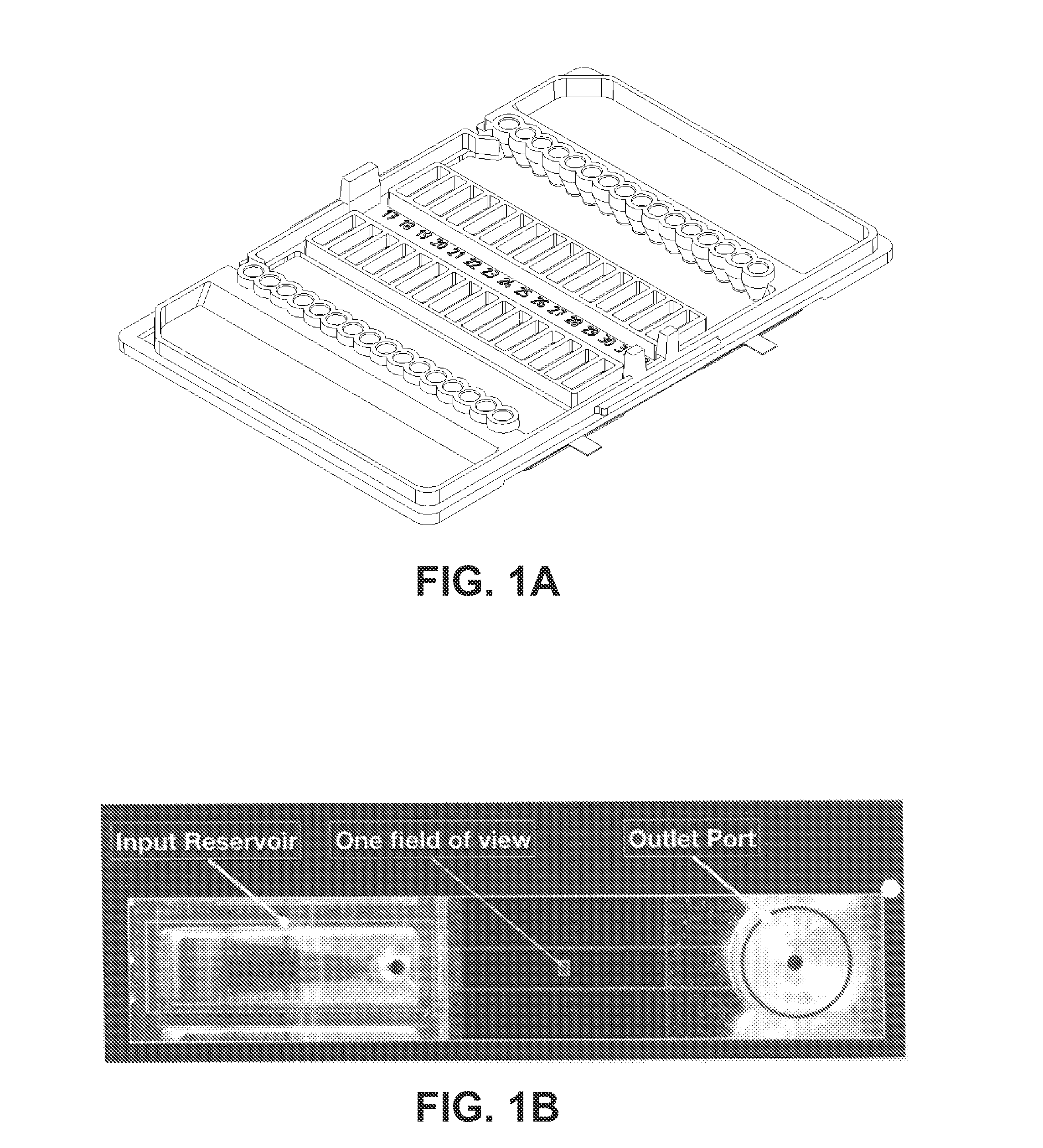
[0061]29 aliquots of 10 mL each were taken from two short-fill CPD blood bank bags. Each sample received isolate spikes to make nominal 5 CFU / mL of bacterial target species. These included 14 Staphylococcus aureus (SA), and 3 Pseudomonas aeruginosa (PA) plus 12 non-target Gram-negative bacilli. Each sample was diluted 4-fold to promote growth. 35° C. incubation for 4 hours was followed by centrifugation, with final resuspension in 1 mL of electrokinetic buffer. 16 flowcell channels in a multichannel fluidic cassette each received 20 μL of sample, followed by 5-minute electrokinetic concentration and surface capture. Liquid (40° C.) Mueller-Hinton agar with and without antimicrobials was then exchanged through each channel and gelled. Automated microscopy acquired images at 10-minute intervals for 3 hours. Image analysis detected clonal growth, identified SA and PA, and simultaneously performed resistance phenotype tests using 32 μg / mL amikacin, 8 μg / mL imipenem, 6 μg / mL cefox...
example 2
Effect of Protease Concentration Adjustment for Higher Cell Concentration Sample on Digestion on Sample Debris Concentration
Introduction
[0071]Enzyme concentration can be optimized for shorter or longer digests to accommodate different requirements for specimen analysis. High bacteria concentration specimens similar to those taken from positive blood culture bottles can be processed with higher concentration of protease for a shorter period of time to enable rapid analysis. For such an analysis, minimal growth is required since the bacteria are in sufficient concentration without growth. However, some debris removal may be desired to allow viewing of live bacteria using time-lapse darkfield or other imaging techniques. This experiment illustrates a method for detecting the bacteria within the debris field using fluorescence in-situ hybridization, using comparison of darkfield and fluorescent images to determine which objects are bacteria.
Methods
[0072]A mock blood culture specimen was...
example 3
Protease Digestion with Centrifugal Concentration of Bacteria and Conventional Slide-Based FISH Detection
[0074]Certain detection methods are not amenable to analysis of the specimen without further purification. Slide-based FISH staining is such an assay when the unpurified specimen is dried onto a slide as is typical with published methods on positive blood culture bottles. This example illustrates a method in which a single centrifugation step is sufficient to remove residual blood debris, purifying the bacteria for analysis, and allowing visualization of bacteria using only darkfield imaging.
[0075]A mock blood culture specimen was created by addition of 10 mL of blood to a BioMerieux BacT / Alert Standard Aerobic bottle. A 1.0 mL portion of this specimen was used for each condition. Each sample was treated with saponin to a final concentration of 4 mg / mL and SPS to a final concentration of 0.96 mg / mL. Samples were then spiked with E. coli bacteria to a final concentration of 1×10⁴ ...
PUM
| Property | Measurement | Unit |
|---|---|---|
| concentration | aaaaa | aaaaa |
| concentration | aaaaa | aaaaa |
| concentration | aaaaa | aaaaa |
Abstract
Description
Claims
Application Information
 - R&D
- Intellectual Property
- Life Sciences
- Materials
- Tech Scout
- Unparalleled Data Quality
- Higher Quality Content
- 60% Fewer Hallucinations
Browse by: Latest US Patents, China's latest patents, Technical Efficacy Thesaurus, Application Domain, Technology Topic, Popular Technical Reports.
© 2025 PatSnap. All rights reserved.Legal|Privacy policy|Modern Slavery Act Transparency Statement|Sitemap|About US| Contact US: help@patsnap.com