
Oncolytic vaccinia virus cancer therapy

a technology of vaccinia virus and cancer treatment, applied in the field of oncolytic vaccinia virus cancer therapy, poxviruses, etc., can solve the problems of affecting normal tissue cells, side effects of chemotherapy drugs, and few effective options for many common cancer types, so as to reduce cell viability and cell viability

Active Publication Date: 2012-11-01
SILLAJEN BIOTHERAPEUTICS
View PDF1 Cites 16 Cited by
  • Summary
  • Abstract
  • Description
  • Claims
  • Application Information

AI Technical Summary

Benefits of technology

[0037]FIGS. 6A-6C—Burst ratio of vaccinia strains, cytopathic effect, and systemic delivery of viral strains to tumors. (FIG. 6A) Burst ratio of vaccinia strains in tumor to normal cells. Different vaccinia strains were used to infect both primary normal cells (NHBE) and a tumor cell line (A2780) at a Multiplicity of infection (MOI) of 1.0 Plaque Forming Unit (PFU)/cell. Virus collected 48 h later was titered by plaque assay and the ratio of virus produced (per cell) in tumor to normal cells is represented. (FIG. 6B) Cytopathic effect produced by viral infection. Western Reserve, Adenovirus serotype 5 and Adenovirus strain d11520 (ONYX-015) (in some assays) were added to cell lines at ranges of MOIs (PFU/cell), and cell viability measured after 72 hours using MTS (Promega). The MOI of virus (PFU/cell) needed to reduce the cell viability to 50% of untreated control wells

Problems solved by technology

Currently, there are few effective options for the treatment of many common cancer types.
However, if the cancer has metastasized and is widespread, surgery is unlikely to result in a cure and an alternate approach must be taken.
However, a major side effect of chemotherapy drugs is that they also affect normal tissue cells, with the cells most likely to be affected being those that divide rapidly in some cases (e.g., bone marrow, gastrointestinal tract, reproductive system and hair follicles).
Other toxic side effects of chemotherapy drugs can include sores in the mouth, difficulty swallowing, dry mouth, nausea, diarrhea, vomiting, fatigue, bleeding, hair loss, and infection.
Unfortunately, the response typically is not sufficient to prevent most tumor growth.
Such methods, while showing some promise, have demonstrated limited success.
However, major limitations exist to this therapeutic approach.

Method used


Image

  • Oncolytic vaccinia virus cancer therapy
  • Oncolytic vaccinia virus cancer therapy
  • Oncolytic vaccinia virus cancer therapy

Examples


example 1

Material and Methods

[0278]Viruses and cell lines. The panel of wild type poxvirus strains (Wyeth, Western Reserve (WR), USSR, Tian Tan, Tash Kent, Patwadangar, Lister, King, 1HD-W, 1HD-J and Evans) was kindly provided by Dr Geoff Smith, Imperial College, London. Human Adenovirus serotype 5 (Ad5) was obtained from ATCC. The Viral growth factor (VGF) deleted strain of WR (vSC20) was kindly provided by Dr Bernie Moss, NIH. The thymidine kinase deleted strain of WR (vJS6) and the TK-, VGF-double deleted strain of WR (vvDD) are described in Puhlmann et al. (2000) and McCart et al. (2001). WR strain expressing firefly luciferase was kindly provided by Dr Gary Luker, (Uni Michigan).

[0279]Vaccinia strain JX-963 was constructed by recombination of a version of the pSC65 plasmid containing the E. coli gpt and human GM-CSF genes (under the control of the p7.5 and pSE / L promoters respectively) into the thymidine kinase gene of the vSC20 (VGF deleted) strain of WR. Further selection of white pla...

example 2

Rat Tumor Model

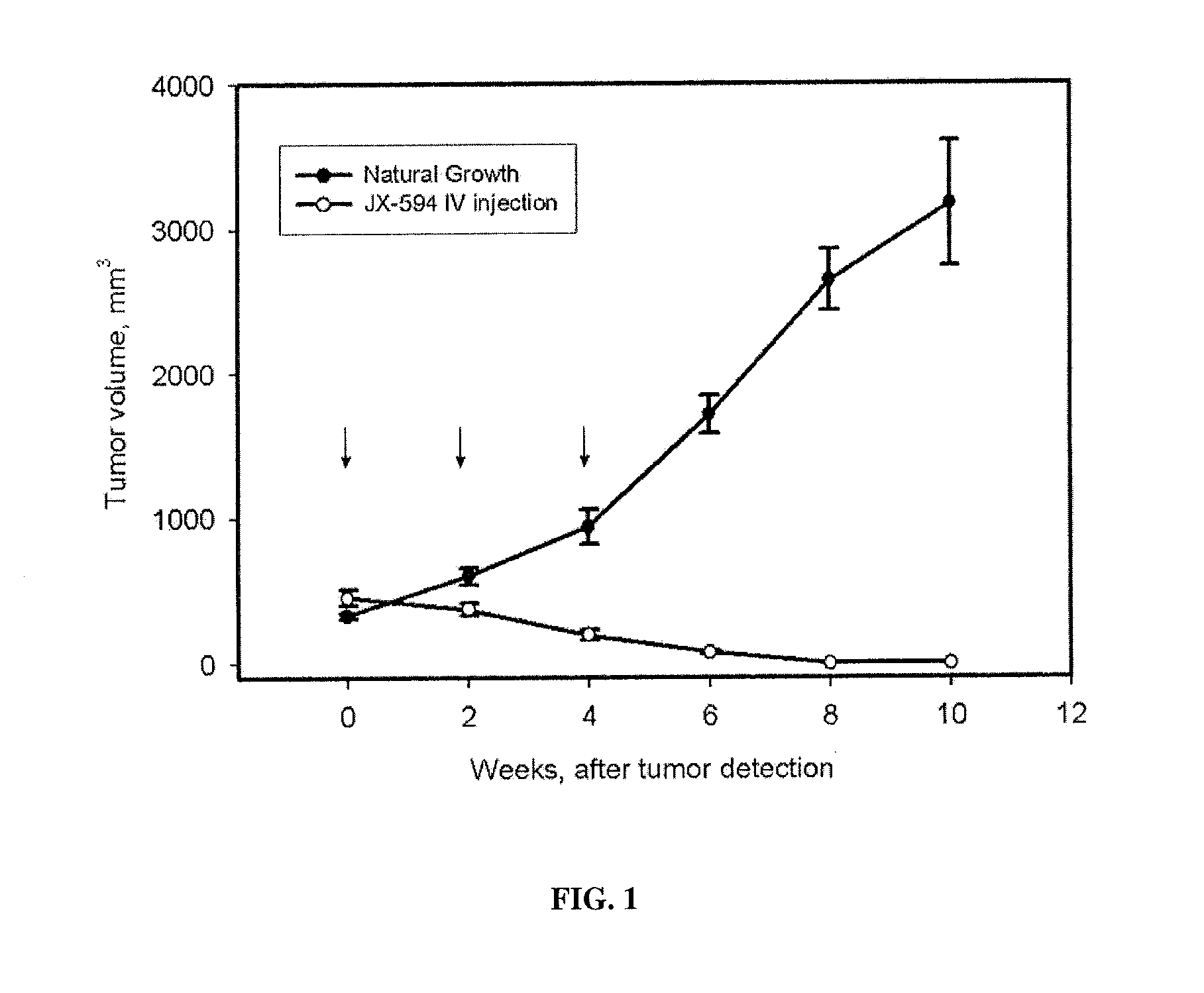
[0293]Rats (Sprague-Dawley, Males) were exposed to carcinogen (N-Nitrosomorpholine, NNM) in their drinking water (175 mg / L) for a period of 8 weeks, during which time liver cirrhosis developed, followed by in situ development of tumors (hepatocellular carcinoma or cholangiocarcinoma) within the liver between weeks 16-20 on average (model previously described in Oh et al., 2002). Tumor detection and evaluation was performed by an experienced ultrasonographer using ultrasound imaging. Tumor sizes were approximately 0.75-1.5 cm. in diameter at baseline immediately prior to treatment initiation; tumor volumes were not significantly different at baseline between the control and treatment groups (estimated mean volumes were 400-500 mm3) Control animals (n=17) received no treatment, whereas treated animals (n=6) received intravenous injections (via tail vein) with a poxvirus (Wyeth strain; thymidine kinase gene deletion present) expressing human GM-CSF from a synthetic early...

example 3

Rabbit VX2 Tumor Model

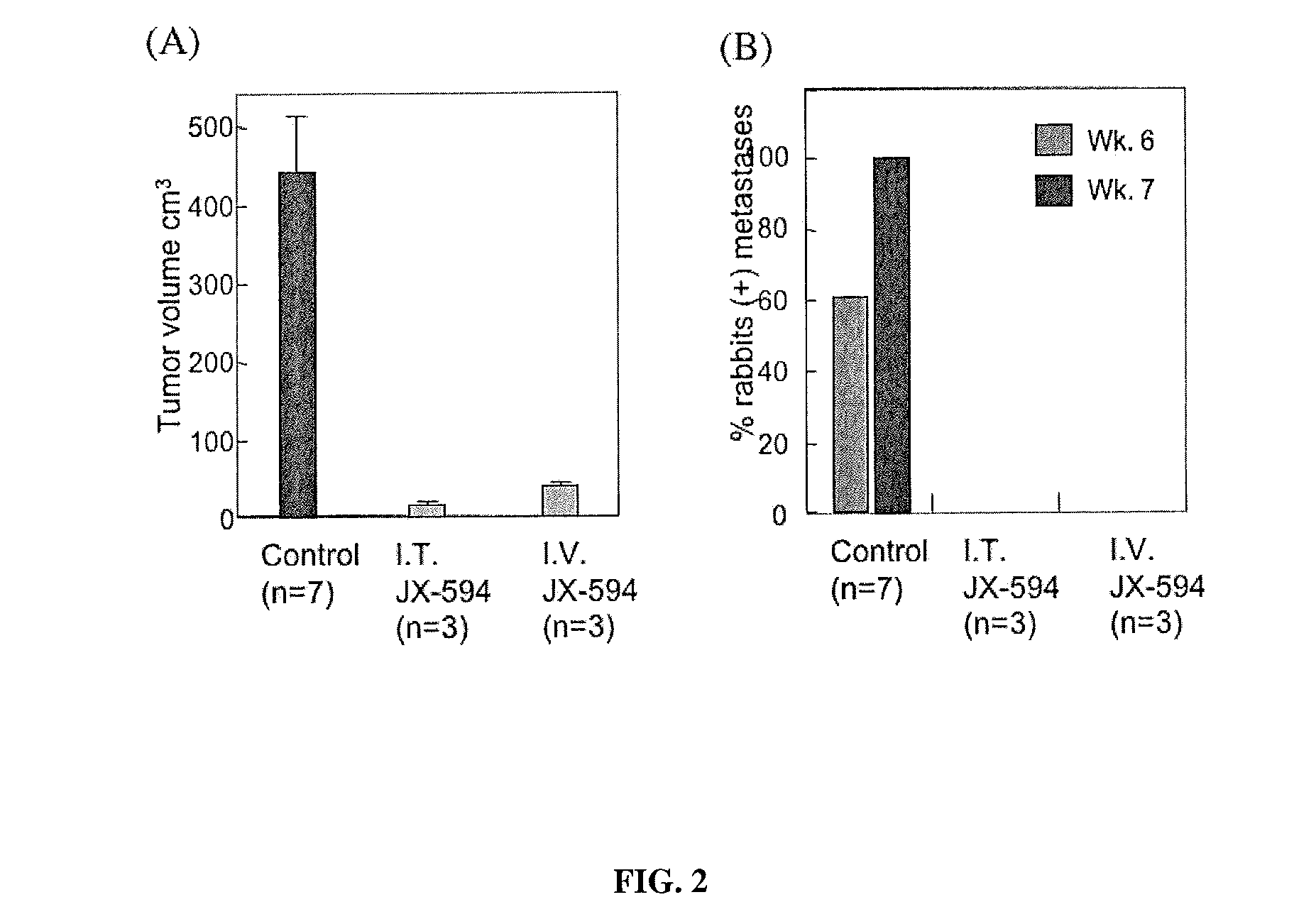
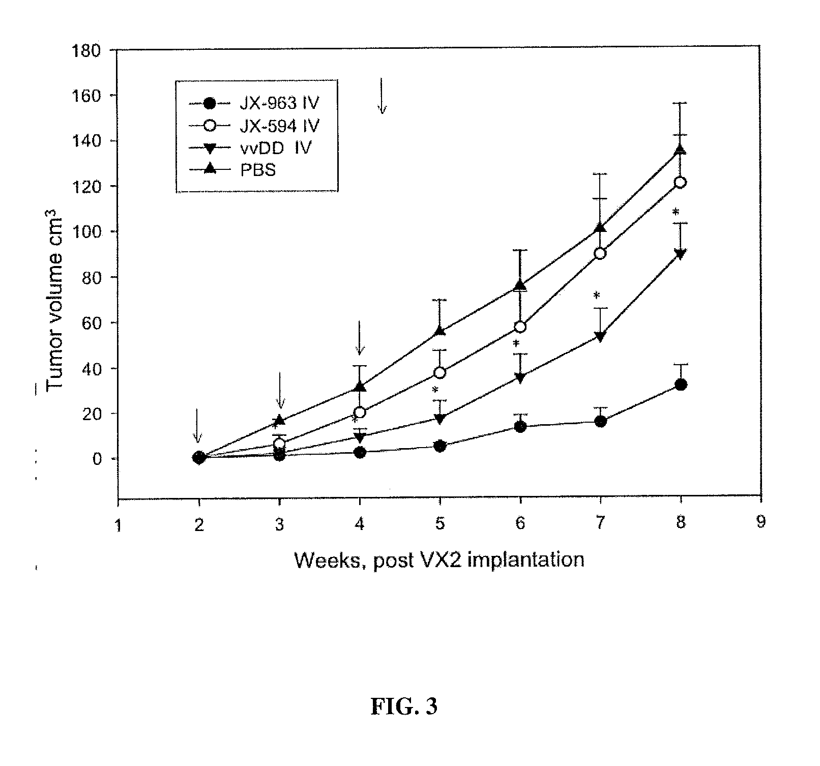
[0295]A study was performed in a VX2 rabbit carcinoma model (as described in Paeng et al., 2003). Rabbit was selected as a species because human GM-CSF was previously demonstrated to have significant biological activity in rabbits (in contrast to mice). VX2 tumors were grown in muscle of New Zealand white rabbits and cells from a 1-2 mm3 fragment of tumor were dissociated, resuspended in 0.1 ml normal saline and were injected beneath the liver capsule (21 gauge needle; injection site covered with surgical patch with a purse-string tie) and allowed to grow for 14 days until primary tumors were established (mean diameter, 1.5-2.0 cm; est. volume 2-4 cm3). VX2 cells were demonstrated to be infectable by vaccinia poxvirus ex vivo in a standard burst assay. Tumor sizes were monitored over time by CT scanning and by ultrasound. Over the following seven weeks, control (untreated) animals (n=18) developed tumor progression within the liver, with estimated mean tumor vo...


PUM

PropertyMeasurementUnit
Fractionaaaaaaaaaa
Fractionaaaaaaaaaa
Timeaaaaaaaaaa

Abstract

Embodiments of the invention are directed methods that include a thymidine kinase deficient vaccinia virus. The methods include administering the vaccinia virus at increased viral concentrations. Further aspects of the invention include methods for inducing oncolysis or collapse of tumor vasculature in a subject having a tumor comprising administering to a subject at least 1×10⁸ infectious viral particles of a TK-deficient, GM-CSF-expressing, replication-competent vaccinia virus vector sufficient to induce oncolysis of cells in the tumor.

Description

I. CROSS-REFERENCE TO RELATED APPLICATIONS[0001]This application is a continuation-in-part of U.S. patent application Ser. No. 12 / 531,353, filed May 11, 2010 which is the U.S. national stage of International Application PCT / US08 / 57257, filed Mar. 17, 2008 and which claims the benefit of U.S. Provisional Patent Application No. 60 / 894,932, filed Mar. 15, 2007 and is a continuation-in-part of U.S. patent application Ser. No. 11 / 470,951, filed Sep. 7, 2006 which claims the benefit of U.S. Provisional Application No. 60 / 714,979, filed Sep. 7, 2005, the disclosures of which are incorporated herein by reference in their entireties.II. FIELD OF THE INVENTION[0002]The present invention relates generally to the fields of oncology and virology. More particularly, it concerns poxviruses, specifically including oncolytic vaccinia viruses suitable for the treatment of cancer.III. BACKGROUND OF THE INVENTION[0003]Normal tissue homeostasis is a highly regulated process of cell proliferation and cel...

Claims


Application Information

Patent Timeline
IPC IPC(8): A61K48/00A61P35/00
CPCA61K38/193A61K35/768C12N2710/24132A61K2300/00A61K38/191A61K38/2013A61K38/208A61P35/00A61K9/0019A61K45/06A61K48/00A61K48/0066A61K48/0075A61N5/10
Inventor KIRN, DAVID
Owner SILLAJEN BIOTHERAPEUTICS
Learn More