Stabilization of RNA in intact cells within a blood sample

Inactive Publication Date: 2011-05-12
STRECK INC
View PDF105 Cites 96 Cited by
  • Summary
  • Abstract
  • Description
  • Claims
  • Application Information

AI Technical Summary

Benefits of technology

[0008]The present invention provides a unique approach to the preservation, isolation, and analysis of nucleic acids. One aspect of the invention involves use of a unique protective agent composition, which includes at least one preservative agent which may include a formaldehyde donor. The nucleic acid may be DNA, RNA, or any combination thereof. The samples from which the nucleic acids may be isolated include any blood sample. The nucleic acids may be cellular nucleic acids (e.g., nucleic acids that are located within cells in vivo as opposed to cell-free nucleic acids found outside of cells in vivo). The method disclosed herein allows for efficient preservation and isolation of cellular nucleic acids while avoiding contamination with undesirable globin mRNA and cell-free nucleic acids that originate at extra-cellular locations in vivo (as compared to cellular RNA that becomes cell-free RNA due to cell metabolism and cell lysis post-blood draw).
[0009]In a first aspect, the present invention contemplates a screening method for the identification of a disease state. The screening method includes the step of contacting a drawn blood sample that includes a plurality of blood cells with a protective agent in an amount and for a time sufficient so that RNA synthesis is inhibited for at least two hours. The contact time with the protective agent and amount of protective agent used may also be sufficient so that the blood cells of the drawn blood sample are fixed to substantially prevent contamination of the cellular RNA with cell-free RNA or globin mRNA. Further, any cellular RNA that is within the blood cells at the time of the blood draw may be substantially preserved to freeze the mRNA expression pattern of the blood cells substantially as of the time of the blood draw (e.g., no longer than 10 minutes post-blood draw or even no longer than 5 minutes post-blood draw). The screening method may also include the step of isolating white blood cells from the whole blood by lysing the red blood cells and isolating the white blood cells. The isolated white blood cells may then be treated to extract cellular RNA from the isolated white blood cells.
[0016]The screening method of the present invention provides a process for preserving a blood sample containing diagnostically useful RNA so that the RNA can be effectively isolated and tested. The preservation technique results in short term inhibition of metabolism (i.e., RNA synthesis), fixation of the cellular RNA within the blood cells to freeze the mRNA expression pattern of the blood cells, protection of the RNA that is in the blood cells from nucleases and proteases, prevention of unwanted interference from globin RNA and cell-free RNA, and fixation of blood cells to prevent the loss of cellular RNA leaked from blood cells during transportation or storage of blood specimens.

Problems solved by technology

However, such tissue biopsies may not be readily available and sampling often requires highly invasive procedures of the human body.
Some issues inherent to gene profiling in blood cells have the significant potential to influence data interpretation.
One such issue is related to the handling of blood samples ex vivo prior to the extraction of mRNA.
Expression levels for many genes in blood cells can be adversely effected by ex vivo incubation because of the metabolic stress brought on by the lack of oxygen and glucose sources.
Another issue is related to the widely used method for obtaining total RNA from blood cells, which includes density-gradient centrifugation to isolate white blood cells.
This method needs equipment beyond what is available in the typical clinical setting and may require shipment to another site for the necessary processing.
This causes delays in sample processing and may create significant changes in gene expression profiles.
However, there are some inherent disadvantages in these blood collection devices.
Since all blood cells are lysed at the point of collection, there is a significant introduction of α- and β-globin mRNA that is released from reticulocytes, which interferes with microarray and real time PCR detection methodologies.
Another significant disadvantage of these devices is the inability to utilize molecular beacon technology, where it is imperative to have intact cells so that one can visualize the gene-specific fluorescence staining by histology or by flow cytometry.
As a result, additional costs are incurred and there is increased time required for sample preparation.
While the use of formaldehyde-donor preservatives for the fixation of cells and tissues is known, formaldehyde-donors have been shown to be less effective in completely inhibiting cell metabolism at least during the first 24 hours of post phlebotomy.
Further, the use of formaldehyde-donor preservatives alone have not shown to stabilize mRNA expression patterns in cells within a blood sample post phlebotomy.

Method used


Image

  • Stabilization of RNA in intact cells within a blood sample
  • Stabilization of RNA in intact cells within a blood sample
  • Stabilization of RNA in intact cells within a blood sample

Examples


example 1

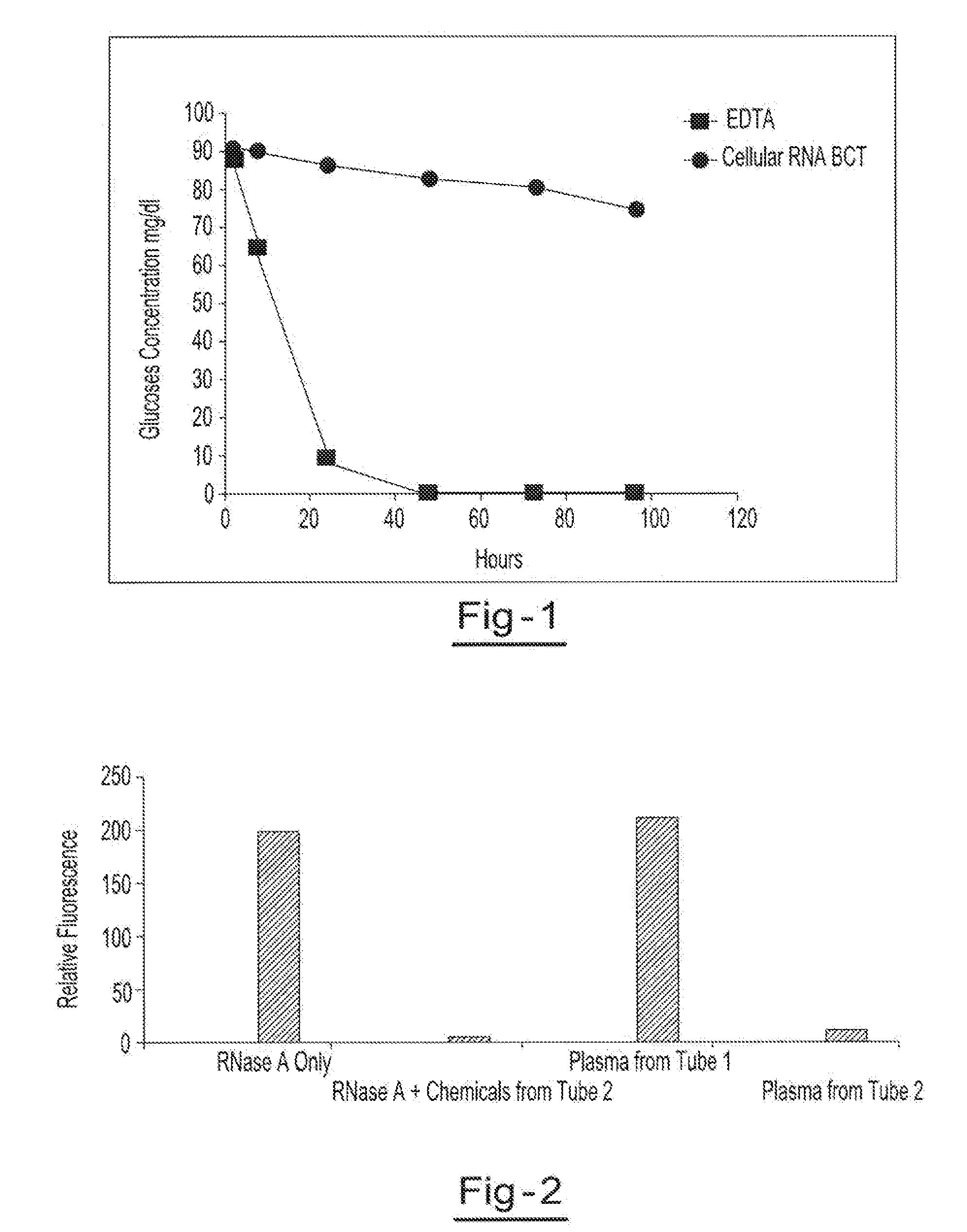
[0052]Blood samples from the same donor are drawn into two separate blood collection tubes (tube 1 (EDTA tube) and tube 2 (RNA BCT tube)). Tube 1 contains only EDTA. Tube 2 contains DU, EDTA, ATA, glyceraldehyde and sodium fluoride. Both tubes are stored at room temperature and 1 ml aliquots of blood are removed from each tube at hours 1.5, 8, 24, 48, 72 and 96. The blood glucose levels of each sample are measured using a YSI blood glucose meter available from YSI Life Sciences (Yellow Springs, Ohio). The blood glucose concentration of samples from tube 2 were the only samples that maintained relatively consistent glucose levels over the test period, indicating that the combination of EDTA, DU, ATA, glyceraldehyde and sodium fluoride provided reduced levels of cell metabolism. The results of this example are shown in a graphic format at FIG. 1.

example 2

[0053]Blood samples from the same donor are drawn into two separate blood collection tubes (tube 1 and tube 2). Tube 1 contains EDTA. Tube 2 contains DU, EDTA, ATA, glyceraldehyde and sodium fluoride. Both tubes are stored for 2 h at room temperature before plasma was separated. RNase activity of plasma from tube 1 and tube 2 was measured using a commercially available RNase activity detection kit, RNaseAlert® Lab Test Kit (Applied Biosystems, Foster City, Calif.). Two additional control experiments were also carried out with purified RNase A enzyme alone and RNase A treated with chemical mixture present in tube 2. RNase activity is presented as relative fluorescence. Results of this example are illustrated in a graphic format at FIG. 2.

example 3

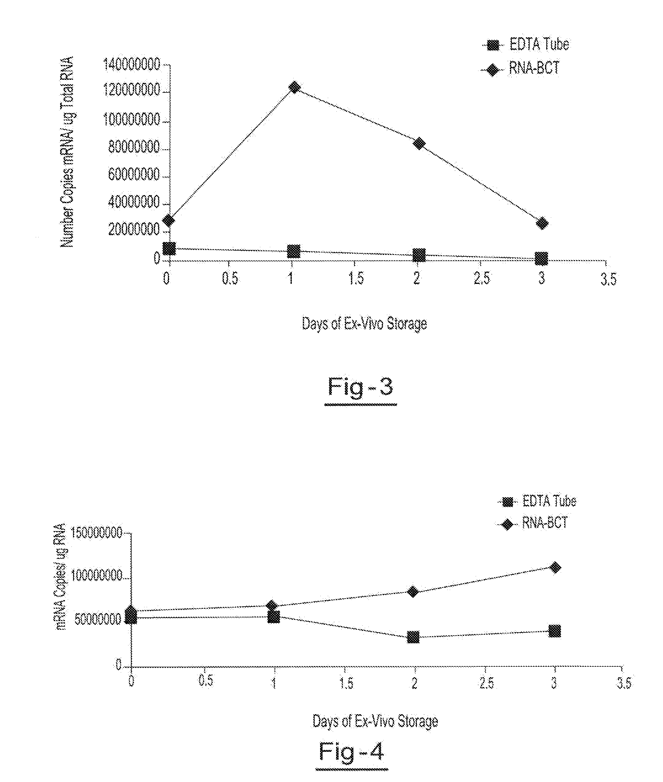
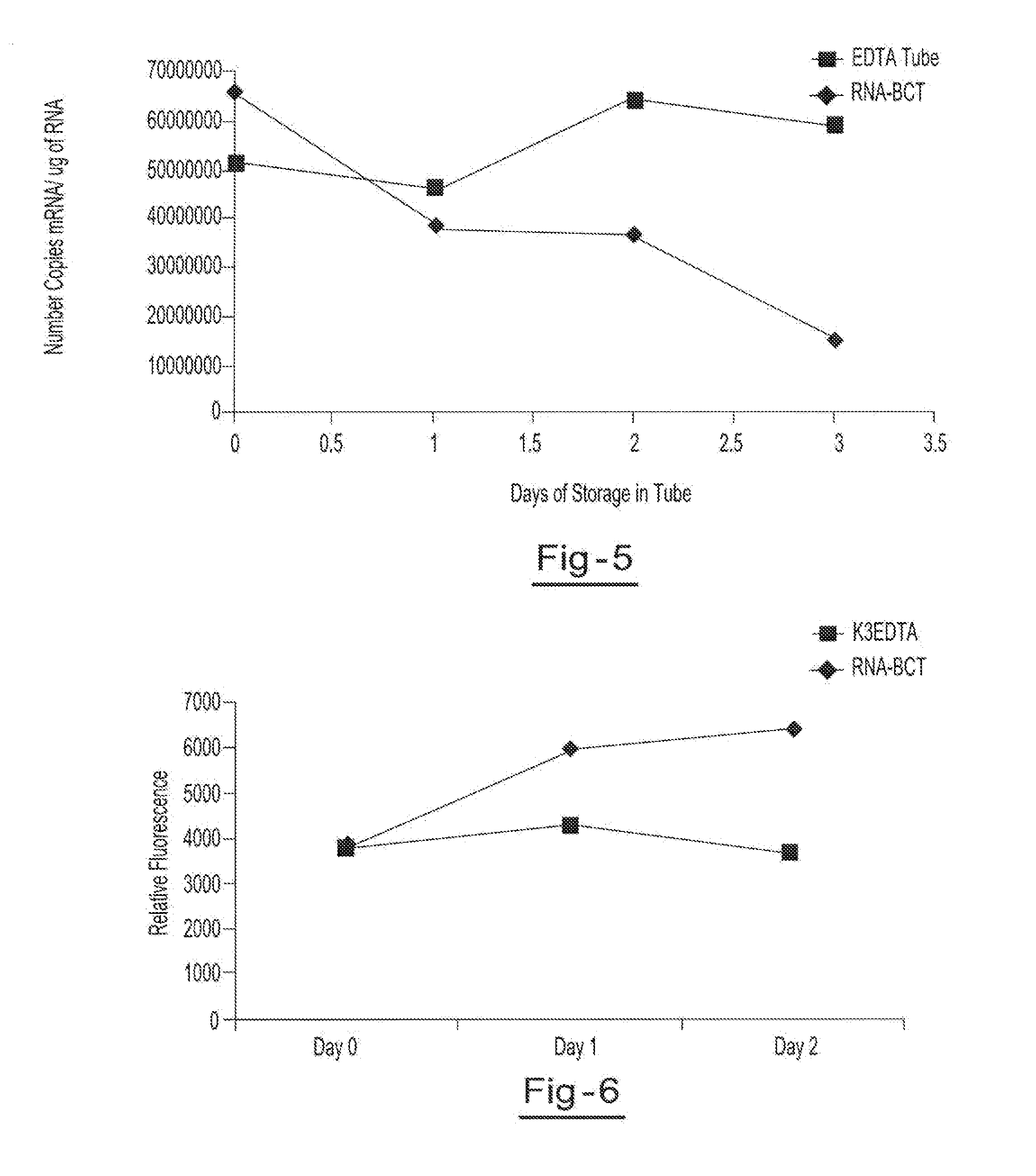
[0054]Two blood samples from the same donor are drawn into two separate blood collection tubes, tube A (RNA BCT) and tube B (EDTA). Tube A contains DU, EDTA, ATA, glyceraldehyde and sodium fluoride. Tube B contains only EDTA. Both tubes are stored at room temperature and 5 ml aliquots of blood are removed from each tube on day 0, day 1, day 2, and day 3 and plasma is separated. All samples are centrifuged at 800 g for 10 minutes at room temperature to separate the plasma. The plasma is then transferred into new tubes and centrifuged at 1500 g for 10 minutes at room temperature. Free circulating RNA is purified using the QIAamp circulating nucleic acid kit available from Qiagen Inc. (Valencia, Calif.). RNA is extracted from each plasma sample. The samples are then amplified by Real Time PCR (using TaqMan® RT PCR reagents available from Applied Biosystems, Foster City, Calif.) to identify the c-fos mRNA copy number per ml of plasma. Results showed a consistent copy number of c-fos mRN...


PUM

PropertyMeasurementUnit
Temperatureaaaaaaaaaa
Timeaaaaaaaaaa

Abstract

A method for preserving and processing nucleic acids located within a blood sample is disclosed, wherein a blood sample containing nucleic acids is treated to reduce both blood cell lysis and nuclease activity within the blood sample. The treatment of the sample aids in increasing the integrity and amount of cellular nucleic acids that can be identified and tested while avoiding contamination of the isolated nucleic acids with cell-free nucleic acids.

Description

CLAIM OF PRIORITY[0001]This application claims the benefit of the filing date of U.S. Provisional Application Ser. No. 61 / 259,363, filed Nov. 9, 2009, the entirety of the contents of this application being hereby expressly incorporated by reference.FIELD OF THE INVENTION[0002]This invention relates to the identification and isolation of nucleic acids in blood samples and more particularly to the stabilization of cellular RNA within a blood sample.BACKGROUND OF THE INVENTION[0003]Messenger RNA (mRNA) in a cell is a snapshot of the real time activity of its genome, depicting what genes are expressed and to what extent. Profiling of cellular mRNA expression patterns is typically done by use of microarrays, quantitative reverse transcriptase real time PCR and molecular beacons. Profiling of cellular mRNA is becoming important in disease diagnosis, prognosis and in clinical trials for biomarker discovery. Such cellular mRNA profiling has relied on tumor and other biopsy material from aff...

Claims


Application Information

Patent Timeline
IPC IPC(8): C12Q1/68C12N5/00
CPCC12N15/1003C12Q1/6806C12Q2527/127C12Q2527/137C12Q2527/101
Inventor RYAN, WAYNE L.FERNANDO, M. ROHAN
Owner STRECK INC
Why Eureka
Try Eureka