
RF coil for use in magnetic resonance imaging in integrated spect and mr imaging

a magnetic resonance imaging and integrated spect technology, applied in the field of magnetic resonance imaging methodologies, can solve the problems of inability to perform temporal coregistration in the time domain of combined dynamic studies using both imaging techniques simultaneously, inability to use conventional motors in high magnetic fields used in magnetic resonance imaging (mri), and inability to accurately perform other less rigid body parts. , to achieve the effect of improving image accuracy, optimizing wiener filtering, and improving image accuracy

Inactive Publication Date: 2011-03-17
RGT UNIV OF CALIFORNIA
View PDF6 Cites 2 Cited by
  • Summary
  • Abstract
  • Description
  • Claims
  • Application Information

AI Technical Summary

Benefits of technology

[0013]Single-photon emission computed tomography (SPECT) can provide specific functional information while magnetic resonance imaging (MRI) can provide high-spatial resolution anatomical information as well as complementary functional information. In this disclosure, we utilized a dual modality SPECT / MRI (MRSPECT) system to investigate the integration of SPECT and MRI for improved image accuracy. The MRSPECT system consisted of a cadmium-zinc-telluride (CZT) nuclear radiation detector interfaced with a specialized radiofrequency (RF) coil that was placed within a whole-body 4 T MRI system. The importance of proper corrections for nonuniform detector sensitivity and Lorentz force effects was demonstrated. MRI data were utilized for attenuation correction (AC) of the nuclear projection data and optimized Wiener filtering of the SPECT reconstruction for improved image accuracy. Finally, simultaneous dual-imaging of a nude mouse was performed to demonstrate the utility of coregistration for accurate localization of a radioactive source.
[0022]The step of performing SPECT imaging includes the step of performing SPECT imaging using the collimated nuclear radiation detector which includes a parallel-hole collimator and the step of simultaneously performing MR imaging includes the step of simultaneously performing MR imaging using a RF birdcage coil into which the parallel-hole collimator is arranged and configured to avoid attenuation of radiation by the MRI coil and to allow for adjustment of the distance from the collimator to the object being imaged.

Problems solved by technology

High magnetic fields used in magnetic resonance imaging (MRI) do not allow the employment of conventional motors due to various incompatibility issues.
Although such co-registration approaches work on rigid organs such as the head it is impossible to perform them accurately on other less rigid body parts such as the abdomen.
Even if the spatial co-registration could be achieved, temporal coregistration cannot be done in the time domain for combined dynamic studies using both imaging techniques simultaneously.
Another difficulty that arises when the detector has to undergo rotational scanning for collecting different angular views is the availability of a motor that can operate close to the magnet bore without affecting the MR image quality or endangering the patient.
Although the motor is MR compatible the amount of torque that can be generated limits its use to low torque applications that are more along the line of medical robotics.
The problem of providing a practical motor for use in an MRI environment is not completely solved for applications that require a large torque to rotate heavy components such as nuclear detectors with collimators.
None of the solutions described above satisfactorily resolve this need.
However, the relatively poor spatial resolution of radionuclide techniques can make unambiguous localization of the probes extremely difficult, especially when the images lack significant anatomical detail for reference.
Limited spatial resolution can also hamper quantification of the probe concentration, especially when localized in small volumes.
While the integration of SPECT and MRI offers numerous advantages and new opportunities, it also presents many technological challenges.
Due to these challenges, the development of a combined SPECT and MRI system (henceforth called MRSPECT) is in its infancy, and a very limited amount of research has been reported to date.
While they investigated the effects of the SPECT and MRI components on each other, they confined their disclosure to the use of a 57Co point source for SPECT imaging and were not able to acquire simultaneous SPECT and MRI experimental data.

Method used


Image

  • RF coil for use in magnetic resonance imaging in integrated spect and mr imaging
  • RF coil for use in magnetic resonance imaging in integrated spect and mr imaging
  • RF coil for use in magnetic resonance imaging in integrated spect and mr imaging

Examples


Embodiment Construction

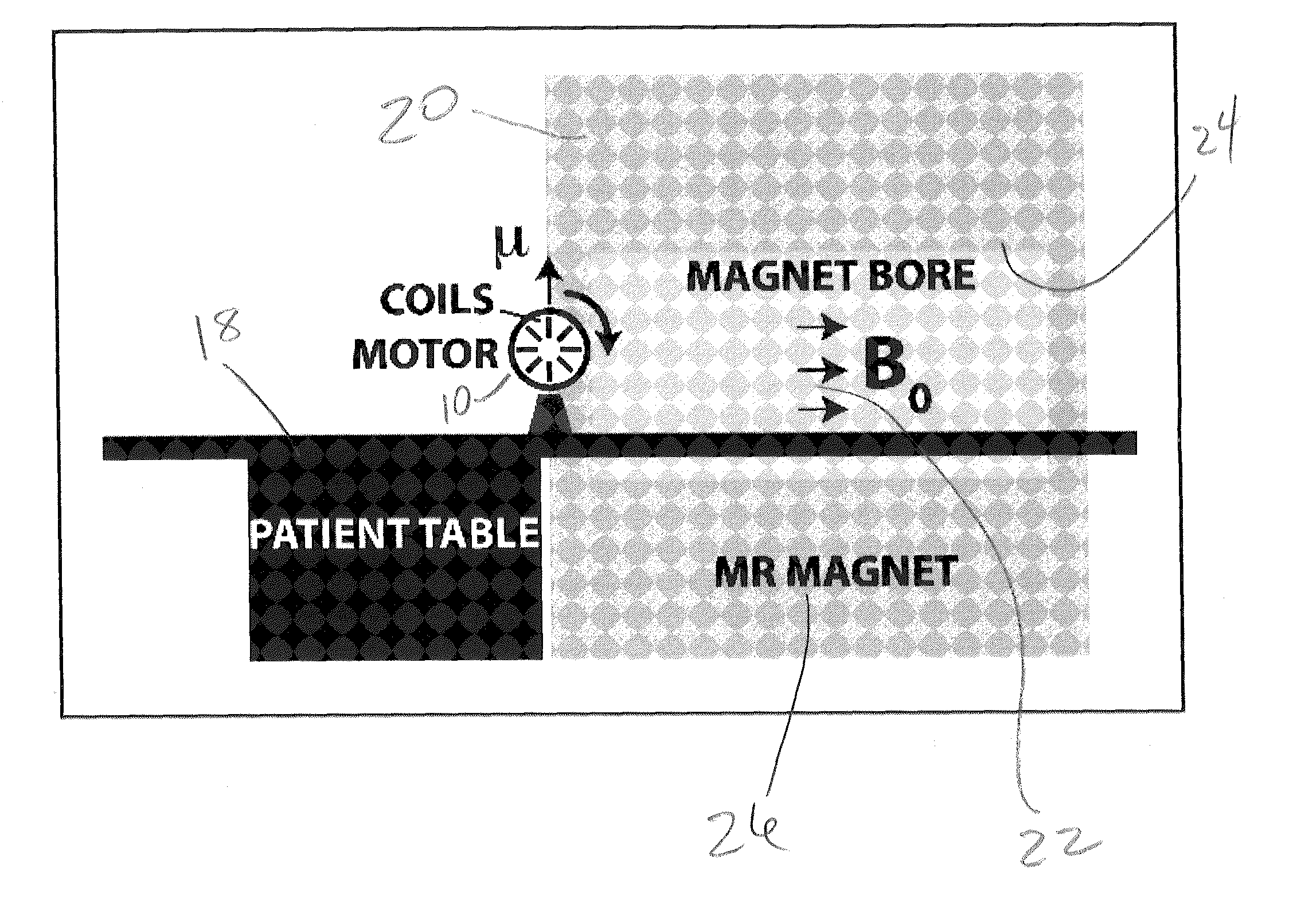
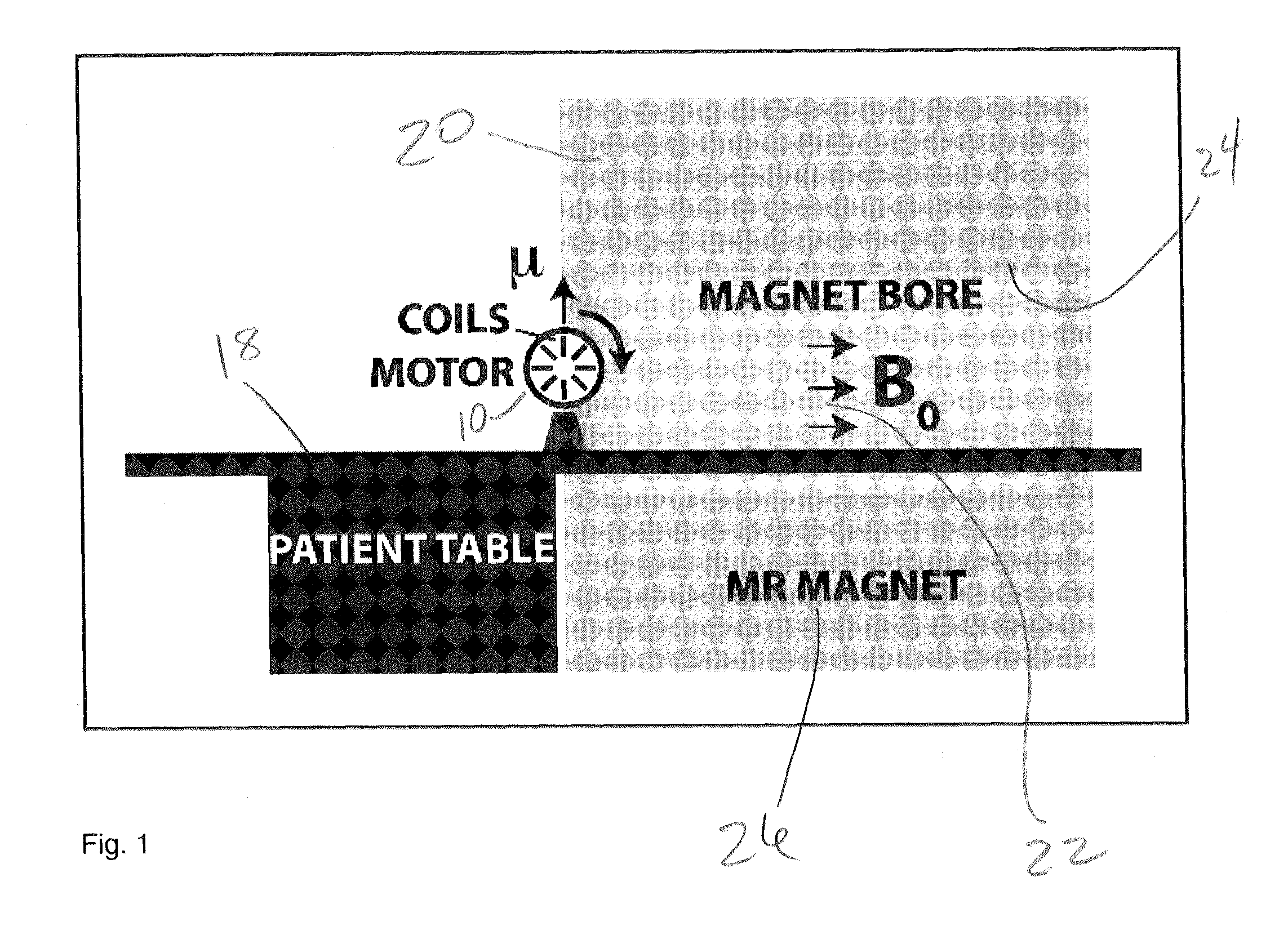
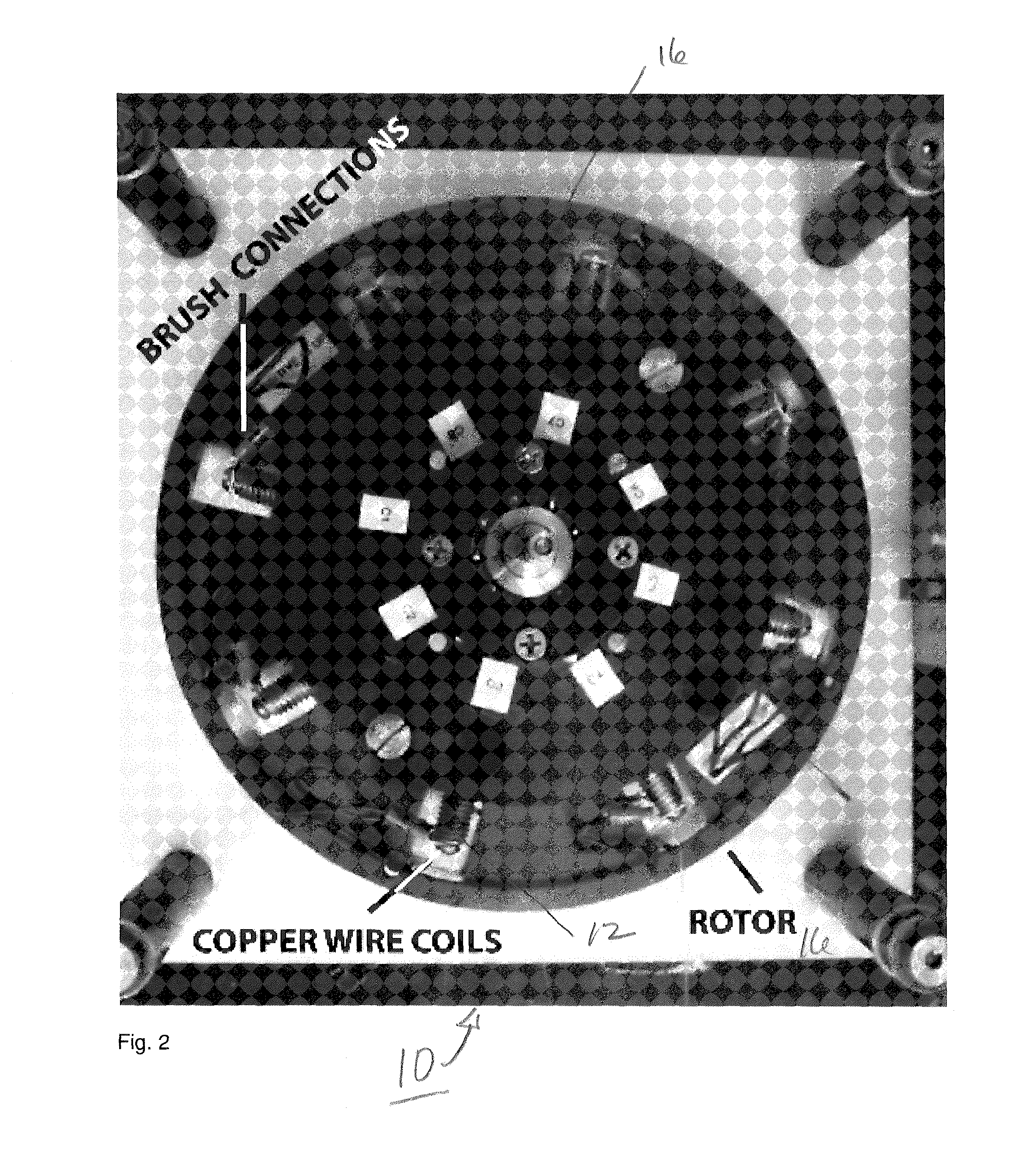
[0038]An illustration of a prototype motor 10 and the experimental set-up is shown diagrammatically in FIG. 1. The motor assembly 10 is constructed with MR compatible plastic material and screws and bolts made out of brass. The motor assembly 10 shown in the photograph of FIG. 2 includes eight MR compatible copper coils 12 wound onto a plastic bobbin (not shown) placed radially within the rotor body 16 and separated by 45 degrees from each other as shown in FIG. 2. The outer diameter of the main rotor body 16 is 20.3 cm. Directional rotary motion is achieved by passing electric current through opposing coils 12 by means of a pair of dedicated brushes (not shown) under computer control and hence creating a torque due to the interaction of the magnetic field induced in coils 12 carrying current with the magnetic field of the MR system as shown in FIG. 1.

[0039]As well known from basic electromagnetics the torque is linearly related to the local magnetic field of the MR system and the c...


PUM


Abstract

An apparatus and method for performing dual modality SPECT / MRI imaging on an object in combination with a whole-body MRI system includes a collimated nuclear radiation detector for receiving radiation from the object, and a radiofrequency MRI coil enveloping the object and interfaced with the collimated nuclear radiation detector. The MRI coil and collimated detector are arranged and configured for disposition within the whole-body MRI system.

Description

RELATED APPLICATIONS[0001]The present application is related to U.S. Provisional Patent Application Ser. No. 61 / 171,238 filed on Apr. 21, 2009, which is incorporated herein by reference and to which priority is claimed pursuant to 35 USC 119.BACKGROUND OF THE INVENTION[0002]1. Field of the Invention[0003]The invention relates to the field of magnetic resonance imaging methodologies in which dual imaging modes are employed and to electric motors used in the MRI environment.[0004]2. Description of the Prior Art[0005]High magnetic fields used in magnetic resonance imaging (MRI) do not allow the employment of conventional motors due to various incompatibility issues. Magnetic resonance imaging (MRI) is a well-accepted tomographic technique that can be used to acquire high-resolution anatomical three dimensional images of the human body. Although MRI could also provide functional or metabolic information its sensitivity is low compared to other medical imaging techniques such as positron...

Claims


Application Information

Patent Timeline
Patent Type & Authority Applications(United States)
IPC IPC(8): A61B5/055
CPCG01R33/481
Inventor ROECK, WERNER N.NALCIOGLU, ORHANHA, SEUNG-HOON
Owner RGT UNIV OF CALIFORNIA
Learn More